MRI Week 4 - Brain/Orbits/Base of Skull w/ Pulse Sequences
 Call Kai
 Learn
Learn Practice Test
 Spaced Repetition
 Match
Match Flashcards
 Knowt Play
1/15

There's no tags or description
Looks like no tags are added yet.
Name | Mastery | Learn | Test | Matching | Spaced | Call with Kai | Chat |
|---|
No analytics yet
Send a link to your students to track their progress
16 Terms
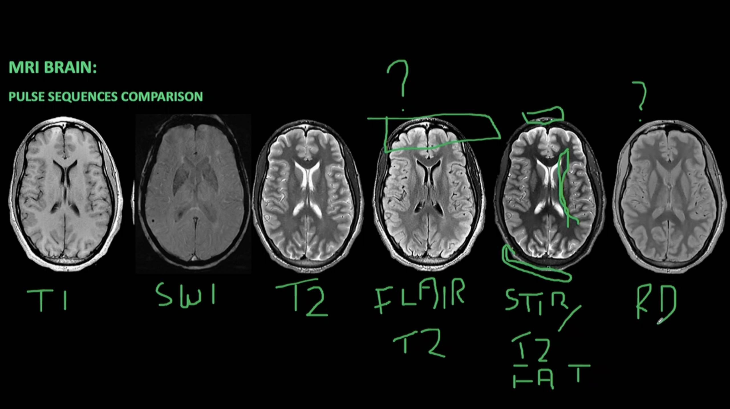
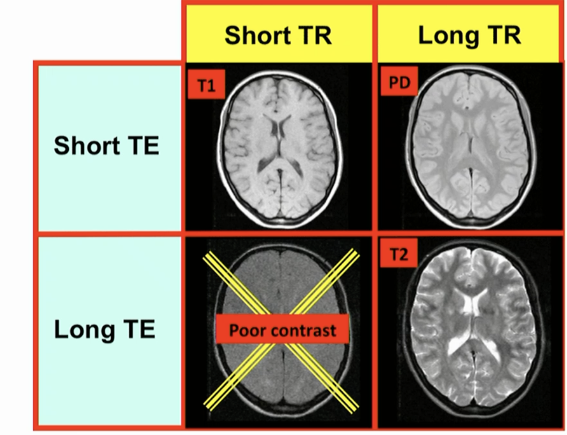
Different Sequences
T1 + T2 + PD + FATSAT + INVERSION RECOVERY (IR) + STIR (Short Tau Inversion Recovery)
Spin Echo + Fast Spin Echo + Gradient Echo
T1
White matter = White
Grey Matter = Grey
CSF = DARK (Not the best to Differentiate)
Additional:
Use Gadolinium (contrast)
We can see another part of the tumour demonstrates contrast enhancement
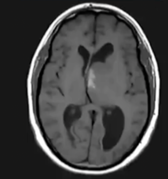
T1
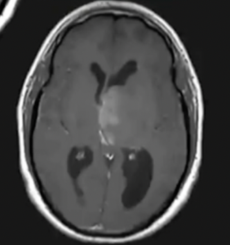
T1 Post Gad
T2
White Matter = Darker
Grey Matter = Brighter
CSF = Bright (Not the best to Differentiate)
Additional:
FLAIR (Fluid Attenuated Inversion Recovery) --> Dark CSF
Most common modifier for T2
Same Tumour stands out more against white matter
White matter = STILL Darker
Grey Matter = STILL Brighter
Even though CSF is Hypointense
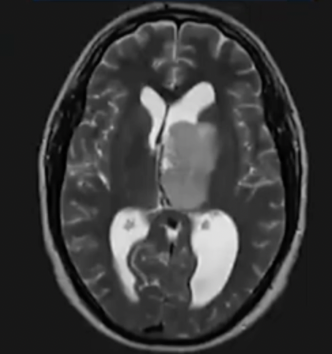
T2
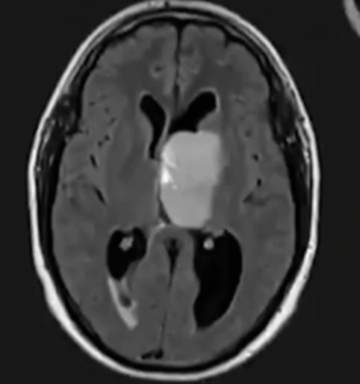
T2 FLAIR
Proton Density
Intermediate Sequence
Used to be used for White matter lesions
Taken over by Flair imaging for brains
Mainly used for MSK
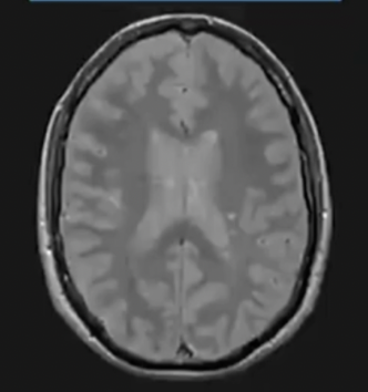
Proton Density
SWI
Susceptibility Weighted Imaging
Good for stagnant Blood
More sensitive
Gradient Echo
White Matter = Darker
Grey Matter = Brighter
CSF = Bright (Not the best to differentiate)
used for:
bleeds
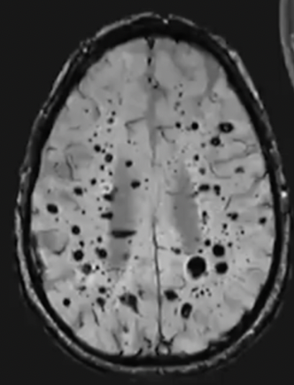
SWI
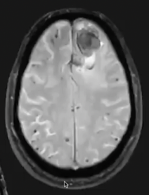
Gradient Echo