Exam 1: RBC/Anemia
1/59
There's no tags or description
Looks like no tags are added yet.
Name | Mastery | Learn | Test | Matching | Spaced |
|---|
No study sessions yet.
60 Terms
platelets
(thrombocytes) clotting factors
anemia
oxygen carrying capacity of blood is reduced
factors that could indicate anemia
-decreased hemoglobin
-decreased hematocrit
-decreased RBC
anemia can be caused by
increased destruction or RBC
decreased production of RBC
excessive blood loss
aplastic anemia
caused by aplasia of bone marrow or its destruction by chemical agents or other physical factors
aplasia
failure of an organ or tissue to develop normally
-penia
decrease in number of cells
pancytopenia
deficiency of all types of blood cells
-cytosis or -philia
increase in number of cells
hemolysis
destruction of RBC in vivo or in vitro
hemolytic anemia
caused by increased desctruction or shortened life span of RBC
intracorpuscular vs extracorpuscular
intra: inside causing factor
extra: outside causing factor
hematopoiesis
process of blood cell production and development
a decrease in oxygen leads to an _____ in blood cell production
increase
EPO
comes from the kidney; signals for RBC production; stimulated from low O2
extramedullary hematopoiesis
hematopoiesis outside bone marrow (e.g., spleen); kidney usually the problem
where does hematopoiesis occur
in utero: in the yolk sac
after birth: bone marrow
long bones are the site of hematopoiesis until
20 years of age
After 20 years of age hematopoiesis
occurs more in the flat bones, vertebrae, pelvis
stem cells
pluripotent; high self-renewal capacity
2 types of wbcs
Granulocytes
Agranulocytes
How long does it take reticulocytes to mature?
erythrocytes mature in 1-2 days in the peripheral circulation
what two RBCs are in the peripheral blood pool
mature erythrocytes
polychromatic erythrocyte (reticulocyte)
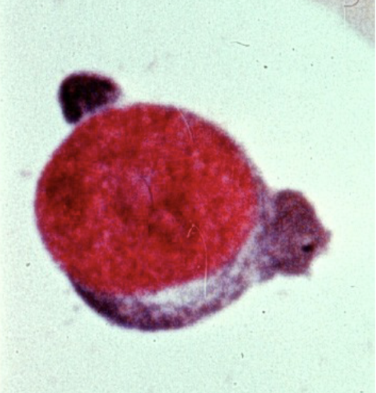
Pronormoblast (rubriblast)
largest precursor cell; large nucleus!!
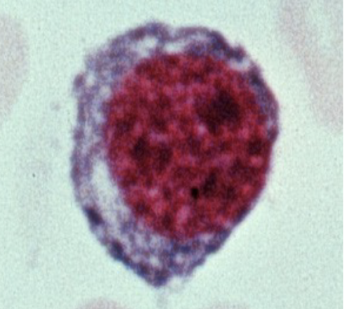
Basophilic Normoblast (Prorubricyte)
smaller nuclei; slightly more cytoplasm
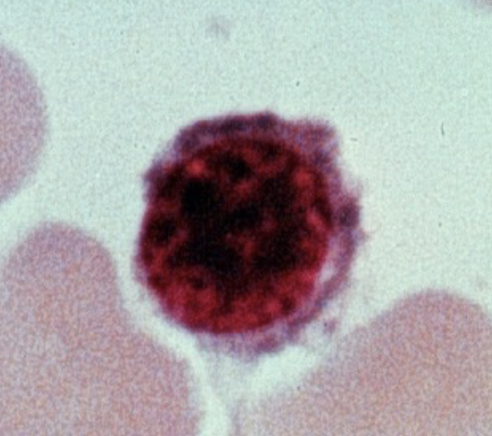
Polychromoatophilic Normoblast (Rubricyte)
cytoplasm pink, darker middle, Hgb is evident; eject nucleus
mature erythorcytes
flat middle
RBC feedback control
tissue hypoxia -> absence of RBC -> bodily need -> signal → kidney→ EPO→ bone→ RBCs
life span of RBC
120 days
what tube is used for a peripheral blood smear
tubes w/ anticoagulant EDTA
benefit of Wrights stained smear
cells are easier to count
fishbone
shorthand way of displaying labs and normal ranges
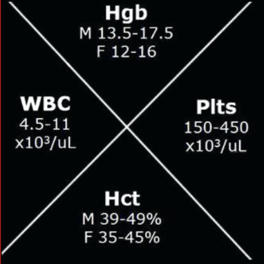
MCV
size of cell (80-100)
MCH
weight of Hgb per cell
MCHC
concentration of Hgb per cell
RDW
range of size of cells (immature vs mature)
T/F anemia is an isolated diagnosis
F : there is always a cause of anemia
Types of anemia
microcytic (<80)
normocytic (80-100)
macrocytic (>100)
hypochromic
less red
most common normocytic anemia cause
acute or chronic blood loss
thalassemia
structural problem
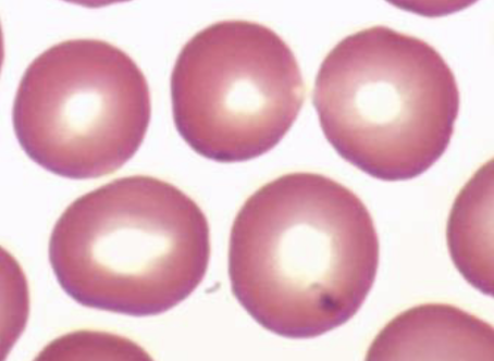
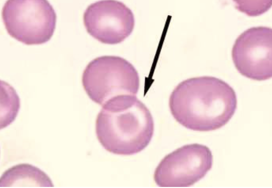
anisocytosis
cells having a variety of sizes
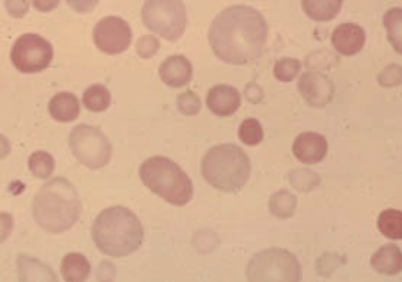
poikilocytosis
variety of shapes
hemoconcentration can indicate sever dehydration when
more RBC than normal
reticulocytes (special stain)
dark blue dots or thin irregular linear structures
Heinz bodies
small, scattered blue doglike structures in RBC
pappenheimer bodies
small, irregular magenta granules that occurs in small groups
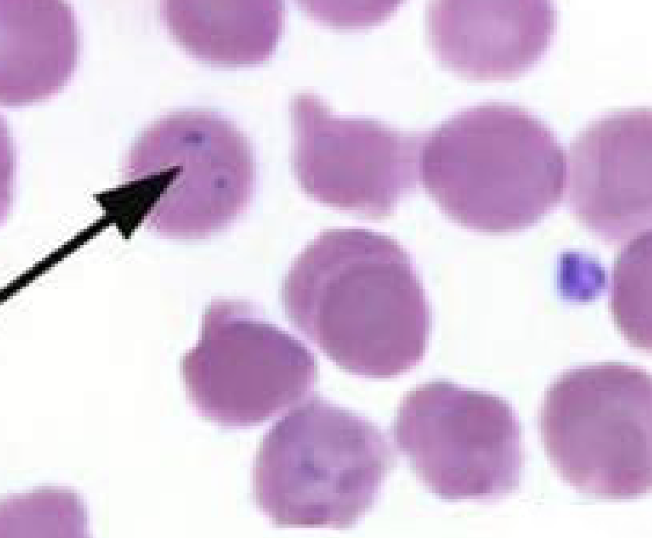
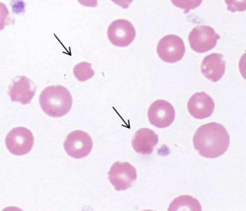
Howell-Jolly bodies
small, round, blue/black inclusions that occur singly or doubly in an eccentric position
Basophilic stippling
small dark blue doglike structure scattered uniformly throughout the RBC
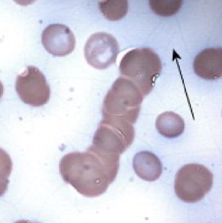
rouleaux
cells partially adhering to eachother; stacking apperance
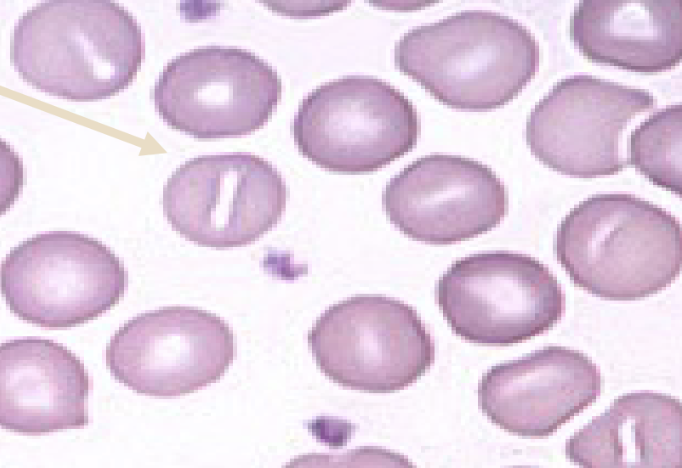
stomatocytes
cells that have a rectangular or slit-like central pale area
target cells
banded
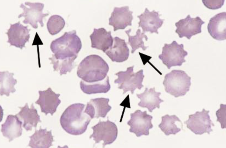
acanthocytes
cell with normal or slightly reduced size possessing 3-8 finger like projections distributed irregularly along membrane
schistocytes
small bizarre shaped cell fragments
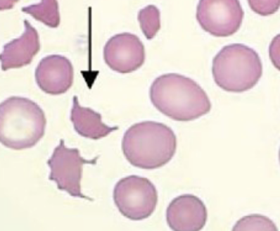
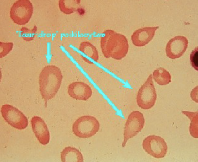
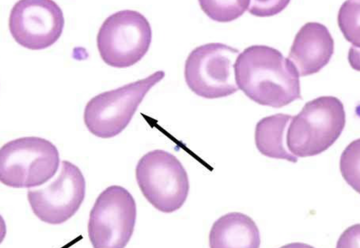
tear drop cells
look like tear drops
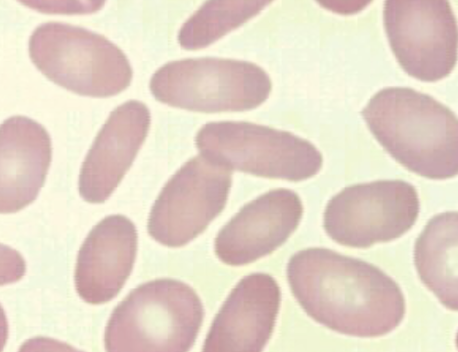
ovalocyte/elliptocyte
oval/egg shaped/elongated cells
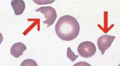
helmet (bite cells)
cells with distinctive projections (usually 2) surrounding an empty area
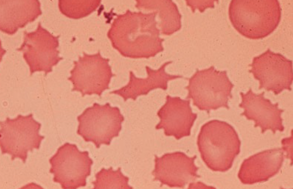
burr cells
cells with 10-30 rounded spicules evenly spaced over cells surface
spherocytes
spherical cells with decrease surface membrane which appear microcytic and hyperchromic