BIOC221 Module 1
 Call Kai
 Learn
Learn Practice Test
 Spaced Repetition
 Match
Match Flashcards
 Knowt Play
1/95

Earn XP
Description and Tags
These flashcards cover essential vocabulary and concepts related to the molecular anatomy of genes and genomes, as introduced in the lecture.
Name | Mastery | Learn | Test | Matching | Spaced | Call with Kai | Chat |
|---|
No analytics yet
Send a link to your students to track their progress
96 Terms
Genome
An organism's complete set of DNA, including all of its genes.
Prokaryotic Genes
Genes found in organisms without a defined nucleus, such as bacteria.
Eukaryotic Genes
Genes found in organisms with a defined nucleus, such as plants and animals.
Promoter
a stretch of DNA upstream of the first exon of a gene, where regulatory sequences are present, and transcription factors and RNA polymerase will bind.
5' UTR
The untranslated region at the 5' end of mRNA that is important for regulation of translation.
Exons
regions of genes that are retained in the mature mRNA. Most exons will encode amino acids/proteins, but some exons will encode 5′UTR and 3′UTR sequences.
Introns
regions of the genes that are transcribed but are not protein-coding. These bases are spliced out from the pre-mRNA in a controlled manner by the spliceosomebefore the mature mRNA is formed.
Splicing
the regulated process performed by the spliceosome to remove introns from a premRNA transcript. Bases at the exon/intron boundaries are critical for accurate splicing. Spliceosome has > 100 proteins and RNA within the complex.
Capping
A modification of the 5' end of mRNA to enhance stability before translation.
Polyadenylation
The process of adding multiple adenosine bases to the 3′ end of almost all mRNA molecules in eukaryotes. The polyA tail is this tract of AAAAAAAAA…..
Nucleosome
A structural unit of chromatin formed by DNA wrapped around histone proteins.
Transposon
A genetic element that has or had the ability to move within the genome, sometimes referred to as a 'jumping gene'.
Open Reading Frame (ORF)
A continuous stretch of codons between the Start and Stop codons that all code for DNA.
Non-coding RNA (ncRNA)
RNA molecules that do not encode proteins but have functional roles in the cell. (tRNA and rRNA)
Variant
A genetic alteration that differs from the reference genome.
Post-translational modifications
Chemical modifications to proteins after translation that can affect their function. e.g. Methylation, Acetylation, Ubiqitination.
6 Possible Reading Frames
A stretch of DNA can contain any of six different reading frames for translation. Three on the sense strand, and three on the antisense strand.
UTRs
Untranslated regions of mRNA that are not translated into proteins but regulate stability and translation.
Mutation
Any change in the DNA sequence.
Centromere
The region of a chromosome where the two sister chromatids are joined.
Telomere
The protective end regions of chromosomes that prevent deterioration of coding DNA via cell replication. The length of a telomere is an indicator of how old a cell is, once it fully deteriorates the cell is supposed to destroy itself.

Name The
1. Regulatory Sequence
2. Enhancer/Silencer
3. Open Reading Frame
4. Promoter
5. 5' UTR
6. 3' UTR
8. Proximal
9. Core
10. Start Codon
11. Stop Codon
12. Terminator

How DNA is packaged at its base level in the nucleus and controlled
DNA is wrapped in chromatin fibres wrapped around nucleosomes like beads on a string. These nucleosomes are made of histones, which control gene expression. The structure of chromatin allows for DNA packaging and regulation of access for transcription and replication.
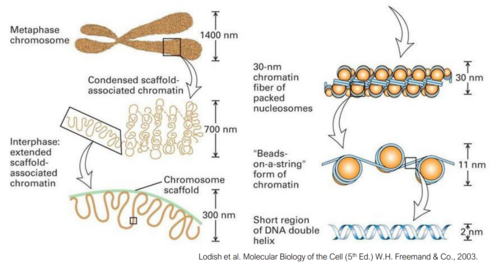
What does Histone H1 do
Histone H1 sits outside the core nucleosome and controls the spacing of nucleosomes. Helps form the higher order structures.
How do four core histones contribute to gene expression
H2A, H2B, H3, and H4 all sit within the nucleosome core. The tails of these proteins are modified by Post-Translational Modifications by enzymes to signal whether a gene should be expressed or not. (PTM's on histone tails are constantly changing.
How do the 'fingers' of the DNA polymerase work with the palm to facilitate nucleotide addition?
The 'fingers' start open and bind to incoming dNTPs in solution. If the correct base pair is bound to, the fingers fold in, bringing the dNTP into the palm of the polymerase, which is the active site, where the DNA strand runs through. The hand twists to make sure there is only one nucleotide to bind to, and the new dNTP binds to its complementary base.
What are the 3 Substrate requirements for DNA polymerase?
1. Template: There must be a template strand to be copied
2. Primer: most DNA polymerases cannot initiate DNA synthesis by themselves - can only extend a pre-existing chain
3. A free 3ʹ OH-end - for the reaction mechanism
How does prokaryotic DNA polymerase perform error correction?
It has a 3’ to 5’ Exonuclease function that, if it detects a mistake in the nucleotide base pairings, backtracks the DNA Polymerase and removes the last few nucleotides, so DNA Pol can go over it again and copy it correctly.
It also has a 5’ to 3’ exonuclease function, which is for filling in nicks.
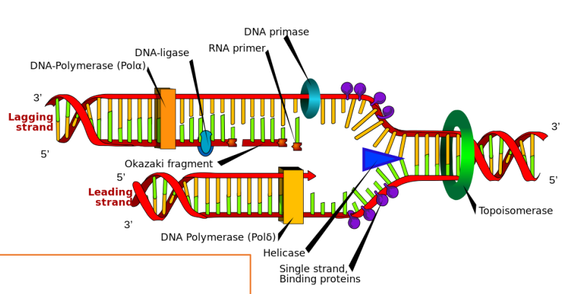
What are the main enzymes and their roles in the replication fork
DNA polymerase.
Helicase unwinds the DNA to create the replication fork; it loads in both directions, so replication extends in both directions.
Primase synthesizes RNA primers, which bind to the lagging strand and allow for DNA to be copied along the lagging strand.
RNA primers are removed by the DNA polymerase exonuclease function.
Ligase seals up any nicks in the DNA where nucleotides may be missing.
Topoisomerase prevents supercoiling of DNA in front of the helicase as it unwinds DNA.
Single-strand DNA-binding proteins (prevent strands from reannealing)
How are only dNTP’s and not rNTP’s bound into DNA by DNA polymerase
One of the requirements for the addition of dNTPs is a free 3’ OH group to bind to. rNTP’s have an additional 2ʹ OH group on rNTPs, which causes steric hindrance, so rNTPs can’t bind to the active site.
What is supercoiling
When DNA strands become to coiled around themselves. Supercoils are removed by topoisomerases (positive supercoil – bad negative supercoil – good)
How is cDNA made from RNA
Enzyme: Reverse Transcriptase, needs primer to make the first strand – so they utilise the polyA tail. Then they degrade RNA by RNase H. 3′ overhang is used to make a primer for second-strand synthesis via DNA polymerase
What polymerase enzymes are used in prokaryotic cell replication and what are their functions?
DNA Pol III is used for most Nucleotide base pairing, whereas DNA Pol I and II are both used for additional error correction and DNA repair.
What proteins are used in prokaryotic DNA to initiate DNA replication and what are their functions? (2)
DnaA binds to oriC – defined point for initiation
DnaA melts DNA (using ATP) to separate the strands at the OriC.
Next, DnaA recruits 2 x DnaB complexes which unwind the DNA like helicase in eukaryotes.
How is error correction for strands that are copying too slowly carried out in Prokaryotic DNA
Termination • Ter sites – one-way ‘valves’ where replication forks can enter but not leave. There are Multiple Ter sites so that slow-replicating DNA strands can be terminated early at a Ter site, and then the strand can be recopied in case mistakes were made.
How do Ter sites stop DNA replication in prokaryotic DNA replication?
The Ter sites stop DNA replication via the Tus protein, which binds to the Ter site and prevents action by the DnaB helicase
Contrast DNA replication in prokaryotic vs eukaryotic cells?
Eukaryotic system:
More complex
Has Multiple origins of replication, with backup systems
Replisome has additional components for error surveillance
Single or double-strand DNA breaks
Fork stalling
Fork reversal
Fork collapse
signals to the cell to pause replication, fire new origins, and initiate DNA damage response
Break down the first stage of Eukaroytic replication Initiation up to helicase loading
The Origin of Replication is not a defined sequence – chromatin landscape (histone PTM marks)
Origin Recognition Complex (ORC) binds across chromosomes before S (Synthesis) phase
Loads on the MCM helicase complex (MCM2-7 helicase)
More inactive MCM loaded than needed – back-up system
Break down the second stage of Eukaroytic replication Initiation and the proteins involved (5)
CDC45 and GINS1-4 get recruited – these activate the MCM helicase
Other proteins – structural support
Also need signals from cell to proceed (via phosphorylation)
Polymerase enters – forms replisome, starts DNA synthesis
RPA – coats single stranded DNA to prevent re-linking of DNA
What are the 3 main Polymerases in Eukaryotic DNA replication and their functions?
In DNA replication:
Alpha = α = synthesis of RNA-DNA primers (with primase), start of Okazaki fragment
Delta = δ = lagging strand polymerase
Epsilon = ε = leading strand polymerase
What Additional proteins are recruited for RNA primer removal on the Okazaki Fragments?
Rnase H1 – removes RNA primer
FEN1 – removes Pol α DNA (no proofreading)
Pol δ (delta) fills in the section
DNA ligase seals the nick
What causes degradation of Telomeres with age?
Since RNA primers are required to be placed ahead of the stretch of DNA being replicated on the lagging strand, since it replicates from 3’ to 5’ direction, the few nucleotides on the end of a chromosome cannot be copied on the lagging strand, so the DNA loses those last few nucleotides. The end of a chromosome is called a telomere and it consists of a motif of T’s and A’s that don’t code for anything, their role is to measure cell age, as every time a cell replicates its DNA, it loses some of the nucleotides on its telomere. When a telomere because very short, this shows the cell is old and it will self-destruct.
What are some of the potential diseases that can arise from an error in DNA replication proteins?
Primordial Dwarfism, where the body is uniformly much smaller than average, this is because cells cannot replicate fast enough to match the size of a full grown human so the body is very small.
It can often cause microcephaly, where the brain and skull is very small, since neurons require the most fast-replicating cells to be turned into neurons from stem cells.
What is Sanger sequencing
An archaic way to determine the exact nucleotide sequence of a stretch of DNA. It is done by mixing DNA polymerase in solution with DNA, dNTP’s and ddNTP’s that have flourescent tags. This should cause a an array of DNA strands to form of length 1-arbitrary base pairs. Then you use gel electrophoresis to sort fragments by size, and the colour that the ddNTP at the end of a strand will tell you what NTP normally lies there.
What is a dideoxynucleotide
A nucleotide base analogue that identical to a deoxynucleotide except it has an 2’ H group instead of a 2’ OH group, so is not able to extend the DNA molecule. This means it can be used to control DNA synthesis, stopping DNA synthesis at any certain nucleotide.
What are the four steps of next-generation sequencing?
Genomic DNA is sheared into small fragments and adaptors are ligated on.
Fragments are attached to a solid surface and amplified by PCR to form clusters
The DNA is extended by DNA pol. and flourescent nucleotides while cameras record the sequence of light.
Sequences of the nucleotides are read and put into BLAST to interpret the data. These can now be used for de novo assembly of DNA or mapped against another genome.
Why are adaptors ligated onto strands of DNA during next-generation sequencing?
The adaptors act as primers for the DNA to be copied/extended. Since they sit at the end of the DNA strand, it allows for the end of the lagging strand to be copied, which usually isn’t possible in natural DNA replication. Making clusters of DNA fragments is necessary to make the light strong enough to detect.
How is the speed of nucleotide addition controlled in next-generation sequencing to ensure accurate recordings of each nucleotide?
Using terminators on each flourescent nucleotide base. These proteins mean that before the next base can be added, the terminator and flourescent tag are removed. This slows down replication and increases clarity between different bases to ensure accuracy in recording.
How is de novo gene assembly accomplished.
Extended DNA strands from different starting strands that overlap are joined together to form larger reads that are called contigs. Thus a larger picture of the overall genome sequence can be pieced together.
How can mapping a tested genome against a template be used to find structural variants in a genome?
Finding a reduced number of read for a gene or DNA stretch could mean it is only present on one chromosome. Finding no reads likely means there is a double deletion for that gene.
How can PCR be used following next-generation sequencing to confirm whether an individual has a genetic deletion?
Check textbook for in-depth
How do we make cDNA from RNA?
Enzyme: Reverse Transcriptase, it Needs a primer to make first strand, this primer binds to the polyA Tail. Rnase H degrades RNA, and then the 3′ overhang is used to make the primer for second strand synthesis via DNA polymerase.
What is qPCR or qRT-PCR?
Quantitative RT-PCR (qRT-PCR, qPCR, real time PCR), is a method of DNA amplification that is similar to RT-PCR but it allows for quantification with fewer cycles, and supplies a reference to compare to, it also doesn’t require gel electrophoresis.
qRT-PCR: how are PCR products detected?
There are two different molecules that are used in this method, usually SYBR green and occasionally Taqman.
A flourescent PCR product is in solution with the cDNA and PCR. With every cycle of PCR, the PCR products fluoresce. Once the fluorescence/brightness of the solution reaches a threshold intensity, we measure how many cycles it took to reach that threshold. This is the CT value, or the cycle threshold. The lower the CT value, the more cDNA was present in the starting sample. This is a technique to quantify how much cDNA is in a sample.

Why are CT values compared to a reference gene to determine the amount of cDNA?
Usually, compared to a reference gene also amplified because the expression level shouldn’t change, regardless of treatment/environment (need to decide on appropriate reference gene(s) for cell type or tissue). This controls for different cDNA / RNA input amounts
Assumption: the reference gene is the same in both, which means the same amount of RNA/cDNA in the reaction as template. Not usually the same, so there is a calculation to follow to adjust for this.

What is qPCR used to measure?
It is used to measure the concentration of a certain RNA molecule, this is used to measure gene expression.
What is the transcriptome?
All the transcripts produced by a cell, tissue or organism. This refers to mature mRNA, rRNA, pre-mature mRNA that has not be spliced or been given a poly-A tail, tRNA or any other transcript of DNA.

Explain how BLAST can be used to measure how related two organisms are genetically.
BLAST is a software that can compare similar regions of DNA between organisms and tell you how much they match. Since most variations occur over millions of years it gives a good indication of how genetically similar and therefore evolutionarily related two organisms are. By comparing against other genomes, we can also see which genes or nucleotides are similar across many very different organisms, so they may be extremely important/sensitive genes that are integral to organism function.
Name 6 tools that are used to understand something about cell function for a research question? And try to name an example of them being used.
Sanger sequencing / next-generation sequencing - To find the nucleotide sequence of any DNA/RNA
RNA-seq - to measure the level of RNA transcripts in a cell and therefore gene expression
Restriction endonuclease digest - Restriction enzymes cut pieces of DNA in a specific place
Electrophoresis - measuring and sorting DNA fragments by their bp length
PCR, molecular cloning, gene synthesis - Amplification of sections of DNA to obtain a lot of a specific piece. qPCR is another kind and is used to measure the expression of one or a few genes.
CRISPR-Cas PCR - targeted mutagenesis/editing, changing the sequence to test gene function or repair a mutation