Deck #3: AS imaging
 Call Kai
 Learn
Learn Practice Test
 Spaced Repetition
 Match
Match Flashcards
 Knowt Play
1/90

There's no tags or description
Looks like no tags are added yet.
Name | Mastery | Learn | Test | Matching | Spaced | Call with Kai | Chat |
|---|
No analytics yet
Send a link to your students to track their progress
91 Terms
What are the 3 main anterior segment imaging procedures?
AS-OCT
Specular Microscopy
Wavefront Aberrometry
Do auxiliary tests replace provider judgment?
No, they are used in conjunction with the clinical picture and provider expertise
What is Optical Coherence Tomography (OCT)?
→ non-contact technique that creates high-resolution, cross-sectional images of ocular structures using reflected light
(+) allows for quantitative analysis of imaged tissues without biopsy
(+) can monitor disease progression
What 4 areas of the eye is OCT used for?
Cornea (e.g., CT)
Anterior chamber angle
Retina
Optic nerve
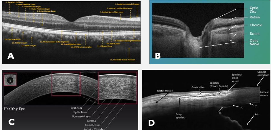
Why is OCT more repeatable than ultrasound biomicroscopy?
lower coefficient of variation (CV)
List 7 specific uses for Anterior Segment OCT (AS-OCT).
Corneal assessment
Thickness
Opacifications
Disorders/dystrophies
Anterior chamber angle
Anterior segment tumors
Planning for anterior segment surgeries
CL fittings/eval
Tear meniscus measurements
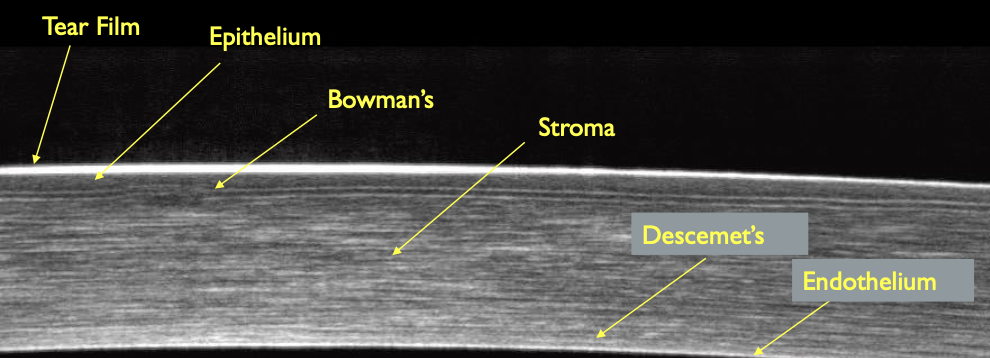
What 5 corneal layers can be seen on AS-OCT?
Epithelium
Bowman’s
Stroma (thickest, where nerves are)
Descemet’s
Endothelium
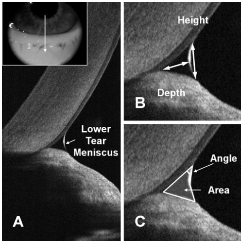
What 3 tear film metrics can AS-OCT quantify?
Tear meniscus height, depth, & area
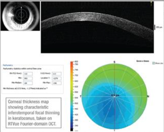
Other than tear meniscus, what else can the AS-OCT measure?
Corneal thickness
epithelium - can detect thickness (helpful for kerataconus)
corneal edema
Note: Measurements don’t always correlate to ultrasound pachymetry
What signs of Fuch’s dystrophy appear on AS-OCT?
Thickened Descemet’s membrane
guttata in endothelium
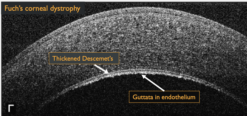
What corneal dystrophy is this an image of?
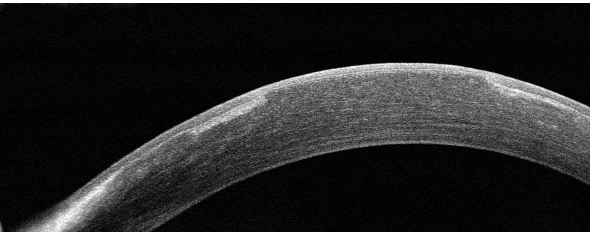
Epithelial Basement Membrane Dystrophy
whitening of tissue is peripheral, so VA isn’t affected
What corneal dystrophy is this an image of?
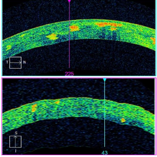
Granular Corneal Dystrophy Type 2
red spots = corneal opacities
useful to determine stroma layer that’s affected
What is this an image of?
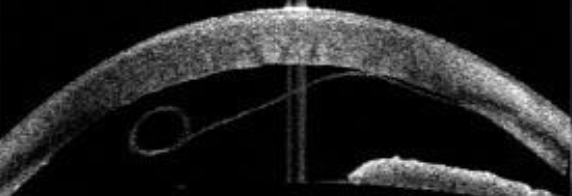
Descemet’s Membrane Detachment (from stroma)
What is this an image of?
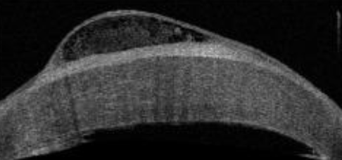
Bullous Keratopathy
What is this an image of?
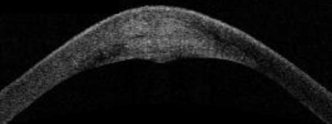
Infective Keratitis
What is this an image of?
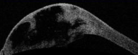
Corneal Hydrops
break in decements → massive influx of aq fluid into cornea → blister on surface that can rupture
Is AS-OCT angle assessment functional or structural?
structural (can’t see how aq is flowing, BUT can see if angle is open/closed)
What angle configurations can AS-OCT visualize?
Narrow angles
Plateau iris
PAS
Pupillary block
Note: Can be observed in dark vs light
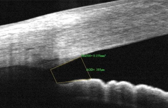
What is the gold standard for measuring angle configuration?
Gonioscopy (AS-OCT can be used in conjunction)
How would you describe this angle?
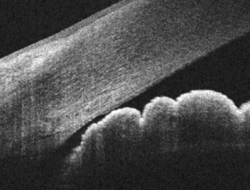
Narrow angle, but not touching (might occlude in dim light)
How would you describe this angle?
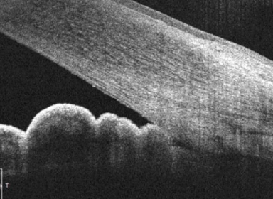
Iridocorneal touch (closed angle)
iris to cornea touch (closed angle)
How would you describe this angle?
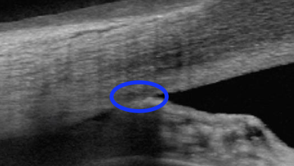
PAS (Peripheral Anterior Synechiae)
iris to cornea touch (closed angle)
What type of iris is this?
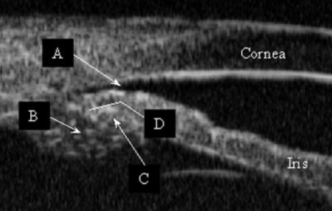
Plateau iris
How does AS-OCT help with iris lesions?
→ distinguishes between tumors and cysts and images the anterior architecture
(+) avoid intraocular biopsy
(+) good in small or hypopigmented lesions
(-) poor analysis of posterior aspect/extension of tumor
What is this an image of?
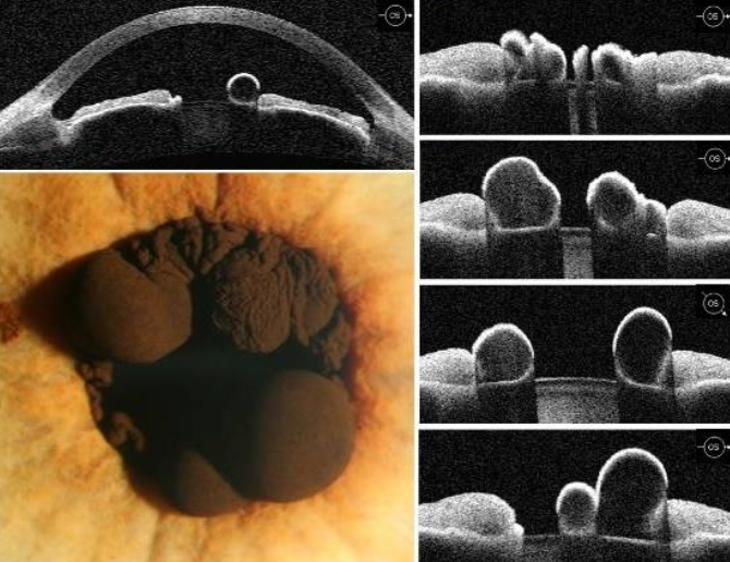
AS-OCT of Iris cysts (clear interior = fluid)
What is this an image of?
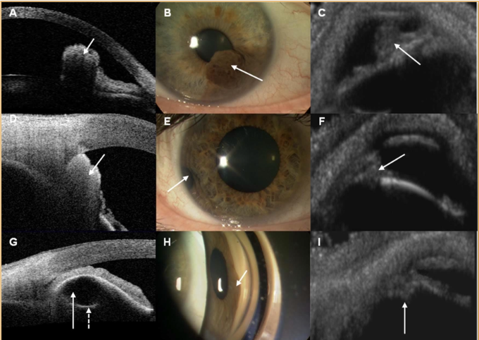
AS-OCT of ant seg tumours
3rd row is a B-scan (which doesn’t give that posterior aspect, you can only see the anterior bowing)
What are the applications of AS-OCT in specialty contact lens fittings?
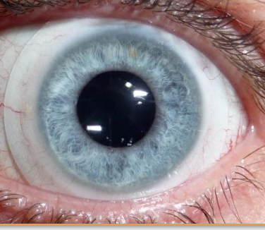
Sagittal depth of the anterior segment
Lens clearance
Posterior lens tear film thickness
Lens bearing in any location
Scleral and limbal interaction
Lens thickness
Lens defects
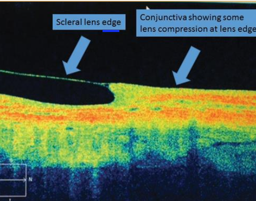
What do each of these arrows represent?
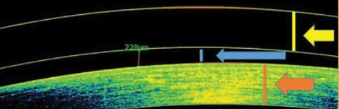
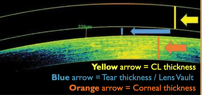
How does AS-OCT assist with soft contact lenses?
Evaluate pachymetry & edema pre-/post-lens wear
What power do you think this soft CL has?
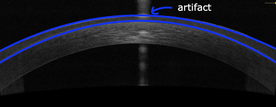
Minus
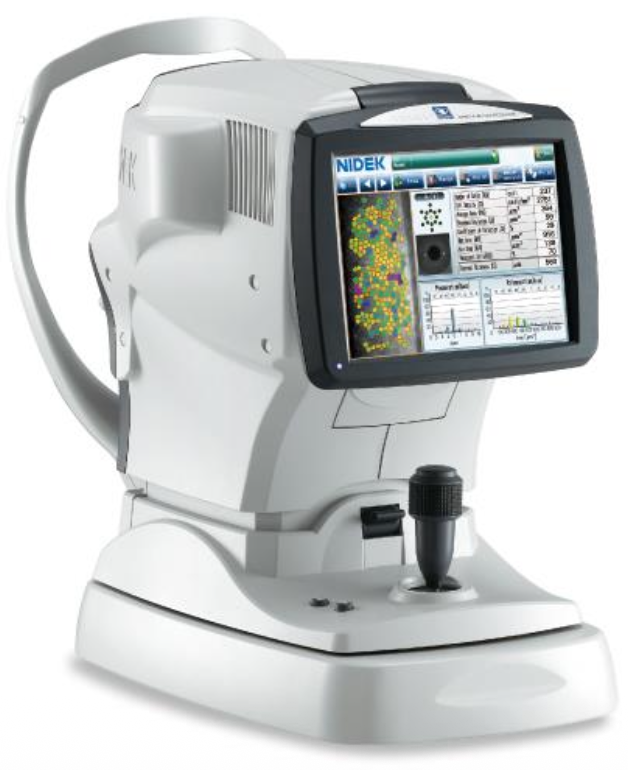
What is specular microscopy? What 3 aspects of the endothelium does it analyze?
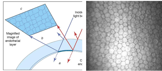
→ Noninvasive, photographic technique that visualizes the endothelial morphology:
Size
Shape
Cell density
List 2 ways to visualize the Corneal endothelium.
Specular reflection
Specular microscopy
What is Specular reflection?
→ Light source & microscope at equal angles from cornea
Focused on the endothelium
Elevations = dark
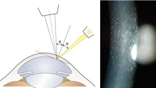
What is Specular microscopy?
Microscopic visualization of endothelial cells in vivo
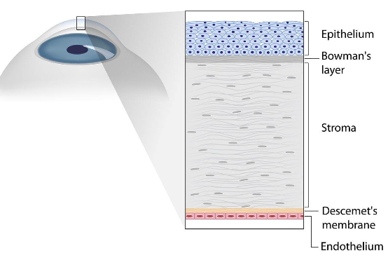
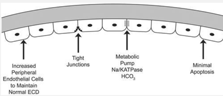
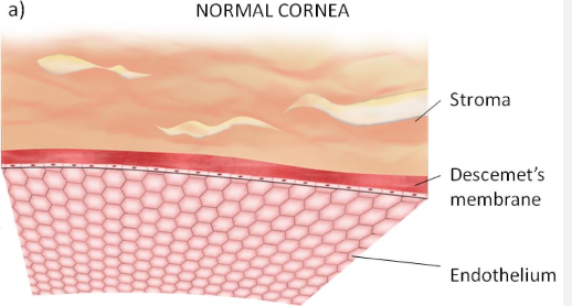
What is the Endothelium & its functions?
→ 1 layer thick
Maintain corneal health & transparency
Semipermeable membrane - facilitate diffusion of nutrients into the cornea from aq
Secrete the Descemet’s membrane
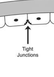
How does the endothelium restrict fluid from entering the cornea?
via tight junctions that b/w endothelial cells
What is the metabolic pump function of the endothelium?
→ Controlled by junctional complex between endothelial cells
Endo → Ant chamber:
Active transport fluid out of the stroma
Translocation of HCO3 across endothelial membrane
Passive transport of H2O into anterior chamber
How many endothelial cells in young patients?
350-550 K
What is the normal ECD for young patients?
~3500 cells/mm2
How many endothelial cell loss is normal? Do endothelial cells regenerate?
100-500 cells/year (slough into anterior chamber)
these DON’T regenerate
What ECD difference between eyes is considered abnormal?
Loss is symmetrical, however >280 cells/mm2 difference in cell loss b/w eyes is abnormal
Once endothelial cells are shed, what happens?
→ Cellular repair
Stretching & sliding of existing cells into new position
Fusion of adjacent cells
What is the critical density range for the endothelium? What occurs if it falls below this range?
300-500 cells/mm2
lose the ability to regulate fluid → corneal edema
How does age contribute to the average cell density?
10-59 = stable cell loss
60-80+ = cell density declines
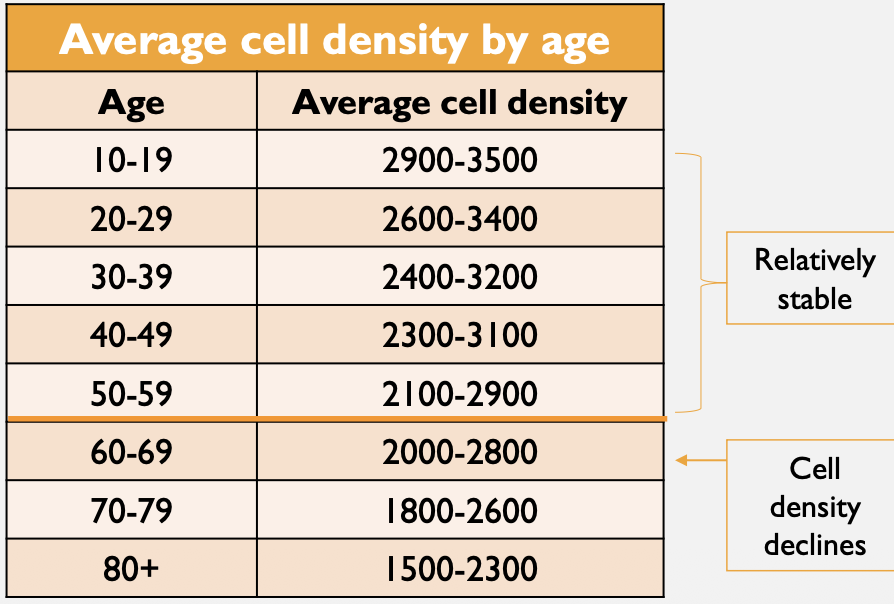
What is the corneal endothelial cell density in newborns, adults and old adults? How does cell diameter vary?
Newborn = 3000-4000 cell/mm2 (18-20 μm)
Adult = 2500 cell/mm2
Old age = 2000 cell/mm2 (40 μm)
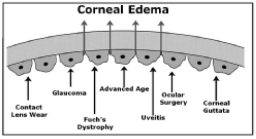
What occurs during Endothelial cell damage? List factors that cause it.
→ Physiologic stress alters normal endothelium, causing the breakdown of fluid barrier function → edema
damage alters central and peripheral cell density + shape
.
Causes
Normal aging
CLs wear
Trauma
Ocular surgeries
Ocular diseases/disorder
Systemic disease
Define pleomorphism. List its causes.
→ Disruption in regular hexagonal pattern of cells (shape change)
↓ cellular stability
.
Causes
Normal aging
CL wear
Physiologic stress
Ocular disease
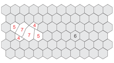
What is Hexagonality (Hex)?
→ Variability in hexagonal shape of cells
measure of pleomorphism (% of cells with 6, >6, <6 sides)
deviation from hexagonal shape
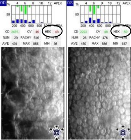
What % of pleomorphism is considered “clinically significant”?
Normal = >50% hexagonal cells (6 sides)
Abnormal / pathology = >50% pleomorphic cells (anything with less/more than 6 sides)
greater than 50% (if more than 50% of cells are not hexagon it's bad → disrupts fluid barrier function)
Define polymegethism. List its causes.
→ change in uniform size of endothelial cells (size change)
1st sign of endothelial disease ⭐
.
Causes
Physiologic stress of cornea
Overactive wound repair
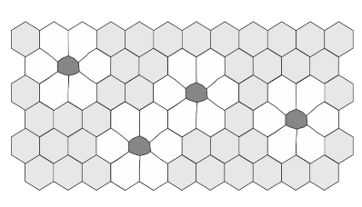
How can you track polymegathism? What are normal, borderline and abnormal values?
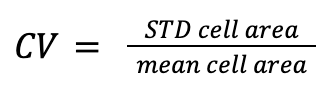
→ via Coefficient of Variation (CV)
Normal CV = 0.22-0.31 (22-31%)
Borderline CV = 0.32-0.40 (32-40%)
Abnormal CV = >0.40 (greater than 40%)
Low CV = similar in size
What is the formula for CV?
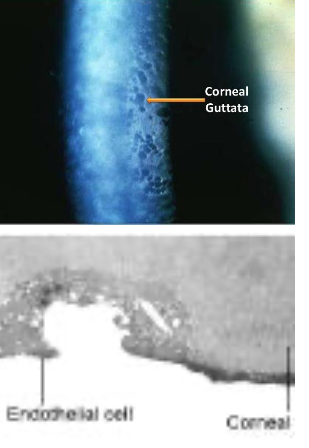
What are guttata?
→ Secretions of abnormal BM that accumulates under cells
made of collagen
nodules covered by endothelial cells
cause thickening of Decemet
can coalesce over time
common in periphery
found in 70% of patients over 40
part of normal aging, but pathologic when there’s loss of fluid barrier function
Compare contact vs non-contact specular microscopy.

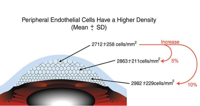
What does the accuracy & precision of measurements taken during Specular microscopy depends highly on?
→ area of corneal image selected for analysis
cell density varies in different endo locations (periphery has more endo cells, than central)
→ presence/absence of defects
List the factors that affect image quality of Specular microscopy.
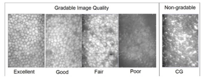
Dry eye
CL use
Keratoconus
Patient compliance
Pt’s age
Training or experience of technician/doctor
What does Specular microscopy measure?
Cell density (CD)
Cells/mm2
Cell morphology
Size (cell area ± SD)
Shape
Variability
Optical pachymetry
CT
List the indications for performing Specular Microscopy.
General corneal health
Diagnose & manage corneal dystrophies/degeneration
Fuch’s dystrophy
Posterior polymorphous dystrophy
Iridocorneal dysgenesis conditions
ICE syndromes
Assessing idiopathic decreased vision in pt
Pre-operative risk assessment & post-operative management (cataracts, LASIK)
CL evaluations & management (e.g., overnight / extended wear lenses, Scleral lenses)
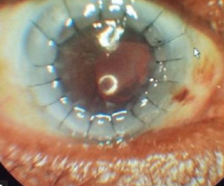
What is the most sensitive measure of corneal health?
Pleomorphism
Polymegethism
🌟 Both can indicate loss of barrier & pump function
What is the least sensitive measure of corneal health?
Cell density
🌟 cornea can still function well at low densities
Diagnose this.
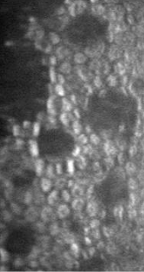
Early Fuch’s dystrophy
Diagnose this.
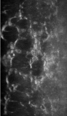
Advanced Fuch’s dystrophy
Diagnose this.
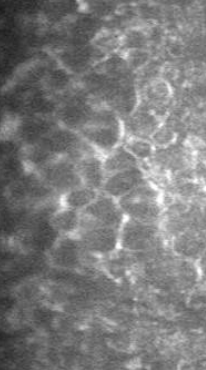
ICE syndrome
Diagnose this.
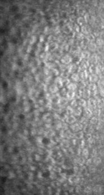
CL changes
Define Wavefront aberrations.
Deviation of a wavefront exiting the pupil after progressing through the optics of an eye compared to a reference unaberrated wavefront free of aberration
What is a Wavefront aberrometer?
→ Instrument used to measure aberrations within an optical system
allows for deeper understanding of aberrations & effect on vision
provides means for improved optics via refractive surgery, custom design CLs
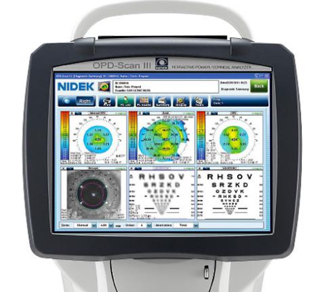
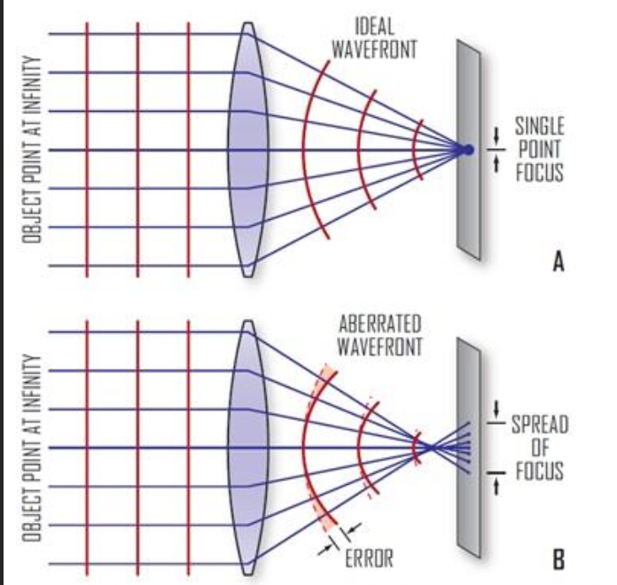
How are ocular aberrations measured?
→ Monochromatic light projected into the eye, off the fundus & back out to the photosensors of an instrument
Ideal eye / optical system = Light from distance focuses sharply → ideal wavefront
Aberrated eye = Light reflected out from the retina → abberated wavefront
List 5 factors affecting ocular aberrations.
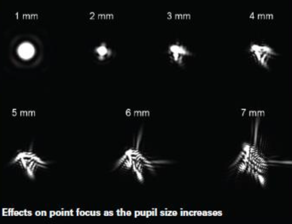
Pupil size (dilated at dark, introducing more abberations)
Accommodation
Lid position
Eye movements
Tear film stability
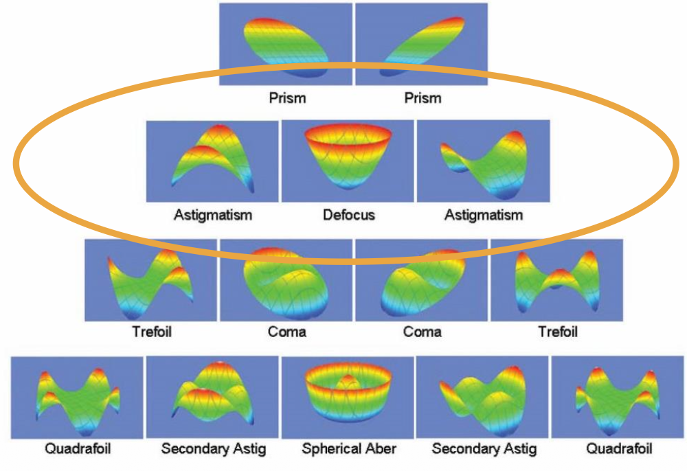
What are lower order abberations? List them.
→ Account for ~80-90% of ocular aberrations
Defocus
Myopia, hyperopia
related to spherical ametropia in RE
describe image postion
Astigmatism
eye can’t produce 1 point image
→ easily corrected with glasses, CL, refractive surgery
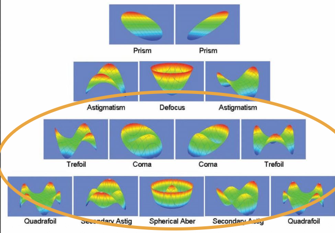
What are higher order abberations?
→ Account for ~10-20% of ocular aberrations (3er order + above)
Coma
Trefoil
Spherical aberration
Quatrefoil
2er astigmatism
→ not easily corrected, worsened by refractive surgery
What is the amount of higher-order aberration proportional to?
pupil size
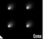
What is Coma?
→ Occurs due to misalignment of the center of the pupil, cornea & lens → creates an image that appears to have a “tail”
Irregular astigmatism
Frequent problem in pts with corneal ectatic or thinning disease (e.g., Keratoconus)
What is Trefoil?
Irregular astigmatism
makes the least amount of image degradation of higher order aberrations
What is Spherical aberration?
→ created by difference in refracting power of lenticular annular rings & pupil size → “haloes” around point images
↑ effect of uncorrected myopia in low light settings
depends on pupil size
2x pupil diameter = 16x SA
affected by accommodation or age-related lens changes
(+) SA = peripheral refracted more than central
(-) SA = peripheral refracted less than central (accom, CL)
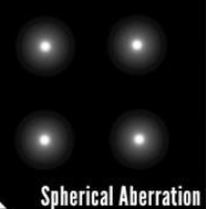
List the visual impact of Haloes created by SA?
Poor night vision
↓ depth of focus for reading
Glare / haloes around lights
Diplopia
Starbursts around lights
What is Root Mean Square (RMS)?
→ calculated using the difference between wavefront surfaces & average of the surface
deviation of the wavefront from a plane wavefront
abberations aren’t directly correlated to visual performance
predictive value for likelihood of increasing HOAs after refractive surgery
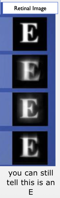
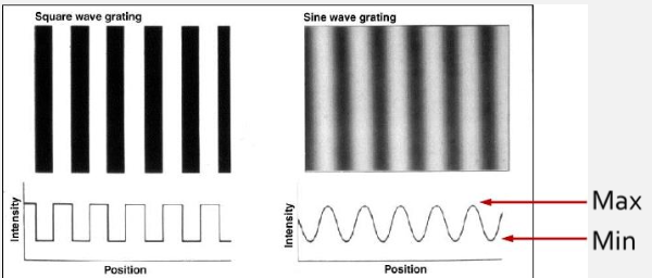
What is Modulation Transfer Function (MTF)?
→ ratio of image contrast as a function of SF
↑ SF = ↓ MTF
Quantitative measure of image quality
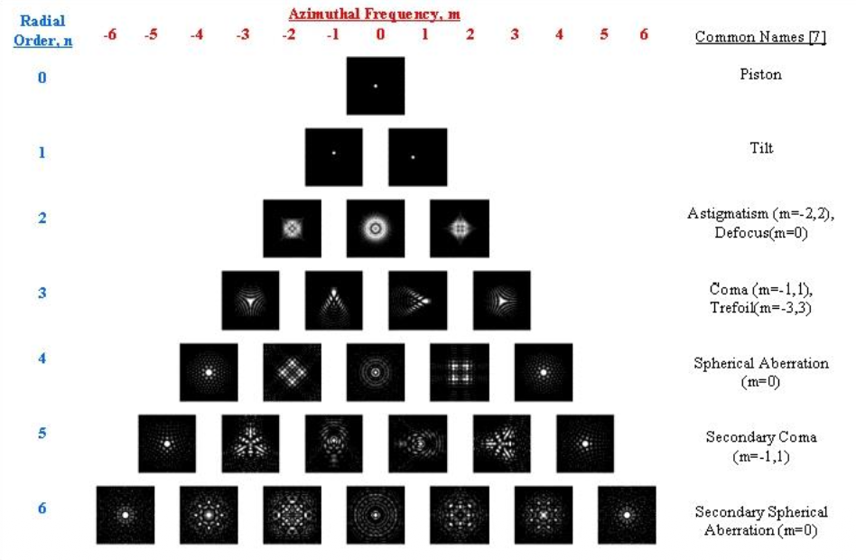
What is Point Spread function (PSF)?
→ Measure of how well an object point is imaged on the retina
shows how aberrations affect a point of light
Ideal optical system: PSF = 0
What are the indications for wavefront analysis (aberrometry)?
Refractive surgery planning
Cataract surgery evaluation
CL fitting
Custom spectacle design
Patients with minimal VA decline
Patients with 20/20 VA but persistent visual complaints
List the clinical uses of wavefront aberrometry.
Potential improvement in vision beyond correction with typical glasses or CLs
Increasing use in refractive & cataract surgery
Objective clinical refraction
Optimizing the sphero-cylindrical refraction
What makes up the majority of aberrations?
LOA (HOA is masked, but becomes noticeable when LOA is corrected)
What’s the difference between Conventional LASIK vs Custom LASIK?
Conventional LASIK (Laser In Situ Keratomileusis)
↓ Defocus (LOA)
↑ Coma & SA (HOA) → night glare, haloes, ghosting
.
Custom LASIK
Aims to ↓ HOAs for better visual quality
What are ODs role in refractive surgery?
Co-management of refractive surgery pt (both pre- & post-op)
Wavefront aberrometry can help detect pts that may not be as good of candidates prior to surgical consult
We know that CL correct LOA. What have many soft lenses brands began to do?
→ incorporate aspheric optics to ↓SA
(+) or (-) SA incorporated into lens to ↓ ocular aberrations
What are the limitations to Abberation control CL?
Fluctuations in pupil size
Expensive
Lens movement/rotation
Disease progression
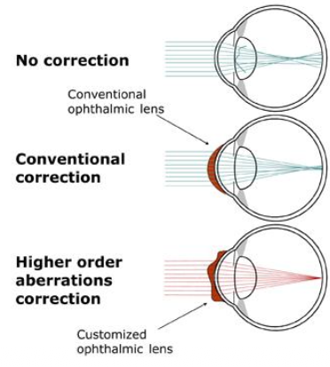
What is Objective wavefront refraction?
→ Process of deriving sphero-cylindrical correction using wavefront aberration measurements
takes into account amount & type of HOAs in an eye to optimize optical correction
can create lenses to ↓ HOAs
What are limitations to wavefront refraction?
→ Wavefront refraction measures optical quality, but perception is not limited to optics alone, it must also consider:
Retinal image quality
How retinal images are processed
Transfer of the image through the entire visual pathway
Why is measuring aberrations with monochromatic infrared light a limitation of wavefront refraction?
→ b/c it doesn’t account for chromatic aberration from white light
White light has multiple wavelengths that focus at different points
What is the gold standard for comparing the success of an objective refraction (e.g., wavefront refraction)? List its limitations.
Subjective refraction
vary between different practitioners
produce different results from the same practitioner at different times/days
How do phoropters and trial lenses limit comparison with wavefront refraction?
quantified in 0.25 D steps → limits precision of subjective correction
Why might different aberrometers give different refractive results for the same patient?
Use different measurement methods
Analyze aberrations differently
Calculate different estimated refractions