Ch 14 bio1191
 Call Kai
 Learn
Learn Practice Test
 Spaced Repetition
 Match
Match Flashcards
 Knowt Play
1/36

There's no tags or description
Looks like no tags are added yet.
Name | Mastery | Learn | Test | Matching | Spaced | Call with Kai | Chat |
|---|
No analytics yet
Send a link to your students to track their progress
37 Terms
How brain contribute to homeostasis
receiving sensory input, integrating information, making decisions, and executing motor responses
Cranial bones
skull
Cranial meninges
Dura, Arachnoid, PIa
Dura Mater
Periosteal layer: Outer layer attached to the inside of the skull
Meningeal layer: Inner layer faces the brain
When these are seperated they form venous sinuses
Venous sinuses
Channels that drain blood and CSF away from the brain
Falx cerebri
Seperates the right and left cerebrum
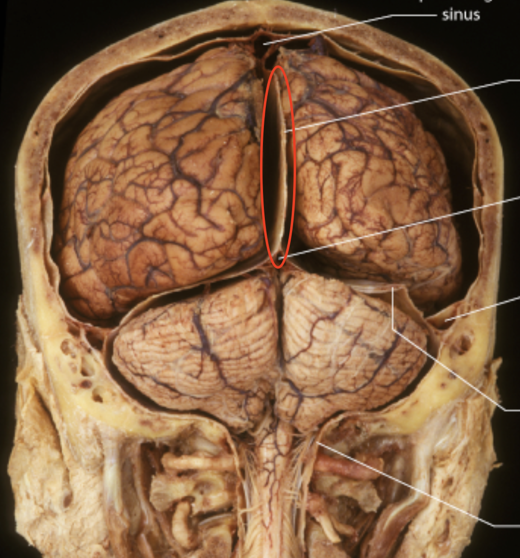
Falx cerebelli
seperates the right and left cerebellum
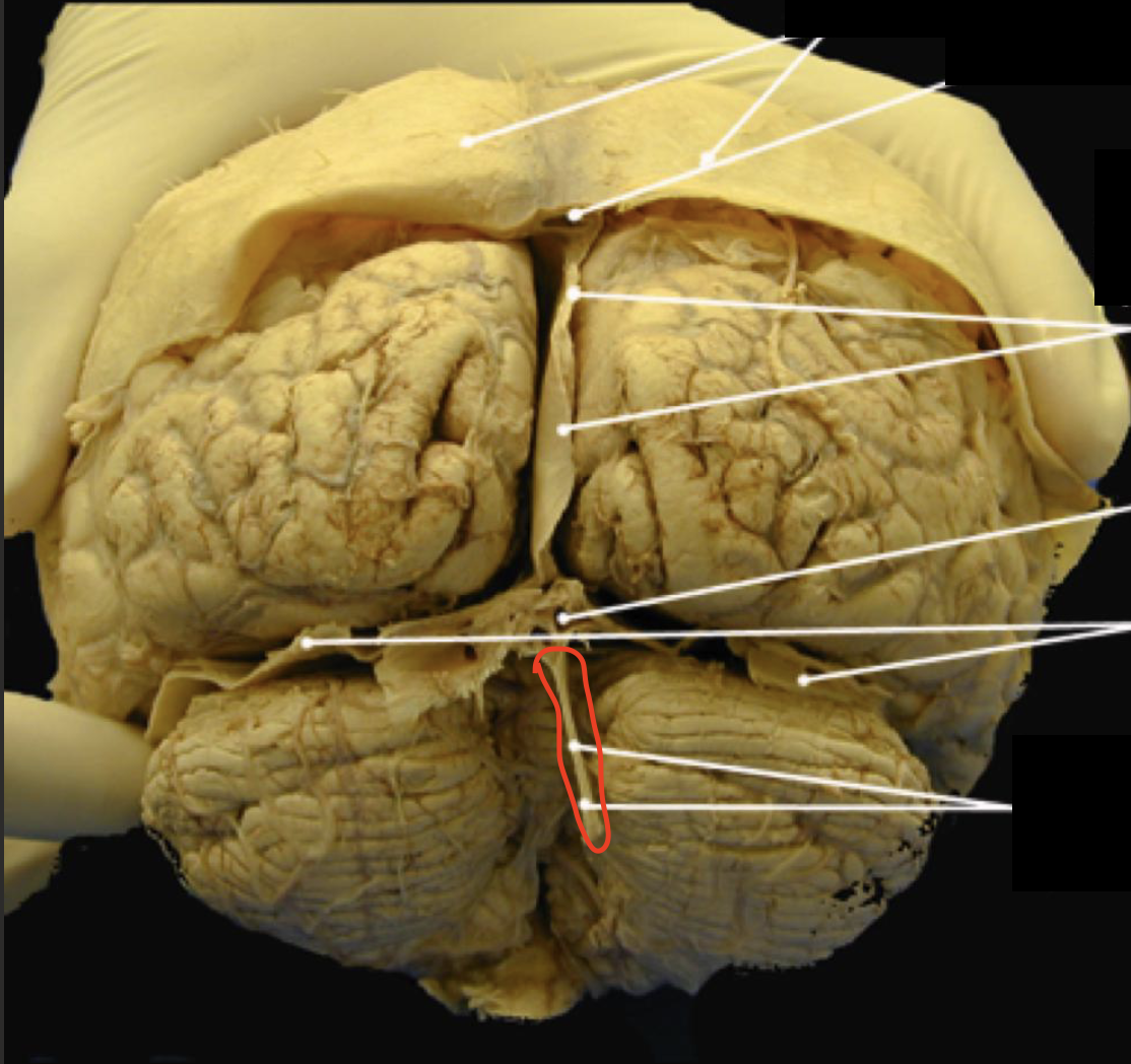
Tentorium cerebelli
Seperates the cerebrum and cerebellum
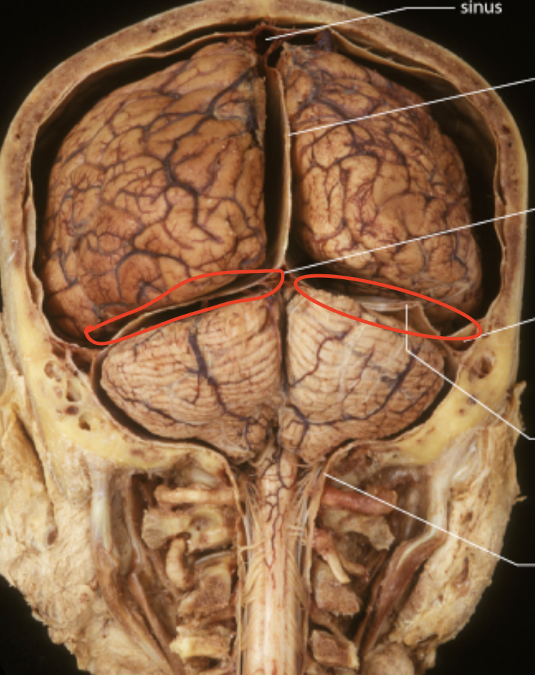
Blood flow to brain
Internal carotid and vertebral arteries
Blood flow away from brain to heart
Dural venous sinuses drain into internal jugular veins
What happens when the brain is deprived of oxygen
weakening, permanent damage, or death of brain cells.
What happens when the brain is deprived of glucose
confusion, dizziness, convulsions, and unconsciousness.
fMRI
When neuron and neuroglia activity increases in a brain region, blood flow to that area also increases
Tight Junctions in BBB
seal endothelial cells tgt which acts like a wall for brain capillaries
Basement membrane in BBB
surrounds endothelial cell (second layer underneath)
Astrocytes in BBB
press up against capillaries – secrete substances that maintain the tight junctions
Blood Brain Barrier (BBB)
Lipid-soluble easy (O2, CO2, alc, nic, caffeine, anesthesia), Water soluble (glucose), most ions transported slowly, big nope
Cerebrospinal Fluid functions
Mechanical Protection – absorbs shock and protects the delicate tissues of the CNS from jolts • Also buoys the brain so it floats in the cranium
Chemical Protection - optimal chemical environment for neuronal signaling (ions!)
Circulation - medium for exchange of nutrients and waste products between blood and nervous tissue
Formation of Cerebrospinal Fluid
Produced in choroid plexuses
Choroid Plexuses
Capillaries covered by ependymal cells
CSF structure
Colourless liquid which is mainly water, contains glucose, ions (Na, Cl), lil bit O2, few WBC
Ependymal cells and making CSF
Filters blood plasma, lets mostly water, dissolved ions, lil bit of protein, and cells thru
Reabsorption of CSF
CSF is absorbed by Aracnoid Granulation, which it then dumps it into the venous sinus to return to blood stream, disposing of it
Hydrocephalus
Blockage of drainage of CSF (tumour,
inflammation, developmental malformation,
meningitis, hemorrhage or injury)
Pyramid (Med oblon)
Control voluntary movement of lumbs and trunk
Nuclei of Medulla func
Cardiovascular center: heart rate, diameter of blood vessels
Respiratory: breathe rhythm
Reflex: cough sneeze, swallow vomit hiccup
Pons
Consists of both nuclei and tracts, the “bridge” that connects parts of brain, help breathing, contains cranial nerves V-VIII
Midbrain
Contains sensory tracts, motor tracts and nuclei (visual and auditory info)
Cerebral peduncles
Contain corticospinal (Cortex → spinal cord), corticopontine (cortex → pons) and corticobulbar (cortex → medulla) motor axons
Tectum superior colliculi
Reflex centers for visual activities (e.g. tracking a moving object, scanning stationary images)
Tectum inferior colliculi
Part of auditory pathway • Relay impulses from receptors for hearing in ear to thalamus – Reflex center for startle reflex
Substantia nigra
Realse dopamine, helps control subcoscious muscle activity, loss of this = parkinsons
Red Nuclei
rich in blood supply, axons from cortex and cerebellum synapse coordinate precise voluntary muscular movements
Nuclei white or gray
gray
Reticular activating system (RAS)
alerts cerebral cortex to sensory signals (like sound, flashlight, BUT NOT SMELL) to awake from sleep, regulate consciousness, stimuli from ear eye skin muscle. COMA IF DAMAGE
Cerebellum structure
10% of brain weight, 50% of neurons in brain
ataxia
loss of ability to coordinate movements damage to cerebellum