Superficial/cutaneous mycoses notes
1/19
There's no tags or description
Looks like no tags are added yet.
Name | Mastery | Learn | Test | Matching | Spaced | Call with Kai | Chat |
|---|
No analytics yet
Send a link to your students to track their progress
20 Terms
What are superficial mycoses?
Fungal diseases that affect only the cornified layers (stratum corneum) of the epidermis. Patients do not show any overt symptoms because the fungal agents do not activate any tissue response of inflammatory reaction
What does Malassezia furfur cause?
Tinea versicolor, a disease characterized by patchy lesions or scaling of varied pigmentation, it is also thought to be a cause of dandruff. Lesions become especially evident in warm months, when sun exposure is more likely. Also been implicated in disseminated infections in patients receiving lipid replacement therapy, particularly in infants.
How is M. furfur identified?
Microscopic examination of skin scrapings from characteristic lesions in a potassium hydroxide (KOH) preparation or by observing yellow fluorescence with wood lamp on examination of the infected body site. Requires lipids for growth and will not grow on routine fungal media that have not been supplemented with a lipid source.
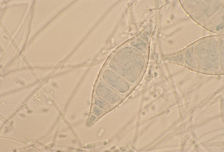
What does Piedraia hortae cause?
Black piedra, an infection of scalp hairs. Produces hard, dark brown to black gritty nodules that are firmly attached to the hair shaft.
Diagnosis of black piedra
Infected hairs are removed and placed in 10% to 20% KOH, the nodules may be crushed open to reveal the asci. P. hortae grows slowly on Sabouraud dextrose agar at room temperature.
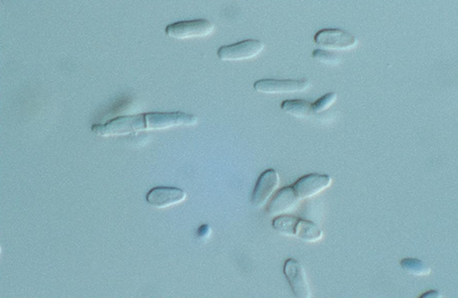
Trichosporon spp.
T. ovoides, T. asteroides, T. inkin and Cutaneotrichosporon cutaneum have been implicated in most cases of superficial mycoses. T. asahii is implicated in severe and frequently fatal disease in immunocompromised hosts. T. mucoides also causes systemic disease (meningitis) and is recovered frequently from CSF.
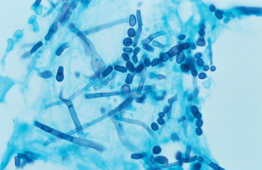
Trichosporon spp, diagnosis
Grow rapidly on primary fungal media and produce arthroconidia, hyphae, and blastoconidia. Straw to cream colored and yeast like. smooth or wrinkled, dry or moist, and creamy or velvety. Identification to the species level is confirmed by absence of carbohydrate fermentation, use of KNO3, assimilation of sugars, and urease positivity. Molecular and proteomic approaches lead to accurate identification of species within this genus.
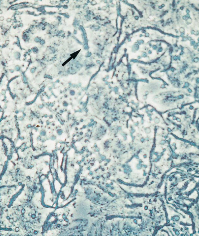
Hortaea werneckii clinical manifestations
Tinea nigra, characterized by brown to black nonscaly macules that occur most often on the palms and soles.
Diagnosis of tinea nigra
Direct examination of skin scrapings placed in 10% to 20% KOH. Microscopic examination shows septate elements and budding cells. Shiny, moist, yeastlike, start with brownish coloration that eventually turns olive to greenish black.
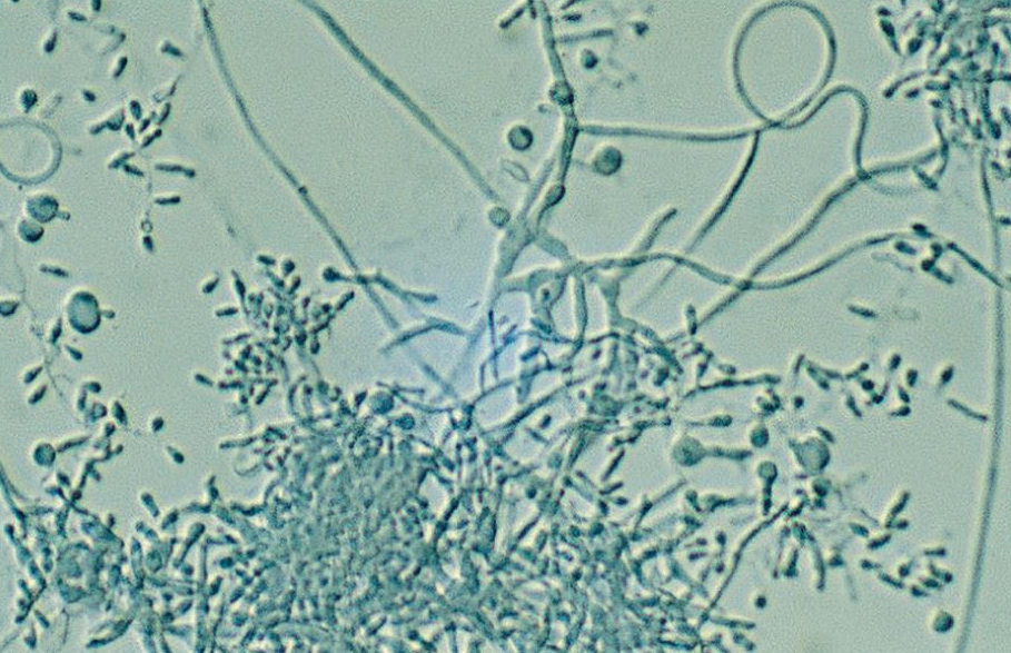
Dermatophytes general characteristics
Trichophyton, microsporum, and epidermophyton. Are keratinophilic, they are adapted to grow on hair nails, and cutaneous layers of skin that contain the scleroprotein keratin. Form two sizes of reproductive cells, macroconidium or microconidium.
Tinea favosa, or favus
Infection of the hair follicle by Trichophyton schoenleinii and progresses to a crusty lesion made up of dead epithelial cells and fungal mycelia
Tinea capitis
Two distinct forms. Gay patch ringworm and black dot ringworm, caused by different species of dermatophytes. Grey path ringworm is a common childhood disease, the lesions are seldom inflamed, but luster and color or the hair shaft may be lost. Black dot ringworm consists of endothrix hair involvement, hair follicle is the initial site of infection and fungal growth continues within the hair shaft causing it to weaken.
Onychomycosis
Infection of the nails, most often caused by dermatophytes. Common agents: Trichophyton rubrum, T. mentagrophytes, and T. tonsurans, and epidermophyton floccosum
Tinea pedis
Athlete’s foot. It is believed that individuals have a genetic predisposition to developing the disease because not everyone encountering infected skin scales becomes infected. Infections within families are common. Various sites on the foot may be involved but usually affects the soles and toe webs.
Microsporum canis
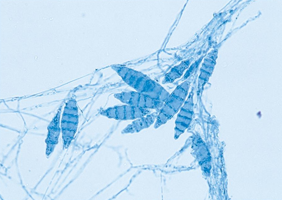
Spindle shaped, with echinulate, thick walls. They measure 12 to 25 um and have 3 to 15 cells. The tapering, sometimes elongated, spiny distal ends are key features that distinguish this species. Colonies are fluffy and white, with the reverse side of the colony usually developing a lemon-yellow pigment, especially on potato dextrose agar. Worldwide distribution.
Nannizzia gypsea
Measure 8 to 15 um and can have as many as 6 cells. Powdery, granular appearance colony surface. Fresh isolates typically form a tan to buff conidial masses, but this species tends to develop pleomorphic tufts of white sterile hyphae in aging cultures and after serial transfers. Abundant brown to red pigment can form beneath some strains. Found in soils worldwide.
Microsporum audouinii
was responsible for most of the gray patch tinea capitis of children> T. tonosurans replaced it as the leading cause of scalp infection. Cottony white and generally form little or no pigment on the reverse.
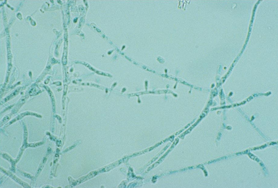
Trichophyon mentagrophytes
Primarily globose but may appear tear shaped and measure 2.5 to 4 um. Microconidia In grape like clusters. Macroconidia are thin walled, smooth, and cigar shaped, with 4 to 5 cells
Trichophyton rubrum
colonies usually remain white on the surface but may be yellow to red. Most strains develop a red to deep burgundy wine colored pigment on the reverse that diffuses into the agar. Worldwide distribution.
Trichophyton tonsurans
Possesses microconidia that are extremely variable in shape, ranging from a round shape to a peg shape. When grown on Sabouraud dextrose agar, colonies usually form a rust colored pigment on the colony’s reverse. Infects skin, hair, and nails, has become the leading cause of tinea capitis in children in many parts of the world.