Ch. 18 LEC - Heart
 Call Kai
 Learn
Learn Practice Test
 Spaced Repetition
 Match
Match Flashcards
 Knowt Play
1/108

Earn XP
Description and Tags
Lecture Exam 2
Name | Mastery | Learn | Test | Matching | Spaced | Call with Kai | Chat |
|---|
No analytics yet
Send a link to your students to track their progress
109 Terms
what is the pericardium?
a multi-layered, fluid-filled sac that encloses and protects the heart
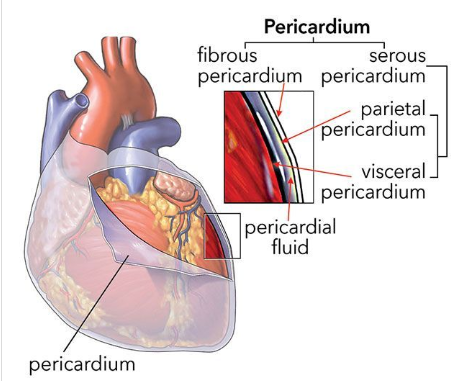
describe the fibrous pericardium
most superficial layer of the pericardium
made of dense connective tissue
protects, anchors, and prevents overfilling of the heart
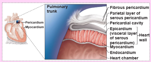
describe the parietal layer of the serous pericardium
2nd most superficial layer of the pericardium
a thin, slippery membrane that produces serous fluid
lines the internal surface of the fibrous pericardium
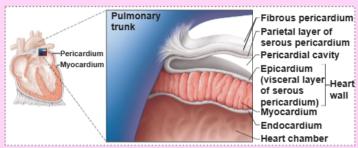
describe the pericardial cavity
fluid-filled space between the parietal and visceral layers of the serous pericardium
reduces friction as the heart beats
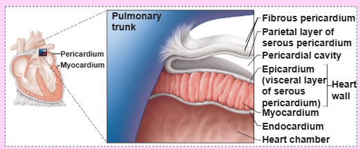
describe the visceral layer (epicardium) of the serous pericardium
layer that sits directly on heart’s surface
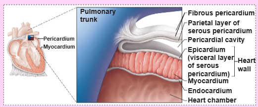
what is another name for the epicardium
visceral pericardium
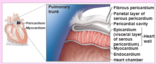
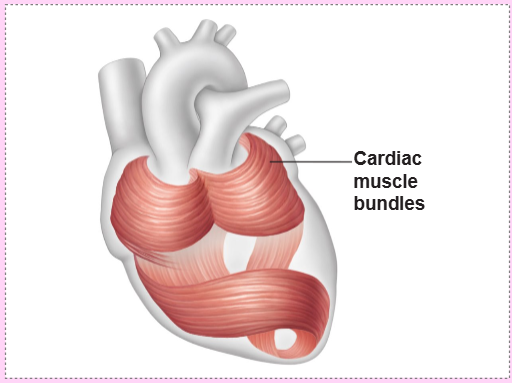
describe the myocardium
histology: spiral bundles of cardiac muscle cells; crisscrossing layer of connective tissue
function:
anchors cardiac muscle fibers
supports great vessels and valves
limits spread of action potentials to specific paths
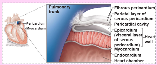
describe the endocardium
thin, inner lining of the heart that covers all 4 chambers and valves
continuous with the endothelium of blood vessels
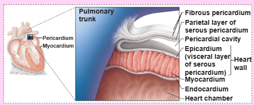
create a flowchart of the layers of the heart (superficial to deep)
fibrous pericardium → parietal serous pericardium → pericardial cavity (fluid) → visceral serous pericardium (epicardium) → myocardium → endocardium → chamber space
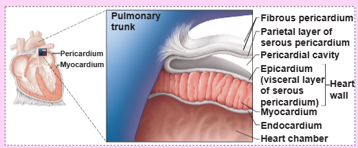
the 2 atria are separated internally by the _
interatrial septum
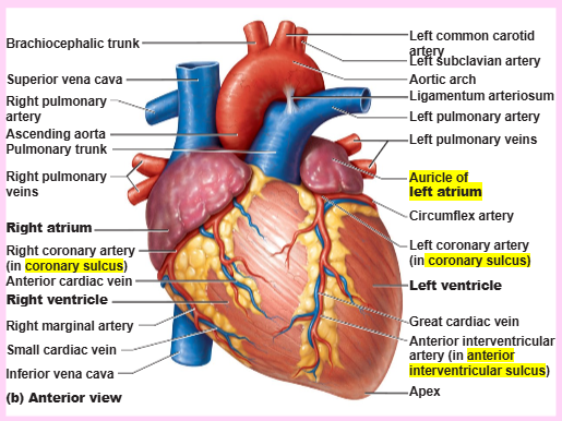
the _ encircles the junction of the atria and ventricles
coronary sulcus (atrioventricular groove)
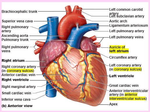
what is the function of the auricles of the heart?
to increase atrial volume
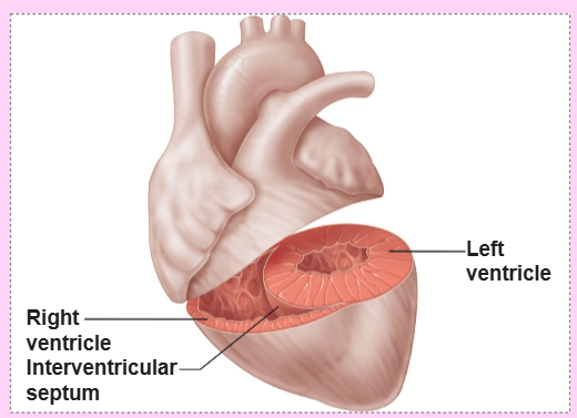
the 2 ventricles are separated by the _
interventricular septum
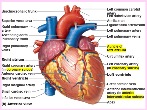
the anterior and posterior _ mark the position of the interventricular septum externally
interventricular sulci
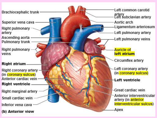
atria: the [RECEIVING/DISCHARGING] chambers
receiving
ventricles: the [RECEIVING/DISCHARGING] chambers
discharging
atria walls are ridged by [MUSCLE]
pectinate muscles
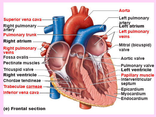
list the vessels that enter the right atrium
superior vena cava
inferior vena cava
coronary sinus
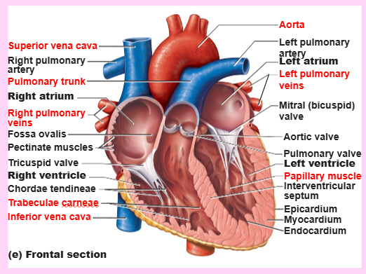
list the vessels entering the left atrium
right and left pulmonary veins
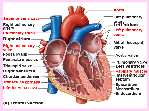
ventricle walls are ridged by [MUSCLE]
trabeculae carneae
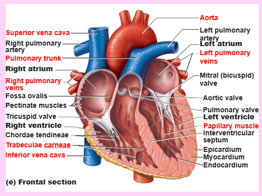
[MUSCLE] project into the ventricular cavities
papillary muscles
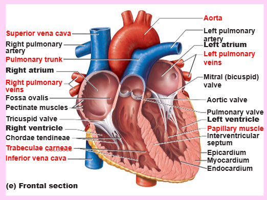
list the vessels leaving the right ventricle
pulmonary trunk
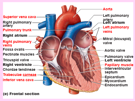
list the vessels leaving the left ventricle
aorta
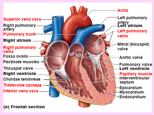
the [LEFT/RIGHT] side of the heart is the pump for the PULMONARY circuit. the vessels carry blood to and from the [LUNGS/ALL BODY TISSUES]
right, lungs
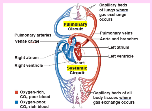
the [LEFT/RIGHT] side of the heart is the pump for the SYSTEMIC circuit. the vessels carry blood to and from the [LUNGS/ALL BODY TISSUES]
left, all body tissues
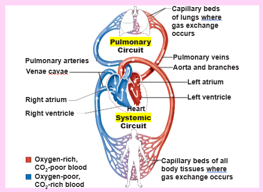
why is the left side of the cardiovascular system (i.e., the SYSTEMIC CIRCUIT) longer and stronger than the right side?
the left side supplies blood to and from all body tissues, which requires more strength and length than the right side that only supplies blood to and from the lungs
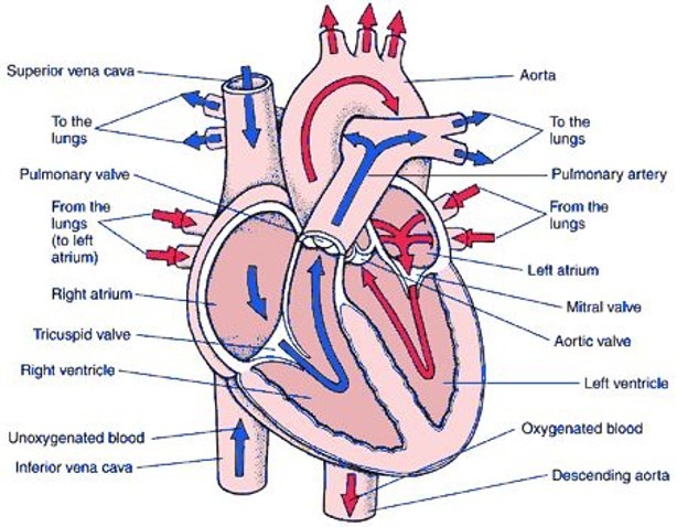
starting with the right atrium, create a flowchart for the blood’s pathway
right atrium → tricuspid valve → right ventricle → pulmonary semilunar valve → pulmonary trunk → pulmonary arteries → lungs → pulmonary veins → left atrium → bicuspid valve → left ventricle → aortic semilunar valve → aorta → systemic circulation → vena cava → right atrium
![<ul><li><p>[EQUAL/UNEQUAL] volumes of blood are pumped to the pulmonary and systemic circuits</p></li><li><p>the pulmonary circuit is a [SHORT/LONG], [LOW/HIGH]-pressure circulation</p></li><li><p>the systemic circuit’s blood encounters [LOW/HIGH] resistance in [SHORT/LONG] pathways</p></li></ul><p></p>](https://assets.knowt.com/user-attachments/1f786500-7514-4f54-bfb7-9a0d03d35d5b.png)
[EQUAL/UNEQUAL] volumes of blood are pumped to the pulmonary and systemic circuits
the pulmonary circuit is a [SHORT/LONG], [LOW/HIGH]-pressure circulation
the systemic circuit’s blood encounters [LOW/HIGH] resistance in [SHORT/LONG] pathways
equal, short, low, high, long
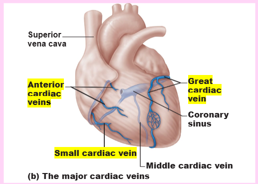
what is the coronary circulation?
the blood supply to the heart muscle itself
![<ul><li><p>coronary circulation</p><ul><li><p>the functional blood supply to the [HEART/LUNGS/BODY TISSUES]</p></li><li><p>arterial supply contains many [NAME] (junctions) among branches</p></li><li><p>[NAME] routes provide additional pathways for blood delivery</p></li></ul></li></ul><p></p>](https://assets.knowt.com/user-attachments/1b7a3871-dd81-4993-9539-baf9b0714707.png)
coronary circulation
the functional blood supply to the [HEART/LUNGS/BODY TISSUES]
arterial supply contains many [NAME] (junctions) among branches
[NAME] routes provide additional pathways for blood delivery
heart, anastomoses, collateral
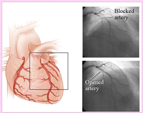
describe angina pectoris
thoracic (chest) pain caused by a fleeting deficiency in blood delivery to the myocardium (temporary blockage); cells are weakened
describe myocardial infarction
also known as a heart attack; prolonged coronary blockage; areas of cell death are repaired with noncontractile scar tissue
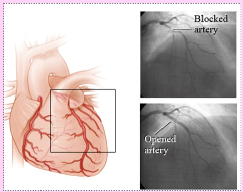
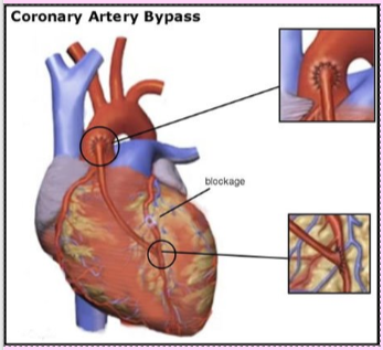
define CABG (coronary artery bypass graft)
a surgery that bypasses/detours a major block in a heart blood vessel by taking another blood vessel from another part of the body
what is the main function of heart valves?
to ensure unidirectional blood flow through the heart
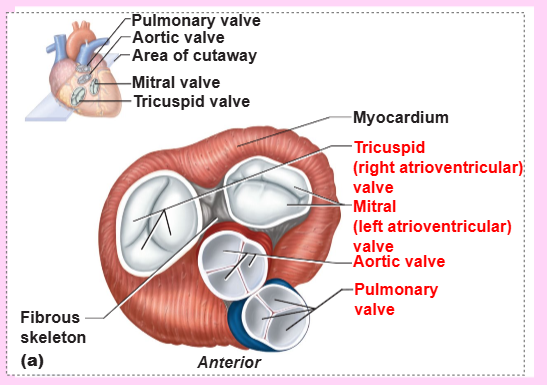
name the 2 atrioventricular (AV) valves
tricuspid valve (right) and mitral valve (left)
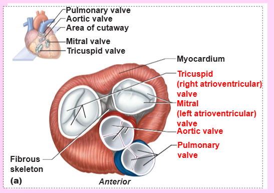
what structure anchors the AV valve cusps to papillary muscles?
chordae tendineae (tendonous chords)
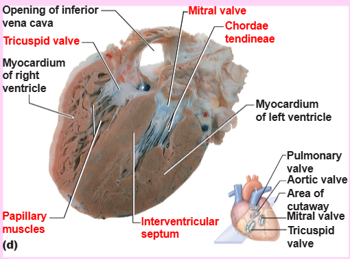
name the 2 semilunar (SL) valves
aortic and pulmonary semilunar valves
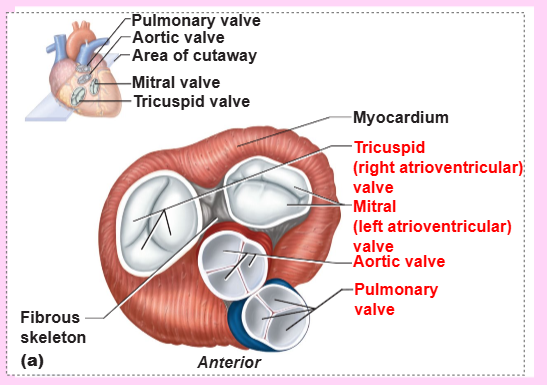
true/false: cardiac muscle cells are striated and interconnected with numerous large mitochondria (25-35% of cell volume)
true - cardiac muscle cells are striated and interconnected with numerous large mitochondria (25-35% of cell volume)
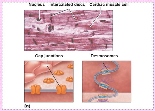
[2 STRUCTURES] allow the cardiac muscle cells to move together in a synchronized way
gap junctions and desmosomes
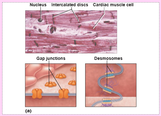
define intercalated discs
junctions between cells that anchor cardiac cells together
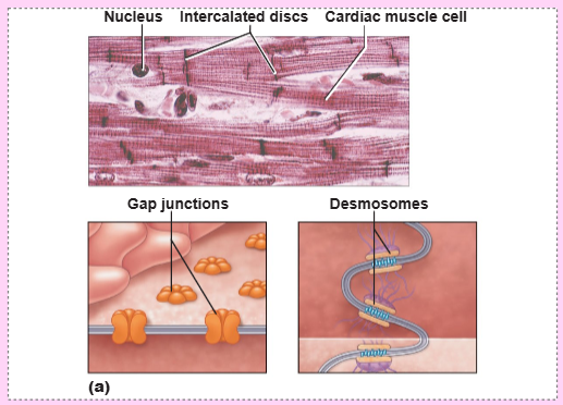
define syncytium (in the context of the heart)
the cells of the heart have distinct cell boundaries, but they are so tightly interconnected that the heart beats in a unified, synchronized rhythm
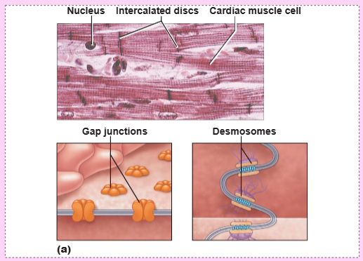
identify the function of desmosomes in the intercalated discs
they prevent cells from separating during contraction
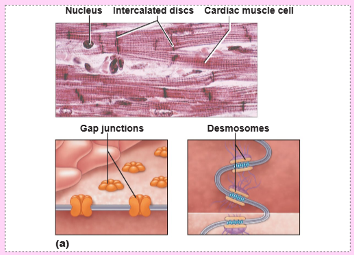
identify the function of gap junctions in the intercalated discs
they allow ions to pass, which electrically couple adjacent cells and ensure that the heart contracts as a unit
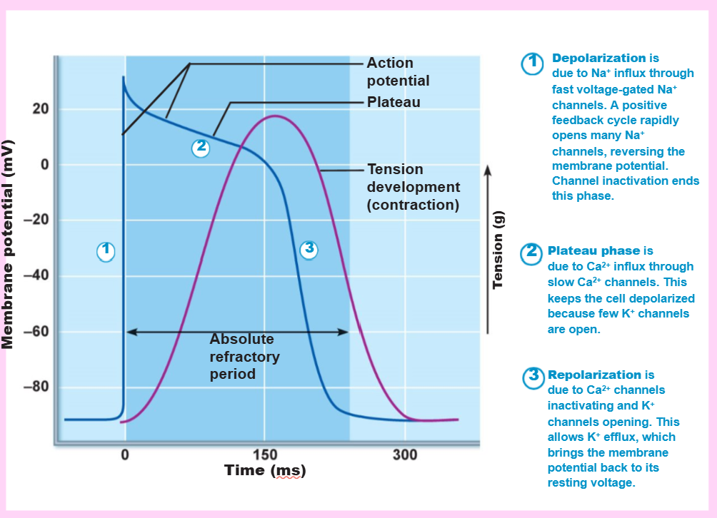
what is the name of the period of time between heart contractions that allows fluid to fill the ventricles before the next contraction?
the long absolute refractory period (250 milliseconds)
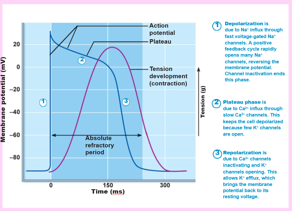
how long is the long absolute refractory period?
250 milliseconds
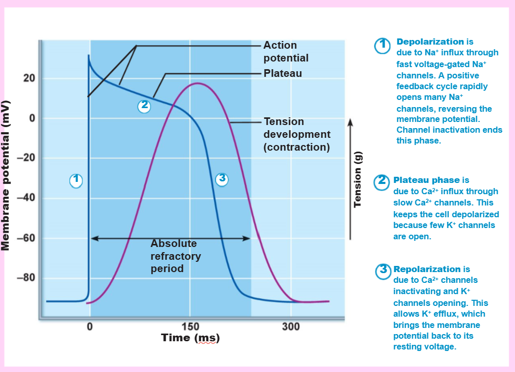
![<p><strong>cardiac muscle contraction</strong></p><ol><li><p>[KEY TERM] opens voltage-gated fast Na+ channels in the sarcolemma</p></li><li><p>reversal of membrane potential from [NUMBER] mV to [NUMBER]</p></li><li><p>depolarization waves cause [TYPE] channels to open</p></li><li><p>Ca2+ surge prolongs the [KEY TERM] phase (hint: shape)</p></li><li><p>[KEY TERM] results from the inactivation of Ca2+ channels and opening of voltage-gated K+ channels</p></li></ol><p></p>](https://assets.knowt.com/user-attachments/098c9298-c53b-4544-b4b7-80a4fd2fac8a.png)
cardiac muscle contraction
[KEY TERM] opens voltage-gated fast Na+ channels in the sarcolemma
reversal of membrane potential from [NUMBER] mV to [NUMBER]
depolarization waves cause [TYPE] channels to open
Ca2+ surge prolongs the [KEY TERM] phase (hint: shape)
[KEY TERM] results from the inactivation of Ca2+ channels and opening of voltage-gated K+ channels
depolarization, -90, +30, Ca2+, plateau, repolarization
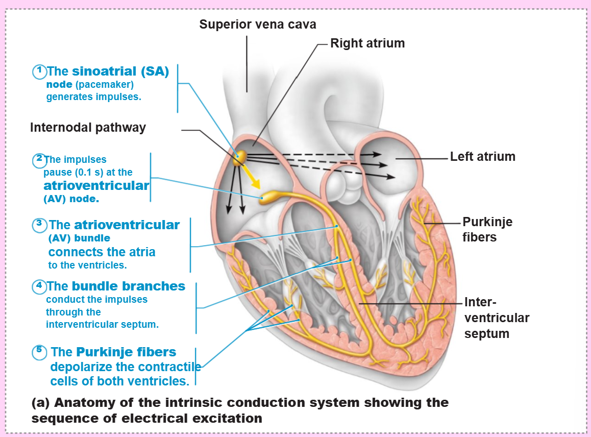
define “the intrinsic cardiac conduction system”
a network of noncontractile, autorhythmic cells that initiate and distribute impulses to coordinate the depolarization and contraction of the heart (without needing external nerve signals)
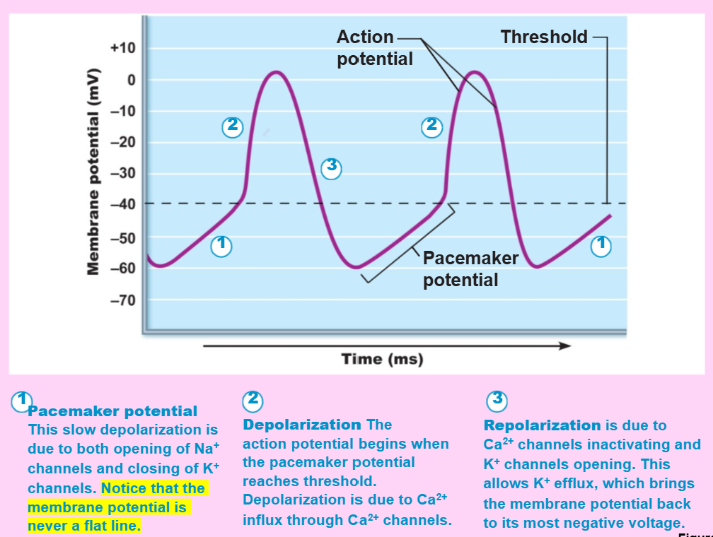
autorhythmic cells (pacemaker cells) have [STABLE/UNSTABLE] resting potentials
unstable
create a flowchart for the heart’s sequence of electrical excitation
SA node → AV node → AV bundle → bundle branches → purkinje fibers
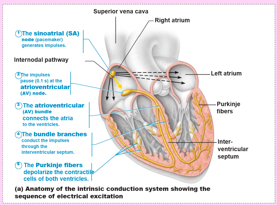
the _ generates impulses in the heart
sinoatrial (SA) node (pacemaker)
how many times per minute does the sinoatrial (SA) node generate an impulse (sinus rhythm)?
75 times/minute (0.8 seconds/beat)
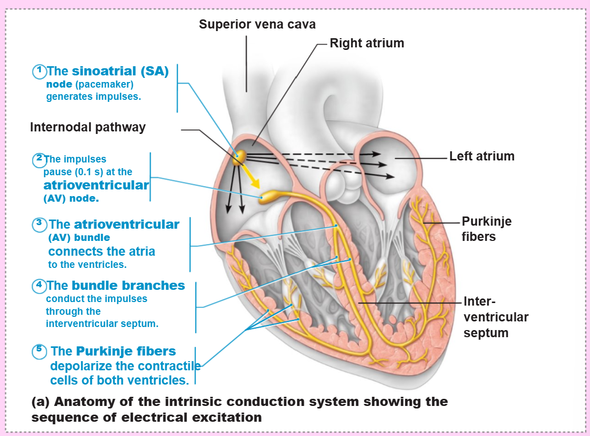
name the structure that fits this description:
delays impulses ~0.1 seconds
has smaller diameter fibers; fewer gap junctions
depolarizes 50 times/minute in absence of SA node input
atrioventricular (AV) node
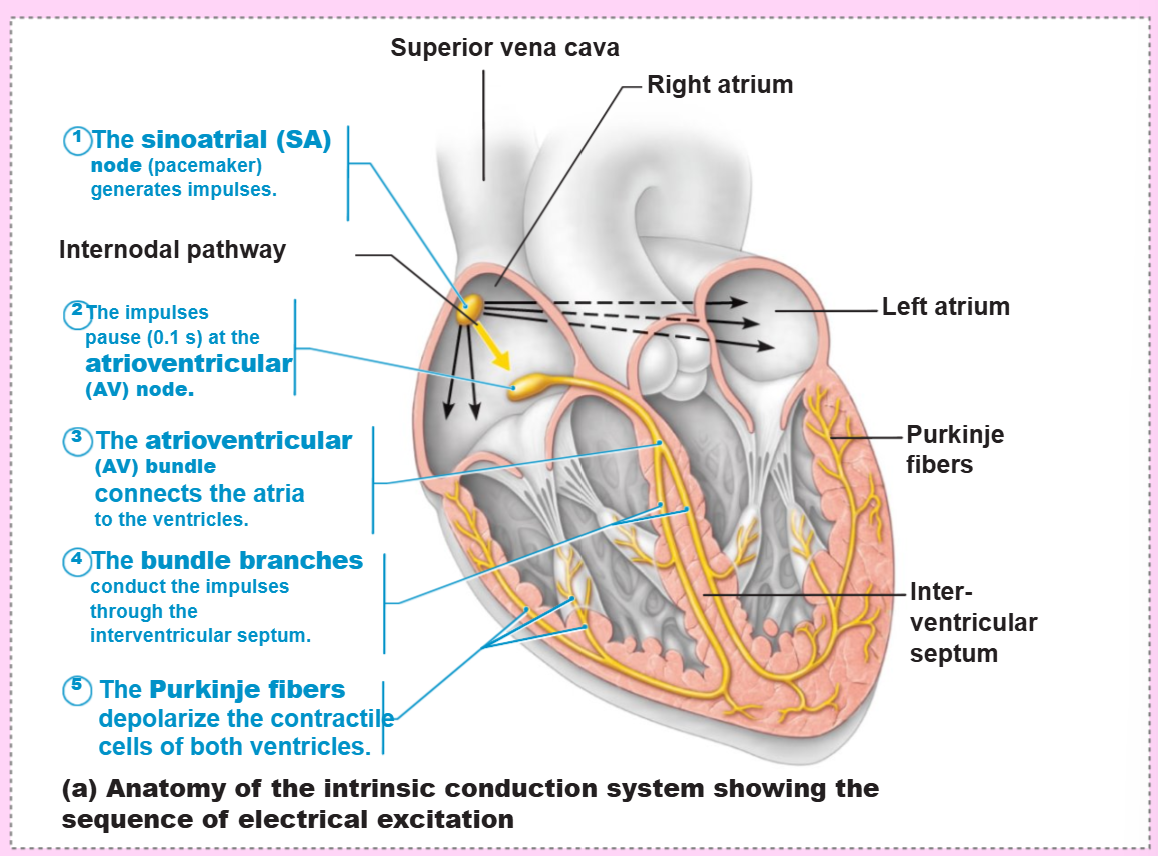
the _ is the only electrical connection between the atria and ventricles
atrioventricular (AV) bundle (bundle of His)
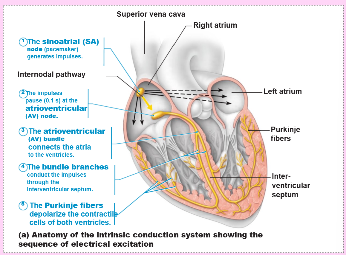
the _ are two pathways in the interventricular septum that carry the impulses toward the apex of the heart
right and left bundle branches
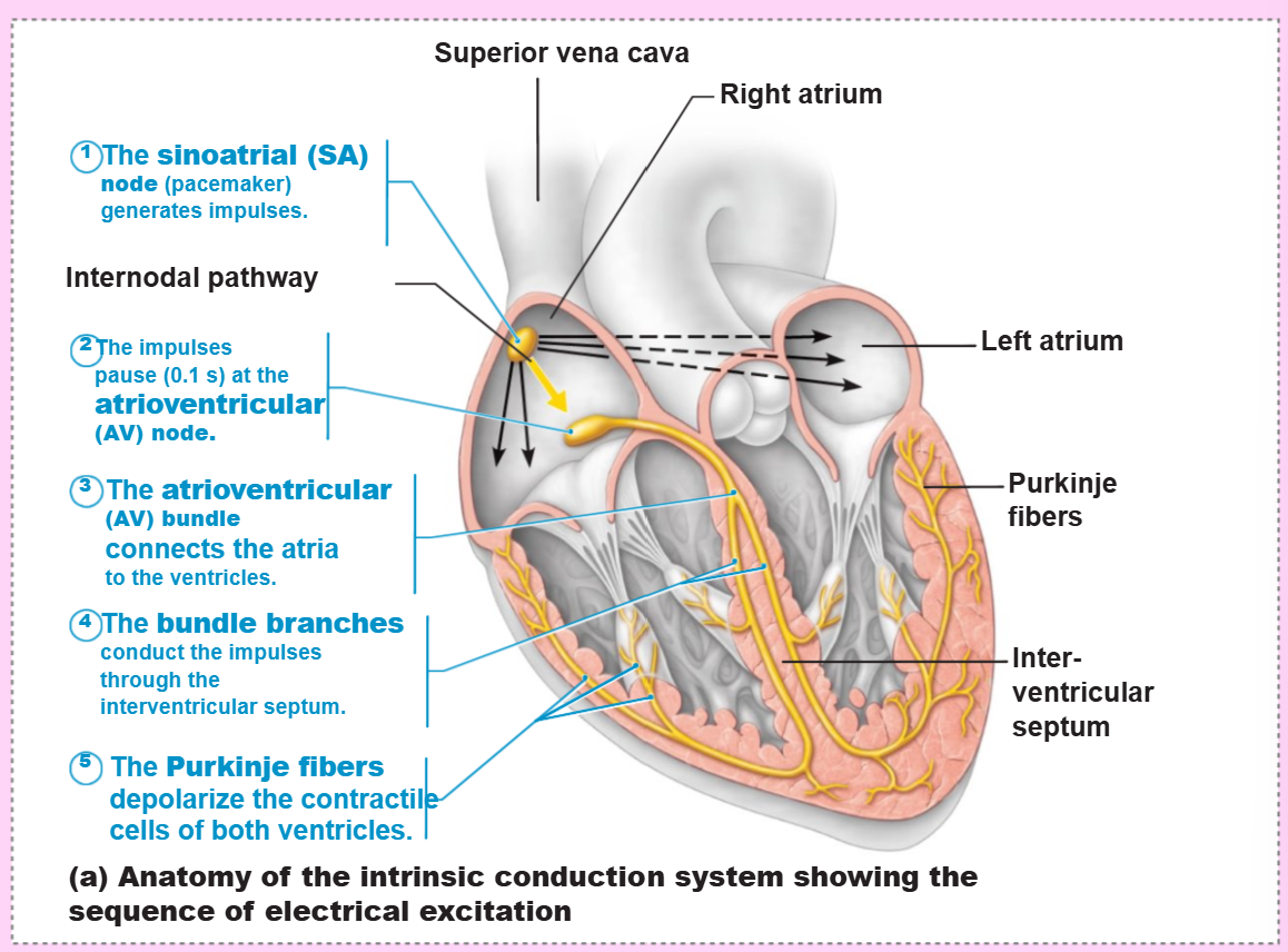
the _ complete the electrical pathway into the apex and ventricular walls
purkinje fibers
what are 3 situations that arise from defects in the intrinsic conduction system?
arrhythmias (irregular heart rhythm)
uncoordinated atrial and ventricular contractions
fibrillation (rapid, irregular contractions that is useless for pumping blood)
a defective SA node may result in _
ectopic focus (abnormal pacemaker cells take over)
the AV node taking over, causing a junctional rhythm (40-60 bpm) (low heart rate)
a defective AV node may result in _
a partial or total heart block
few or no impulses from the SA node reaching the ventricles
what system modifies the heartbeats?
the autonomic nervous system
what part of the brain innervates the SA node, AV node, heart muscle, and coronary arteries through sympathetic neurons?
the cardioacceleratory center of the medulla oblongata
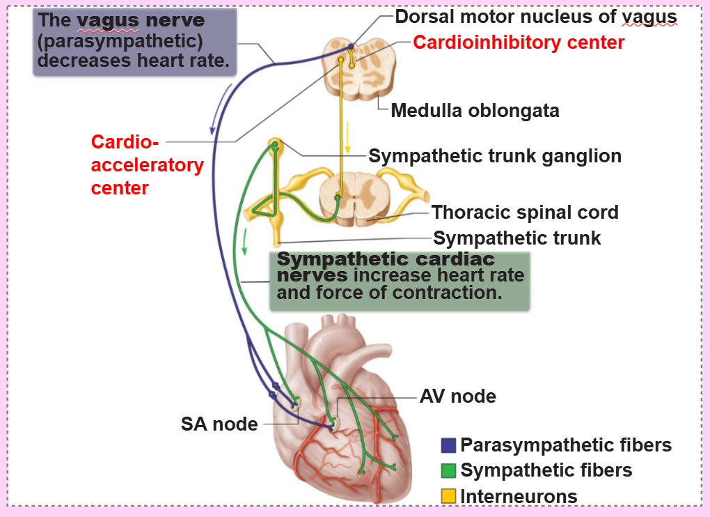
what part of the brain inhibits the SA and AV nodes through parasympathetic fibers in the vagus nerves?
the cardioinhibitory center of the medulla oblongata
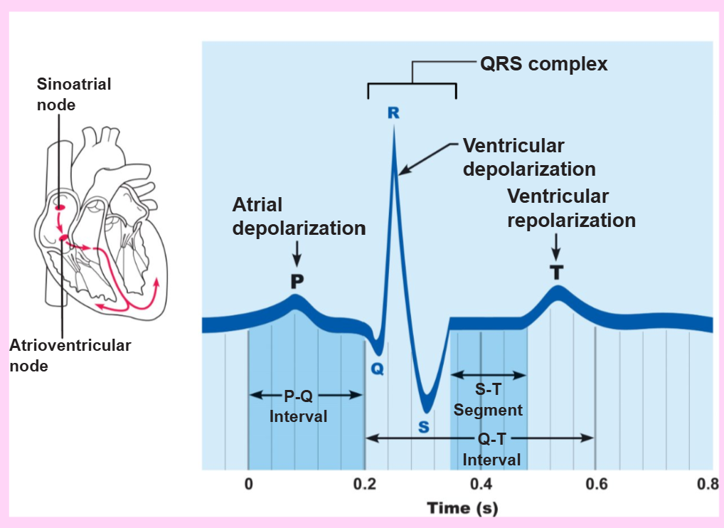
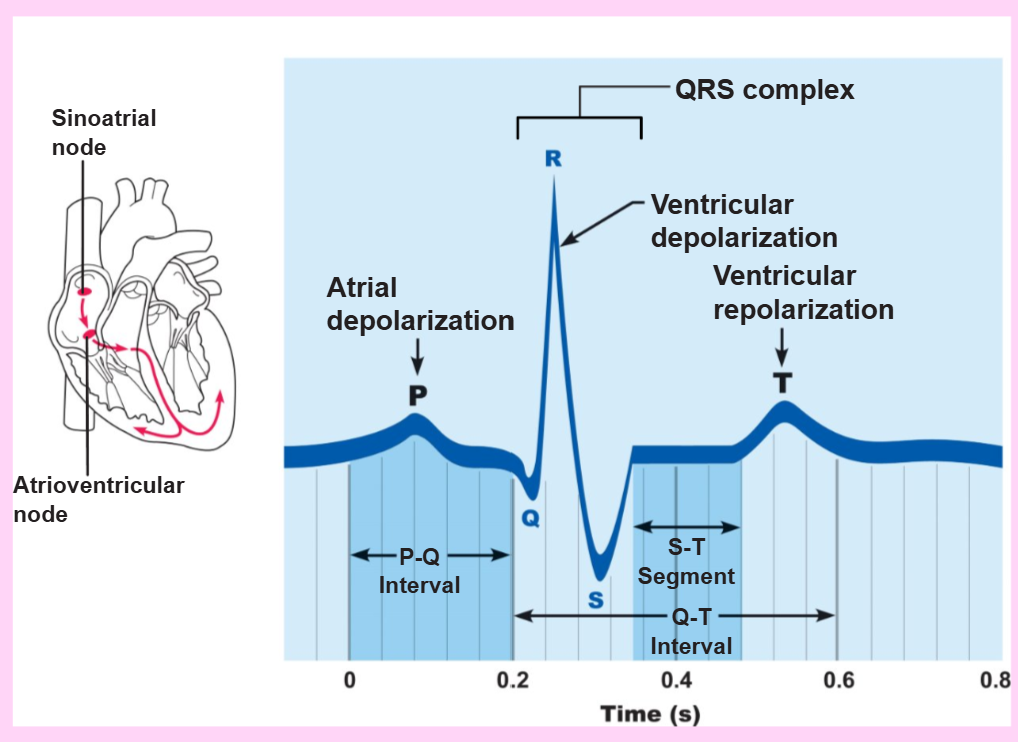
define electrocardiogram (ECG or EKG)
a composite of all the action potentials generated by nodal and contractile cells at a given time; it is a tool to see if the heart is working properly
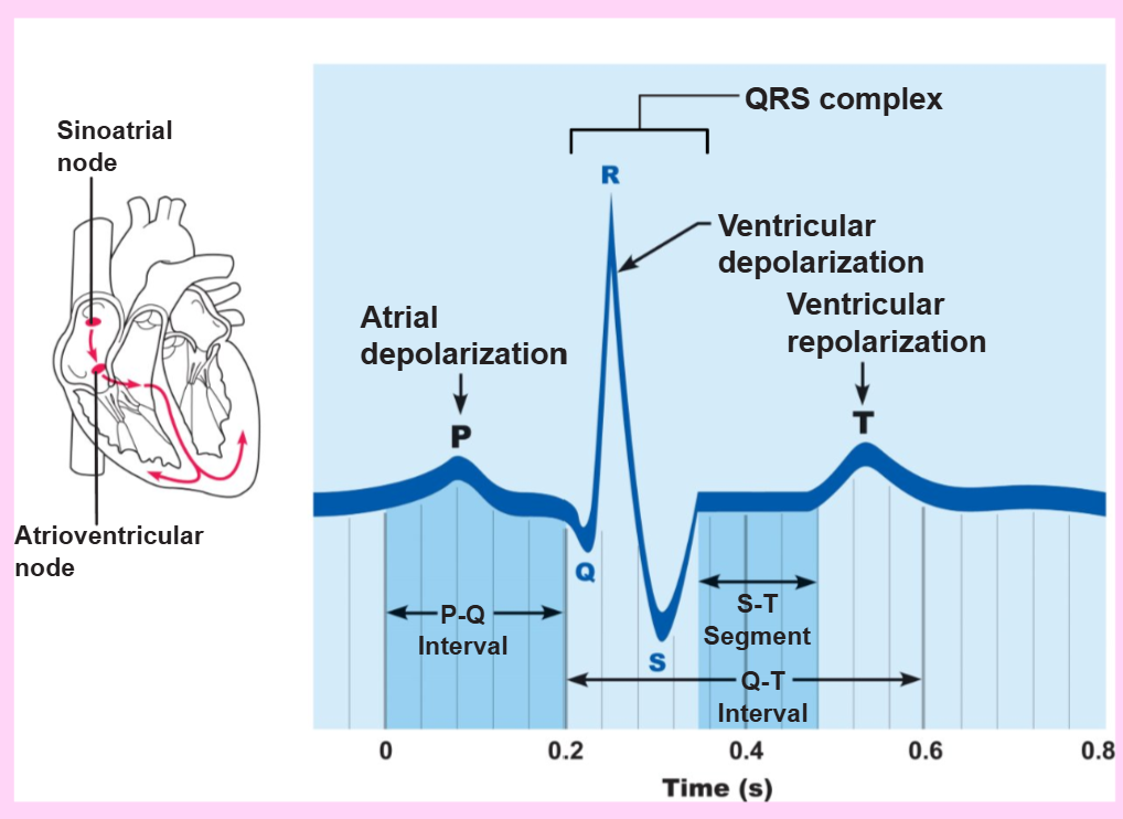
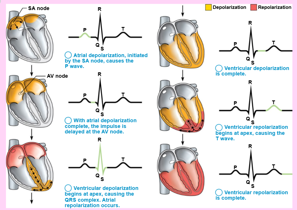
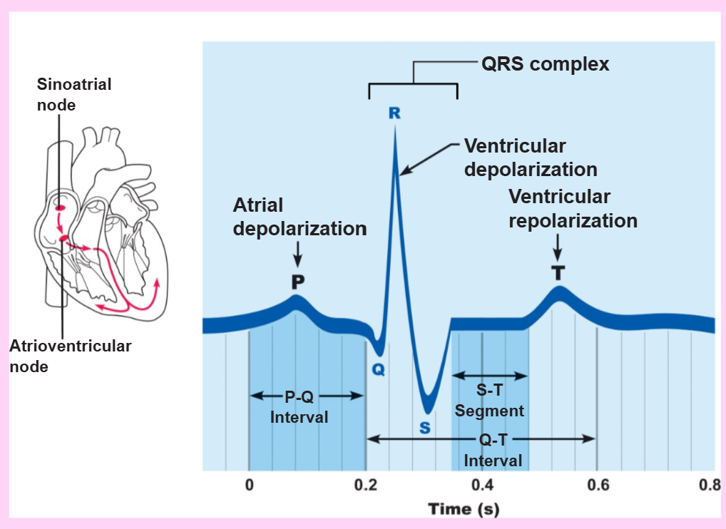
name the waves seen in a typical electrocardiograph (EKG)?
P wave, QRS complex, and T wave
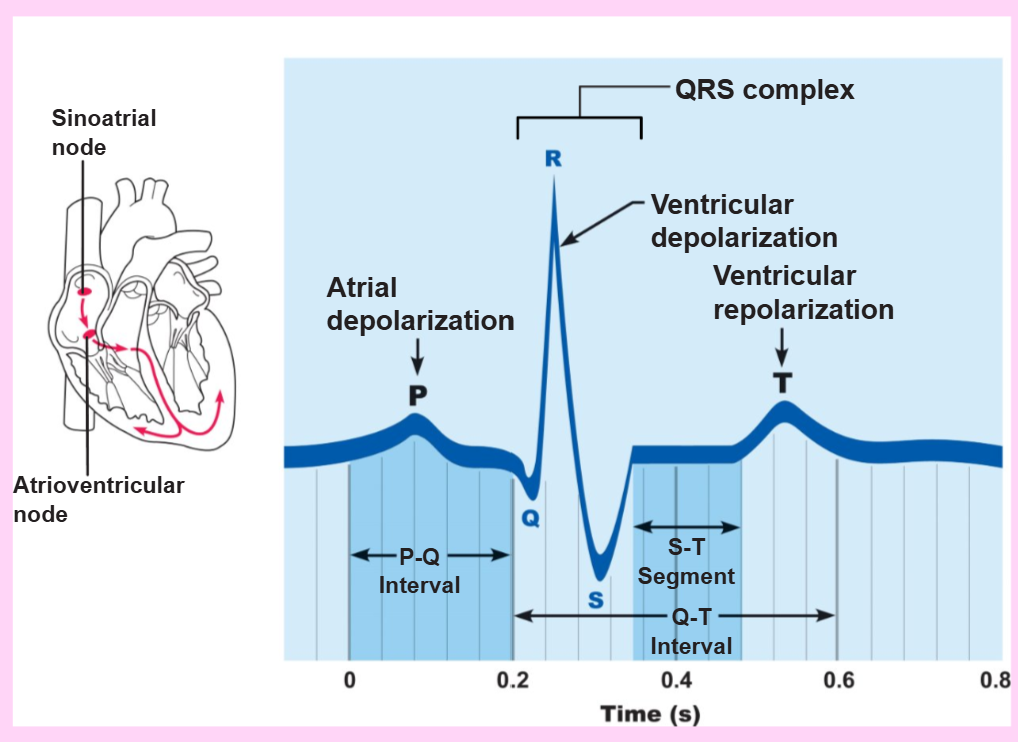
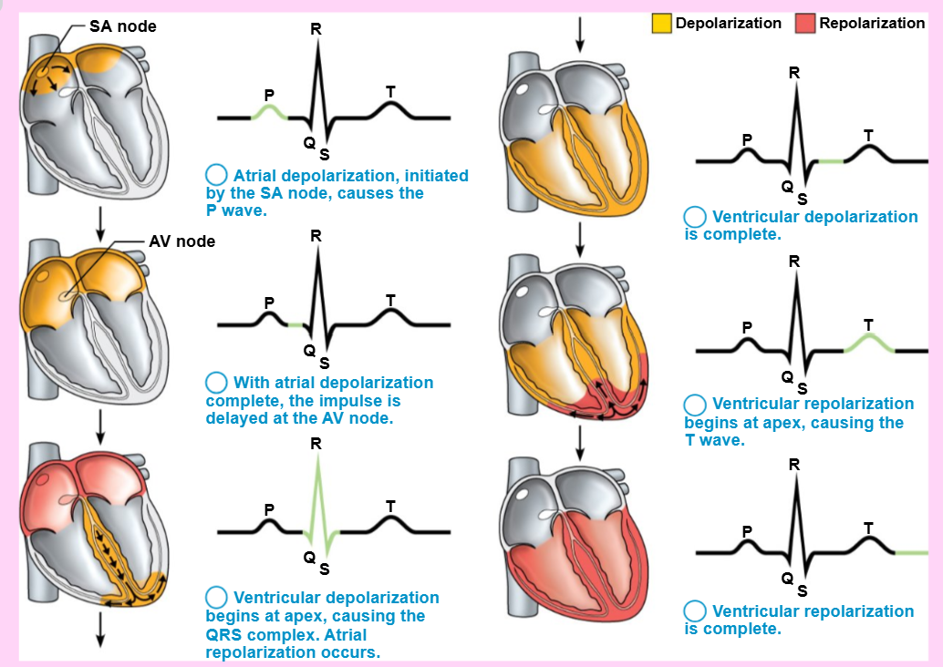
the depolarization of the SA node is during the _ wave
P
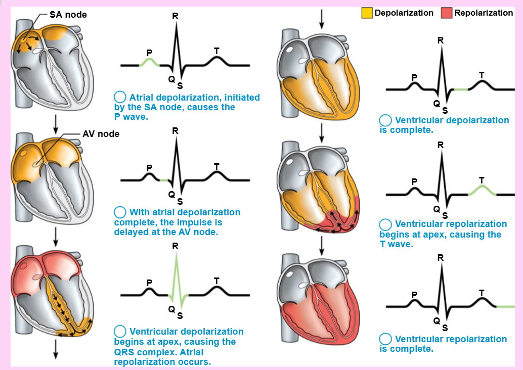
the ventricular depolarization is during the _ complex
QRS
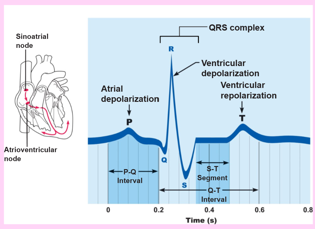
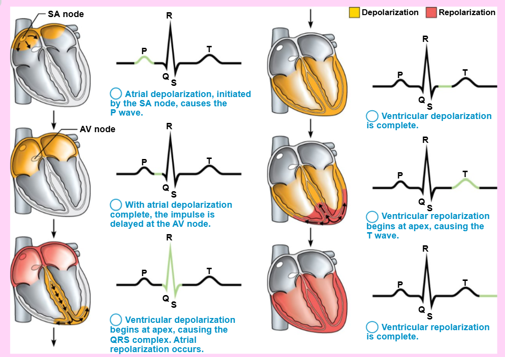
ventricular repolarization is during the _ wave
T
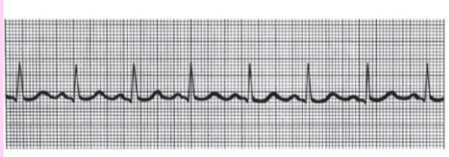
normal sinus rhythm
P wave, QRS complex, and T wave present consistently
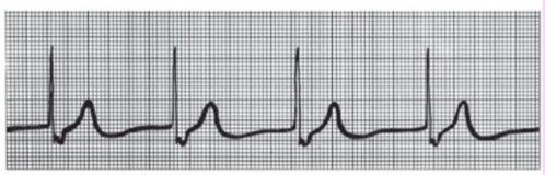
junctional rhythm
SA node is nonfunctional, AV node takes over as main pacemaker (40-60 beats/min), P waves are absent
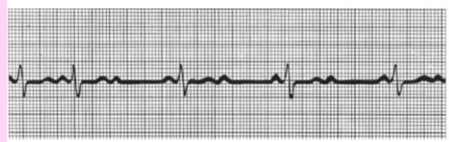
second-degree heart block
some P waves are conducted through the AV node; ratio of P waves to QRS waves is ~2:1; slow heart rate
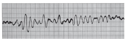
ventricular fibrilation
chaotic, irregular EKG deflections; acute heart attack and electrical shock
what causes the “lub-dup” heart sounds?
the closing of the AV and SL valves, respectively
what are heart murmurs and what causes them?
abnormal heart sounds caused by valve problems
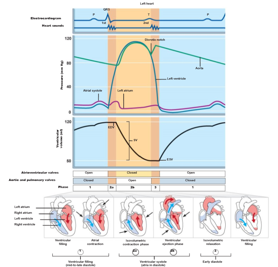
define “cardiac cycle”
all events associated with blood flow through the heart during one complete heartbeat
name the phases of the cardiac cycle
ventricular filling (mid-to-late diastole (rest))
ventricular systole (contraction)
isovolumetric relaxation (early diastole)
define systole
the contraction phase of the heartbeat
define diastole
the resting and refilling phase of a heartbeat
when does ventricular filling occur?
mid-to-late diastole (relaxation)
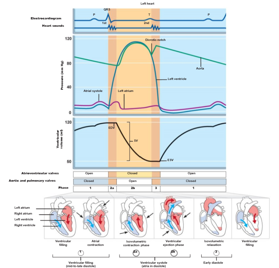
define “end diastolic volume (EDV)”
the volume of blood in each ventricle at the end of ventricular diastole
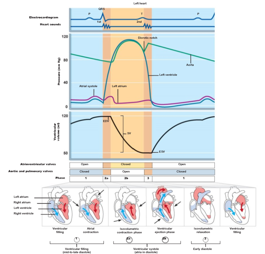
during mid-to-late diastole (rest), [NUMBER]% of blood passively flows into ventricles while [NUMBER]% of blood is delivered during atrial systole (contraction)
80, 20
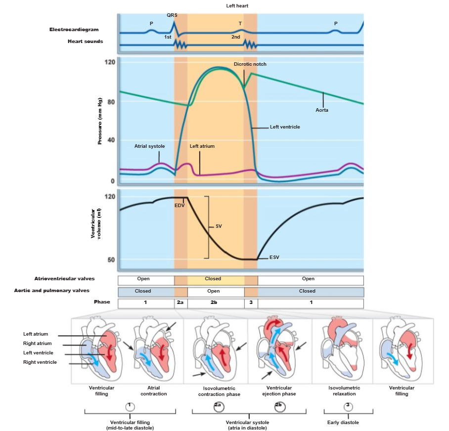
ventricular systole (contraction):
[1ST/2ND/3RD] phase of the cardiac cycle
[ATRIA/VENTRICLES] relax and [ATRIA/VENTRICLES] begin to contract
AV valves [OPEN/CLOSE]
in ejection phase, ventricular pressure [SUBCEDE/EXCEED] pressure in large arteries, forcing the [AV/SL] valves open
2nd, atria, ventricles, close, exceed, SL
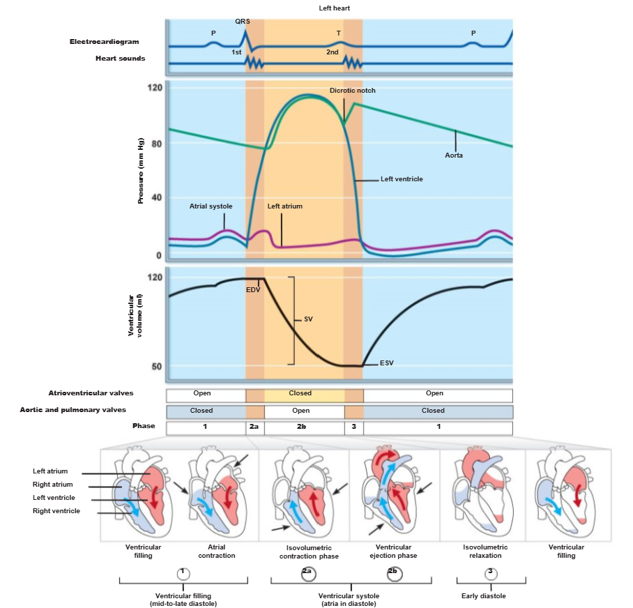
define “end systolic volume (ESV)”
the volume of blood remaining in each ventricle after contraction
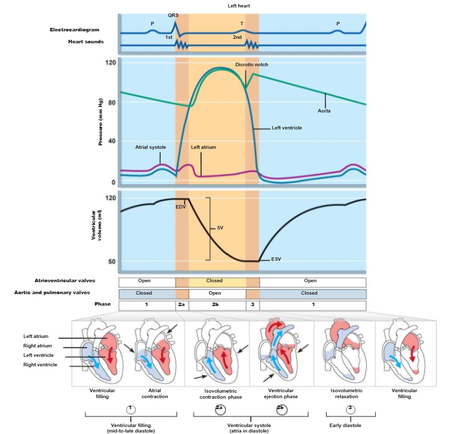
ventricles relax during early diastole, in the 3rd phase of the cardiac cycle called _
isovolumetric relaxation
define “cardiac output (CO)”
the volume of blood pumped by each ventricle in one minute (L/min)
how does one calculate cardiac output (CO)?
heart rate (beats/min) x stroke volume (volume/beat)
define “cardiac reserve”
the difference between resting and maximal cardiac output (CO)
define “stroke volume (SV)”
the volume of blood a ventricle pumps out per beat
maximal cardiac output is [NUMBER RANGE] times the resting cardiac output in nonathletic people
4-5
if Sally has a cardiac reserve of 7.00 L/min and Josh has a cardiac reserve of 6.00 L/min, which person has a stronger heart?
Sally - she has a higher cardiac reserve
how does one calculate stroke volume (SV)?
EDV - ESV
name the factors that affect stroke volume (SV) (how much blood can be pumped out of the heart)
preload
contractility
afterload
increased preload = [DECREASED/INCREASED] SV
increased contractility = [DECREASED/INCREASED] SV
increased afterload = [DECREASED/INCREASED] SV
increased, increased, decreased
define “preload” (related to the Frank-Starling law of the heart)
the degree of stretch of cardiac muscle cells before they contract
at rest, cardiac muscle cells are [SHORTER/LONGER] than optimal length
shorter
define “contractility”
contractile strength at a given muscle length (independent of muscle stretch and EDV)
what causes increased heart contractility?
positive inotropic agents (e.g., increased calcium and hormones like thyroxine, glucagon, and epinephrine)
what causes decreased heart contractility?
negative inotropic agents (e.g., acidosis, increased extracellular potassium, and calcium channel blockers)
define “afterload”
the pressure (of the aorta and the pulmonary trunk) that must be overcome for ventricles to eject blood
how does hypertension (chronic high blood pressure) affect afterload?
hypertension increases afterload (pressure), resulting in increased ESV and reduced SV
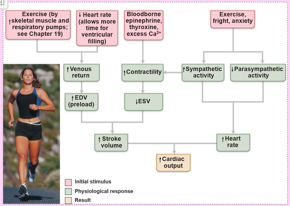
_ factors increase heart rate
positive chronotropic
_ factors decrease heart rate
negative chronotropic