Lecture 4: Synaptic Transmission, Neurotransmitters, and Receptors
 Call Kai
 Learn
Learn Practice Test
 Spaced Repetition
 Match
Match Flashcards
 Knowt Play
1/56

There's no tags or description
Looks like no tags are added yet.
Name | Mastery | Learn | Test | Matching | Spaced | Call with Kai | Chat |
|---|
No analytics yet
Send a link to your students to track their progress
57 Terms
bipolar
neuron structural type
one dendrite, one axon
goes opposite direction in the body
Ex. retina, etc
multipolar
neuron structural type
motor neurons and interneurons
most common type in CNS
unipolar
neuron structural type
psuedounidpolar
peripheral and central nervous branches (off a single process)
peripheral NS is sensory
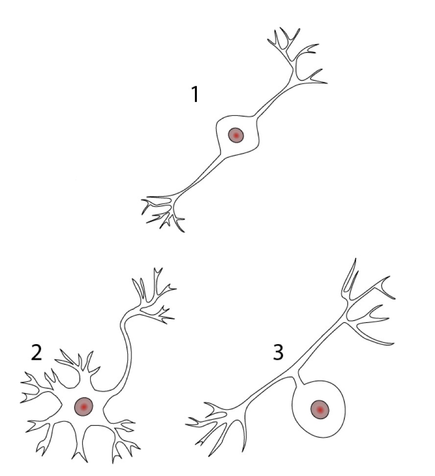
Name the neuron structural type indicated by 1, 2, and 3.
bipolar
multipolar
unipolar
What are the two types of synapses?
electrical and chemical
In an electrical synapse, the current flows between cells via the __________.
gap junctions
Electrical synapse occurs in what types of tissues? are the faster or slower than a chemical synapse?
cardiac and smooth mm.
faster communication and conductivity than a chemical synapse
In a chemical synapse, the current flows between cells via the _________.
synaptic cleft
3 steps of a chemical synapse
an AP in the presynaptic cells causes the opening of VGCCs on the membrane
Ca2+ influx causes the release of neurotransmitter from the presynaptic terminal (into synaptic cleft)
neurotransmitter binds receptors on the postsynaptic membrane
can lead to either hyper- or depolarization depending on whether it is excitatory or inhibitory (can be both)
Neurons are arranged in circuits where _______ cells synapse on _____ cells.
input
output
Neurons are arranged in circuits where input cells synapse on output cells. The output cell may or may not fire an _______ depending on the amount of __________ provided by the input cells.
action potential
excitation
Types of synaptic arrangements
one-to-one synapses
one-to-many synapses
many-to-many synapses
one-to-one synapses
neuromuscular junction
single AP in motoneuron causes a single AP in muscle fiber
neuromuscular junction
single AP in motoneuron causes a single AP in muscle fiber
This describes which synaptic arrangement between neurons?
one-to-one synapses
one-to-many synapses
found in some motoneurons of the spinal cord
one presynaptic neuron synapses with multiple postsynaptic neurons
inputs can be excitatory or inhibitory
found in some motoneurons of the spinal cord
one presynaptic neuron synapses with multiple postsynaptic neurons
inputs can be excitatory or inhibitory
This describes which synaptic arrangement between neurons?
one-to-many synapses
many-to-many synapses
sum of inputs determines whether depolarization is enough to generate an action potential
many presynaptic cells converge on a postsynaptic cell
common
most neurons integrate thousands of excitatory and inhibitory inputs before firing
sum of inputs determines whether depolarization is enough to generate an action potential
many presynaptic cells converge on a postsynaptic cell
common
most neurons integrate thousands of excitatory and inhibitory inputs before firing
This describes which synaptic arrangement between neurons?
many-to-many synapses
excitatory postsynaptic potentials (EPSPs)
presynaptic neuron depolarizes postsynaptic neuron, brings neuron’s membrane potential to threshold, and opens NA and K channels
major excitatory NTS;
glutamate (CNS)
acetylcholine (NMJ)
What is the major excitatory NT for an excitatory postsynaptic potential in the NMJ? CNS?
acetylcholine
glutamate
inhibitory postsynaptic potentials (IPSPs)
presynaptic neuron hyperpolarizes postsynaptic neuron, membrane potential moves away from threshold, opens Cl channels
major inhibitory NTs;
GABA
glycine
What are the major NTs of inhibitory postsynaptic potentials?
GABA and glycine
Many neurotransmitters can be excitatory or inhibitory depending on the receptor _______.
subtype
spatial summation
two EPSPs arrive at the postsynaptic cell simultaneously
the depolarizations summate
temporal summation
two inputs arrive at postsynaptic cell in rapid succession
if they are both inhibitory or excitatory, they will summate
synaptic fatigue
repeated stimulation yields a smaller than expected response
An excitatory stimulus → ____________ → Na+ _______
depolarization
influx
ionotropic receptors
ligand-gated ion channels that usually open in response to the binding of a NT
usually located along dendrites or cell bodies and recieve incoming information form other neurons
effects of ionotropic receptors on the psotsynaptic neuron
may be excited due to Na+ influx → depolarization
may be inhibited due to K+ efflux or Cl- influx (negative charge in cell) → hyperpolarization
Ca2+ influx can contribute to depolarization, but primarily acts in activation of intracellular signalingW
hat are the NTs that activate ionotropic receptors?
glutamate
acetylcholine
GABA
Glycine
Glutamate
NT that activates ionotropic receptors
amino acid
primary excitatory NT in CNS
AMPA and NMDA receptors
Types of glutamate receptors
AMPA
NMDA
AMPA receptors
glutamate receptor
causes depolarization when receptor opens
allows Na+ influx and K+ efflux
gradient caused by Na+ is higher → positive charge in cell → depolarization
NMDA receptors
glutamate receptor
needs both glutamate to bind and a voltage change
Mg block on channel is removed
important for learning and memory centers in the brain
acetylcholine
activate ionotropic receptors
neurotransmitter used at all vertebrate NMJs
also used by ANS (PSNS and SNS)
Glycine and GABA
NT that activates ionotropic receptors
both AA
both used in CNS and both bind to chloride channels
binding of these causes inhibition (an increase of Cl- permeability is inhibitory)
GABAA receptor is associated with chloride
metabotropic receptors
type of NT receptors
located along dendrites or cell bodies and recieve incoming information
slower onset, but longer lasting effects compared to ionotropic receptors
can use GPCRs
G-protein-coupled receptors (GPCR)
G-protein activated when neurotransmitter binds to receptor
G-protein alpha subunit binds GTP
Beta subunit may activate an ion channel (K+) - direct G-protein gating
alpha subunit may activate enzymes that promote second messenger synthesis
The alpha subunit of a GPCR may activate enzymes that promote second messenger synthesis. What are those enzymes, and what second messengers would they promote?
phospholipase C
IP3 and DAG
adenylyl cyclase
cAMP
second messengers will activate a protein kinase to give the desired effect in the cell
What NTs activate GPCRs?
norepinephrine
glutamate (metabotropic receptor)
GABA (GABAB receptor)
norepinephrine (NE)
activates GPCRs
uses alpha- or beta-adrenergic receptors
B, a1, a2
Beta-adrenergic GPCRs
NEpi binds
couple to a stimulatory G-protein subunit → activates cAMP second messenger system
alpha-1 adrenergic receptors
NEpi binds
couple to G-protein Gq → activates the phospholipase C second messenger system
alpha-1 adrenergic receptors
NEpi binds
couple to an inhibitory G-protein → suppresses cAMP system
glutamate (metabotropic receptor)
NT that activates GPCRs
acts on postsynaptic sites in CNS
modulates cell excitability and synaptic transmission via second messenger pathways
3 groups of receptors
1, 2, and 3
group 1 glutamate metabotropic receptors
increase neuron excitability
activates phospholipase C pathway
groups 2 and 3 glutamate metabotropic receptors
suppress neuron excitability
inhibit adenylyl cyclase pathway
GABA (GABAB receptor)
NT that activates GPCRs
found in CNS and autonomic division of peripheral NS
GAGA acting via G-proteins linked to K+ channels → hyperpolarize cell at the end of an AP
biogenic amines
norepinephrine (NE)
epinephrine (Epi)
dopamine (DA)
serotonin (5-HT)
histamine
amino acid NTs
glutamate
glycine
gamma-aminobutyric acid (GABA)
NT biochemical property groups (4)
acetylcholine
biogenic amines
amino acid NTs
neuropeptides
neuropeptides
NTs
synthesized and packaged in nerve cell body instead of axon terminal
includes neuromodulators and neurohormones
neuromodulators
VIP, substance P
subgroup of neuropeptides
may act on presynaptic cell to alter amount of NT released
may be co-secreted with NT to alter response of postsynaptic cell
neurohormones
subgroup of neuropeptides
released from neurons into blood
Compare and contrast the speed, duration, receptor, mechanism, and examples of ionotropic and metabotropic receptors.
ionotropic
speed: fast
duration: short
receptor: ion channel
Mechanism: ion movement
Example: nicotinic ACh
metabotropic
speed: slow
duration: long
receptor: GPCR
Mechanism: second messenger
Example: muscarinic ACh
Most synapses in the NS are _______ synapses, where NTs carry information across a ____________.
chemical
synaptic cleft
__________ alter neuronal communication by modifying neutotransmitter release or the responsiveness of target cells.
Neuromodulators