Distal Limb
 Call Kai
 Learn
Learn Practice Test
 Spaced Repetition
 Match
Match Flashcards
 Knowt Play
1/209

There's no tags or description
Looks like no tags are added yet.
Name | Mastery | Learn | Test | Matching | Spaced | Call with Kai | Chat |
|---|
No analytics yet
Send a link to your students to track their progress
210 Terms
Define the terms:
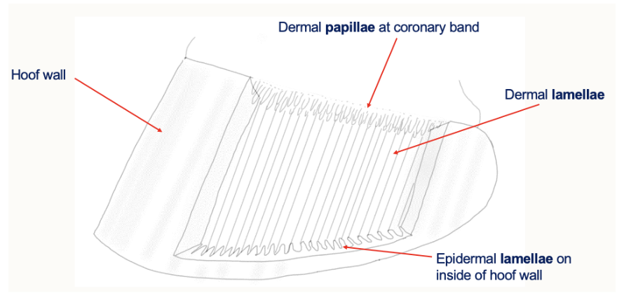
Papillae
Lamellae
Corium
Dermis
Epidermis
Papillae: microscopic, finger-like projections extending from the dermis to the epidermis
Lamellae: folds of tissue which interlock, to suspend the inner coffin bone to the outer hoof wall
Corium: sensitive layer of tissue between the hoof capsule and the internal bones of the foot
Dermis: as above
Epidermis: the outermost protective layer of tissue, forming the hard hoof capsule
Draw out the structure of the equine distal limb

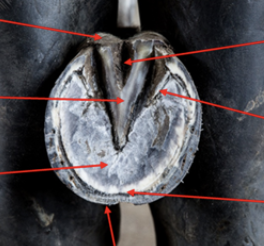
Label the following diagram

Describe the blood supply and venous drainage of the hoof
Supplied by: digital arteries (medial and lateral)
These run within the neurovascular bundles on the palmar/plantar aspects of the distal limb
Venous drainage
The veins run along with the arteries: they are ‘satellite’
There are many venous plexuses around the foot (no valves in these plexuses)
Horses need to be able to move around in order to get the venous blood moving back up the limbs
Draw a diagram to show the blood supply and venous drainage of the hoof

Draw a diagram to show the different structures in a longitudinal cross-section of the hoof
What is the hoof dermis?
Dermis: squidgy, living tissue
Nerves (it has pain sensation)
Blood vessels (it can bleed and become inflamed)
Attached to P3
What is the hoof epidermis?
Epidermis: not squidgy
Thin layer of cells that produce and support the horn
Mostly dead horn
What can be found between the epidermis and dermis of the hoof?
Between the epidermis and the dermis, there is a basement membrane
Describe laminitis
Can be caused by a number of issues
Failure of adhesion of cells to the basement membrane
This results in detachment and mechanical deformation of the dermal-epidermal junction
If this is not managed, this causes rotation of P3
This is a very painful condition that requires prompt, and in some cases, emergency treatment
Describe how the epidermis of the hoof is formed
Squames lost
Gradual keratinization
Loss of nuclei
Division of basal cells
What is the intertubular horn of the hoof made by?
The inter-tubular horn is made by the cells between the bases of the papillae
The dermal papillae are found in the region of the coronary band
Describe how the hoof wall and sole is formed
The hoof wall is produced predominantly by the coronary dermis (dermal papillae)
The perioplic dermis produces a thin, waxy and waterproof cover
The sole is produced by the sole dermis
What is the white line?
The white line is the junction of the sole and the hoof wall
It is important to be able to identify it: seedy toe, white line disease, subsolar abscesses
What are the following structures?
Stratum internum
Stratum medium
Stratum externum
Stratum internum: the layer with the epidermal lamellae
Stratum medium: the bulk of the hoof wall with horn tubules and intertubular horn
Stratum externum: a thin, waxy layer of keratin forming the outer coating of the hoof wall

Label the following image

Describe how the distal deals with weight bearing
P3 is suspended from the hoof wall, rather than bearing weight through the palmar/plantar surface
Collagen fibre bundles run between P3 and the inner aspect of the claw capsule
Describe how the distal limb deals with support and shock absorption
There are internal structures that support the caudal foot:
Hoof/collateral cartilages
Digital cushion
List the joints of the distal limb, including their common name in the horse and cow
Metacarpophalangeal (fetlock in horse and cattle)
Proximal interphalangeal (pastern in horse and cattle)
Distal interphalangeal (coffin in horse and cattle)
Draw a diagram of the equine MCP joint

Draw a diagram of the equine PIP joint

Draw a diagram of the equine DIP joint

Describe the extensor tendons of the digits
Common digital extensor
Located on the dorso-lateral aspect
Receives branches from interosseus
Tendon of insertion is split according to the number of functional digits in the species
Inserts on the extensor process of P3 (distal phalanx)
Describe the flexor tendons of the digits
Located on the palmar/plantar aspects
Superficial digital flexor tendon
Flexes the whole digit and stabilises the fetlock joint
Subcutaneous and palpable
At the distal end of P1, it splits and inserts on P2
Forms a sleeve around the DDFT at the manica flexoria
Deep digital flexor tendon
Runs deep to the SDFT
Passes through the manica flexoria
Inserts on the flexor tuberosity of the distal phalanx of each functional digit
Describe the structure and function of the TIOM
Also known as the TIOM (third interosseous muscle), middle interosseous muscle or suspensory ligament
Varies in structure and function according to the species
Muscular in the dog and cat
Mostly tendinous in the horse
TIOM forms part of the suspensory apparatus of the horse
This supports the fetlock, preventing excessive extension
Describe the pathway of the TIOM
Arises from the proximal metacarpal between splint bones: deep to the SDFT and DDFT
Divides just above the fetlock, before inserting on the abaxial surface of the proximal sesamoids
Extensor branches run to the extensor tendon
Beyond the sesamoid bones, it continues as sesamoidean ligaments to phalanges
Describe the accessory (check) ligaments of the distal limb
The accessory ligament of the SDFT connects to the radius (just above the carpus)
The accessory ligament of the DDFT connects to the carpal region
Function: limiting movement of the tendon to prevent over-stressing, and to support the MCP joint through anchoring flexor tendons
These ligaments are weak/absent in the hindlimb
How do bursae protect the distal limb?
Cushion tendons in areas at risk of damage and distributes pressure
Navicular bursitis can be inflammatory or infectious
How do digital flexor tendon sheaths protect the distal limb?
Encompasses flexor tendons of the fore/hind limbs
Extends from above the fetlock joint to the middle of P2
How do annular ligaments protect the distal limb?
Local thickenings of fascia
Stabilise tendons
List the structures that help to protect the distal limb
Bursae
Digital flexor tendon sheaths
Annular ligaments
Dorsal elastic ligaments
What are the two types of distal forelimb nerve blocks?
Palmar digital nerve block (PDN)
Abaxial sesamoid nerve block
Which nerves innervate:
Forelimb extensors
Forelimb flexors
Extensors: radial nerve
Flexors: median and ulnar nerves
Give the roots, muscles supplied and cutaneous supply of:
Radial nerve
Median nerve
Ulnar nerve
Nerve | Roots | Muscles supplied | Cutaneous supply |
Radial | C7, C8, T1 | Extensors of the elbow, carpus and digits | Craniolateral surface of the forearm Dorsal surface of the digits |
Median | C8, T1 | Flexors of the carpus and digits | Palmar surface of the manus |
Ulnar | C8, T1, T2 | Flexors of the carpus and digits | Caudal surface of forearm and lateral manus |
What do the tibial and fibular nerves supply?
Tibial: extensors of hock, flexors of hindlimb digits
Fibular: flexors of hock, extensors of hindlimb digits
In the forelimb, where does the radial nerve terminate?
The carpus
Where does the dorsal nerve supply of the forelimb come from?
Medial and ulnar nerves
Where does the dorsal nerve supply of the hindlimb come from?
Fibular nerve
Draw a diagram to show the nerve distribution in the forelimbs and hindlimbs

Describe the vasculature of the distal hindlimb
Main supply to the distal hindlimb is the cranial tibial a.
Continues as dorsal metatarsal a.
Describe the vasculature of the distal forelimb
Main supply to the distal forelimb is the median a.
Continues as palmar a.
There is no dorsal a. in the equine forelimb
What are:
Nasalconchae
Ethmoconchae
Nasalchonchae: folds of bone projecting from the side walls of the nasal cavities- towards the midline septum. Has thick mucosa to warm, moisten and protect inspired air
Ethmoconchae: conchae projecting rostrally from the ethmoid bone. Has thick mucosa to warm, moisten and protect inspired air, and are also a site of olfaction
What is the intermandibular symphysis?
Intermandibular symphysis: connection between the two halves of the mandible
What connects the different flat bones of the cranium?
Fibrous joints of the skull
Describe the synovial joints of the skull
Joints in the hyoid apparatus
Temporomandibular joint
Between the condylar process of the mandible and the mandibular fossa of the temporal bone
Contains a joint disc, which divides the joint cavity into two separate chambers
Atlantooccipital joint
Between the occipital bone condyles and C1
How does the skull manage weight?
The wall of the outer skull consists of an outer and inner layer of cortical bone
The middle layer has a reduced spongiosum, and is called Diploe
The surface of all these bones is covered by the periosteum
Major foraminae
Foramen: hole (e.g round foramen- maxillary division V2)
Fissure: slit (e.g orbital fissure- ophthalmic division V1)
Canal: tunnel
What are the ramus and body of the mandible?
Ramus: vertical part of the mandible
Body: horizontal part of the mandible
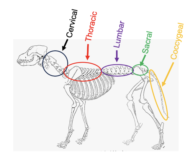
Draw a diagram to show the (rough) vertebral regions of the dog
Draw a table to show the species variations in the number of cervical, thoracic, lumbar and sacral vertebrae. Include the following species
Rabbit
Sheep
Chicken
Dog
Cat
Pig
Horse
Cow
Cervical | Thoracic | Lumbar | Sacral | |
Rabbit | 7 | 12 | 7 | 4 |
Sheep | 7 | 13 | 6 | 4 |
Chicken | 7 | 7 | 14 | |
Dog | 7 | 13 | 7 | 3 |
Cat | 7 | 13 | 7 | 3 |
Pig | 7 | 14 | 6 | 4 |
Horse | 7 | 18 | 6 (Arabs= 5) | 5 |
Cow | 7 | 13 | 6 | 5 |
Draw a diagram to show the bones of the skull and mandible

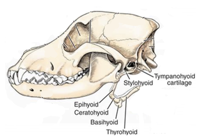
Draw a diagram to show the ones of the hyoid apparatus
What is the function of the hyoid apparatus?
Attachment site for the tongue (cranially)
Attachment site for the airways and gut tube (caudally)
Suspends the tongue, airways and gut tube from the skull
What are the components of:
The tongue
The gut tube
The airway
Tongue:
Glossa (further forward)
Lingua (further backward)
Gut tube:
Pharynx (throat)
Oesophagus (gullet)
Airway:
Larynx (voice box)
Trachea (windpipe)
Describe the functions of the skull
Cranial part: houses and protects the brain and ear
Facial part: houses the nasal and oral cavities and supports teeth
Both contribute to the orbit to protect the eyes
Membranous or flat bones form the roof of the cranium, whereas the primordial bones form the base of the skull
Define the different types of skull
Mesaticephalic: equal proportions
Brachycephalic: short head
Dolichocephalic: long head
What are:
The vertebral foramen
The intervertebral foramen
Vertebral foramen: contains the spinal cord
Intervertebral foramen: in between bones
Describe the different types of a vertebra
Body- mechanical loading
Arch: lamina (layer), pedicle (column/foot)- protects the spinal cord
Processes- attachment for muscles and ligaments
Joint surfaces (facets)- for synovial joints

What type of vertebrae is this?
Cervical

What type of vertebrae is this?
Thoracic
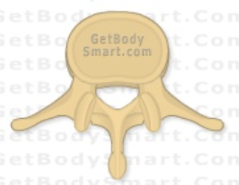
What type of vertebrae is this?
Lumbar
What types of joints are there in the vertebrae?
Fibrocartilagenous joints
Synovial joints
Describe the:
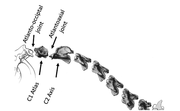
Synovial atlantooccipital joint
Synovial atlantoaxial joint
Draw a diagram to show their location
Synovial Atlantooccipital joint:
Joint between the condyles of the occipital bone, and the cranial articular fovea of the atlas
Allows upward and downward movement only (dorsal and ventral flexion)
Synovial Atlantoaxial joint:
Pivot joint, allows movement and rotation around the axis
Projection (called the dens/tooth) will locate inside bone C1
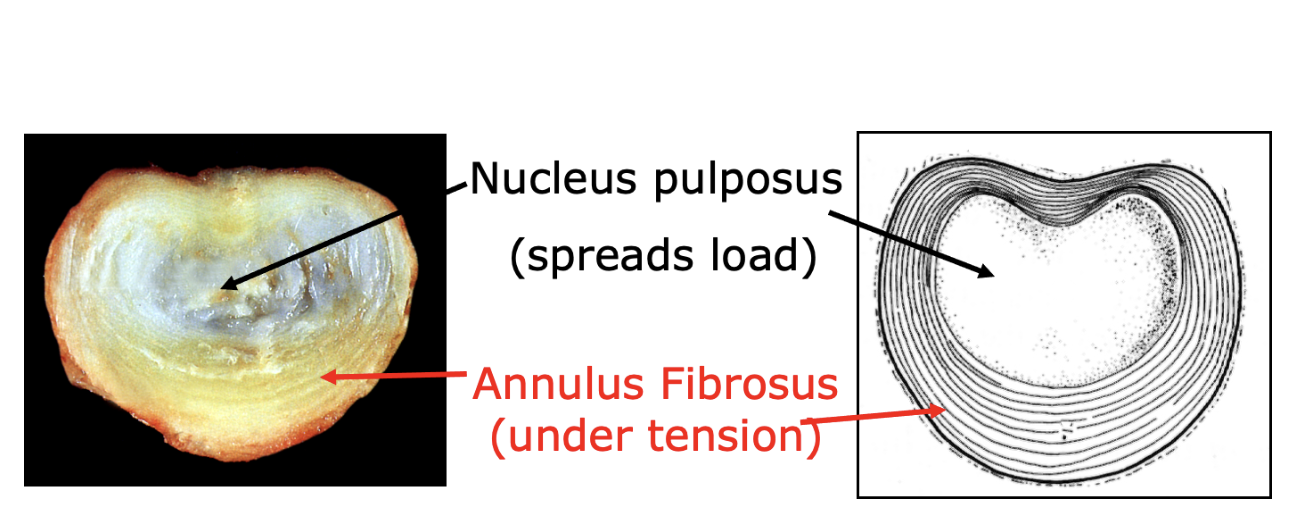
What is the fibrocartilagenous IVD? Draw a diagram to show the different parts
Acts as a shock absorber, and spreads load between bones to avoid overload
Only present between vertebral bodies: not between arches
Parts:
Nucleus pulposus: spreads load
Annulus fibrosus: under tension
List the long vertebral ligaments, including their locations
Nuchal ligament: occipital bone - 3rd/4th thoracic vertebrae
Supraspinous ligament: 3rd thoracic vertebrae - sacrum
Ventral longitudinal ligament: 8th thoracic vertebrae - sacrum
Dorsal longitudinal ligament: inside the vertebral canal, from neck - sacrum
Describe the types of short vertebral ligament
Includes an intertransverse ligament (between the transverse processes)
Must be pierced during paralumbar anaesthesia
Interspinous: between adjacent spinous processes
Interarculate/flavum: inside and between adjacent vertebral arches
Must be pierced during epidural anaesthesia
What is the term for:
Spinal flexion
Spinal extension
Spinal flexion pathology: kyphosis
Spinal extension pathology: lordosis
Describe the two types of muscle group of the spine
Epaxial muscles: will extend the spine if contracting bilaterally
In the body
Iliocostalis (ilium to the first rib)
Longissimus (along the entire back to head)
Transversospinalis (along the entire back)
In the neck
Longissimus
Semispinalis capitis (under splenius)
Hypaxial muscles: will flex the spine if contracting bilaterally
None in the sacrum or thorax
e.g longus colli (in neck)
eg psoas/quadratus (in lumbar region)
Describe how to investigate a lame horse
Clinical examination and history
Palpation of the back, neck and limbs: checking for heat, swelling or pain
Visual evaluation for asymmetry, swelling or abnormal posture
Inspection of the hoof to look for sensitivity, bruising or abscesses
Gait evaluation
Walking and trotting in a straight line
Lunging
Flexion tests
Nerve blocks
Diagnostic imaging
Radiography
Ultrasound
MRI/CT
List the indications and contraindications of performing nerve blocks
Indications
Surgery
Trauma and fractures
Pain management
Diagnostic purposes
Contra-indications
Patient refusal
Infection
Allergies
Lack of resources
Describe the common equine nerve blocks
Palmar digital (heel) block
Targets the caudal third of the foot, including the navicular bone
The nerve runs in the neurovascular bundle, about 1cm proximal to the cartilage of the foot
Abaxial sesamoid block
Desensitises the entire foot, including the proximal interphalangeal (pastern) joint
Numbs the palmar and plantar digital nerves
Low four-point block
Desensitises the fetlock joint and everything below it
What are the issues around nerve blocks?
Injection site: bruising, soreness, bleeding, localised infection
Nerve injury: temporary tingling, numbness or weakness
Inadequate relief: the nerve block may fail to work
What are the most common diseases affecting the ovine foot?
The most important causes of lameness are scald, foot rot and CODD- which are all contagious, and caused by bacteria
Describe the bacteria responsible for causing foot rot in sheep
Dichelobacter nodosus is the essential organism for foot rot
Scald (interdigital dermatitis) is caused by various bacteria that invade the surface layers of the skin between the claws
This usually follows damage by moisture, frost and/or mechanical damage
One common bacteria associated with scald is Fusobacterium necrophorum
However, if D.nodosus is present on the farm, foot rot develops in the first stage as scald, and in the second stage characterised by under-running of hoof horn, and a characteristic smell
F.necrophorum is ubiquitous in the environment, and impossible to eliminate from a farm completely
Both F.necrophorum and D.nodosus are anaerobic bacteria
Describe the spread of foot rot in sheep
D.nodosus is adapted to live and survive on the feet of sheep
Can survive on pasture or bedding for up to 14 days
Can survive in hoof horn clippings for a long time
Infection can spread between sheep if they walk on the same surface
Lame sheep can be treated with a combination of antibiotic injections, and topical antibiotic sprays
In a small number of sheep, the infection can be very difficult to treat- they tend to get repeat infections, and are a source of infection to the rest of the flock

Label the following image
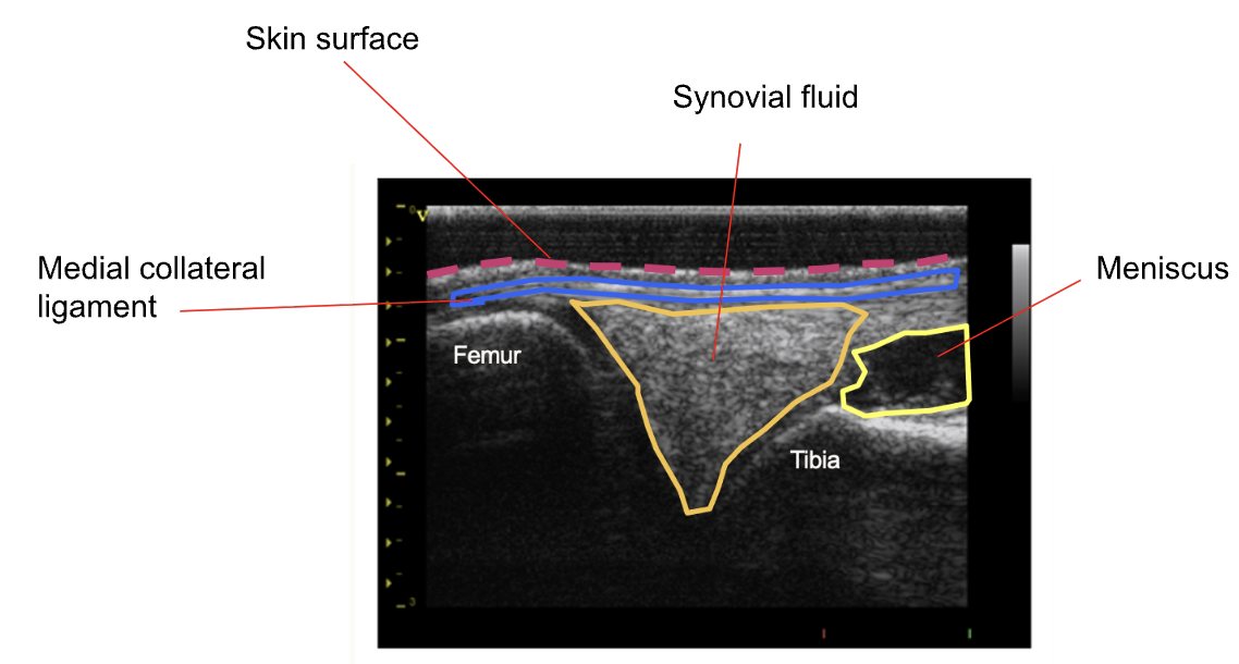
Describe the advantages and disadvantages of ultrasonography in investigating joints
Advantages
No radiation risk
Real-time imaging
Soft tissue detail (cartilage, ligaments, muscles)
Disadvantages
Operator dependent
Can be stressful for the animal
Harder to interpret
Describe the advantages and disadvantages of radiography in investigating joints
Advantages
Cost-effective
Non-invasive
Good for bone and thoracic imaging
Easy to send off for referral
Disadvantages
Radiation exposure
Limited soft tissue detail
Patient movement/artefacts
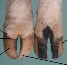
Label the following image


Label the following image
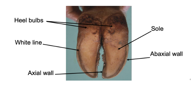
List the equipment used in hoof trimming cattle
Hoof nippers
Hoof knives
Hoof gauge
Hoof testers
Arm guards/wrist protectors
How do you ensure correct toe length when trimming the feet of cattle? Draw a diagram to support
Start with the stabilising claw (medial hind claw or lateral front claw)
Measure an appropriate length from where the claw horn goes palpably hard below the coronary band
Use hoof nippers to cut excess toe length that exceeds 85-90

What is ataxia?
Ataxia: a neurological term, meaning "lack of coordination"
This describes poor, uncoordinated muscle control- which causes clumsy or unsteady movements
Describe the three different types of ataxia
Sensory Ataxia
Caused by the loss of sensory feedback (proprioception), which leaves the brain unable to track where body parts are in space
Cerebellar Ataxia
The most common form, caused by damage to the cerebellum: the part of the brain that coordinates movement
Vestibular Ataxia
Involves issues with the inner ear, which ruins the body's sense of balance
What are the causes of ataxia?
Spinal issues: slipped or bulging discs (IVDD), spinal tumors, or trauma
Ear infections: severe inner or middle ear infections, that disrupt the vestibular system
Congenital or genetic conditions: some breeds (such as Jack Russell Terriers) are predisposed to inherited cerebellar ataxia
Toxins: accidental ingestion of certain drugs, toxic plants, or chemicals
Inflammatory/infectious disease: conditions that cause swelling in the brain or spinal cord
How can you diagnose and treat ataxia?
Treating ataxia depends entirely on pinpointing the root cause
You may perform a physical and neurological exam to locate the issue, which is often followed by blood work, X-rays, an MRI, or a spinal tap
Depending on the diagnosis, treatments can range from medications (e.g., antibiotics for ear infections) to surgery or supportive care
Describe how you can differentiate between lameness and ataxia by giving the pattern, cause and presentation?
Lameness | Ataxia | |
Pattern | Regularly irregular | Irregularly irregular |
Cause | Typically localised to a specific limb, joint or tendon- due to injury or arthritis | Neurological conditions such as spinal cord compression, nerve damage, or vestibular disease |
Presentation | The animal usually tries to shorten the stance phase on the painful leg- shifting weight away from the affected area to avoid pain | Often affects multiple limbs, shows general weakness, and becomes drastically worse when the animal is made to walk in tight circles, back up, or walk with its head elevated |
How is weakness different to ataxia and lameness?
Weakness: the reduced physical capacity to generate force in a muscle- making tasks feel heavy or impossible
What should you cover when taking a history of the lame horse?
Information about the current lameness issue:
Duration
Onset
Severity
Signs shown
Exacerbated by different work/surfaces?
Current management (care, work, farriery)
Previous clinical problems
Concurrent medications
What does the static examination consist of when evaluating lameness of the horse?
Conformation
Assess stance, symmetry and weight distribution (from the front, back and both sides), with the horse standing square on a hard, level surface
Consider where the weight is distributed through each limb/joint
Look at the foot balance, and think about weight distribution and load
Visual evaluation, palpation
Look for asymmetry in muscles, conformation and feet
Palpate all limbs, joints and soft tissues to identify: heat, pain, swelling, injuries
Check the shoes/shoeing
Apply hoof testers as an indirect form of palpation
What does the dynamic examination consist of when evaluating lameness of the horse?
Assess the gait in walk and trot in a straight line
Evaluate and grade the lameness:
Is the animal lame?
If so, which leg is it lame on?
How severe is the lameness?
Perform exacerbation tests: lunging, flexion tests
Describe the lameness scoring system
0: No lameness (sound)
1: Mild or inconsistent lameness
2: Moderate, consistent lameness
3: Obvious, marked lameness
4: Severe lameness
5: Severe, non-weight bearing lameness (horse cannot and should not be trotted)
What kind of tests would you carry out to investigate lameness in the horse?
The most commonly used diagnostic tests are regional anaesthesia (nerve and joint blocks) and diagnostic imaging (X-Rays, ultrasound, MRI and CT)
Laboratory testing of blood/synovial samples are used for some cases
Describe the principles of CT image production
Computed Tomography (CT) image production relies on an X-ray tube rotating around the patient, while detectors opposite the tube measure beam attenuation
A computer processes these attenuation measurements from thousands of angles, using complex algorithms to reconstruct a 3D volume made up of pixels and voxels
How does CT differ to normal radiographic imaging?
While a standard X-ray generates a single 2D overlay of all tissues, CT acquires thousands of angles to compute highly detailed 3D views
Describe the radiation dosage of CT
A single standard radiograph (X-ray) delivers an extremely low patient dose
By contrast, a Computed Tomography (CT) scan administers a radiation dose equivalent to 10x to 50x that of a standard radiograph
What are CT scans good for viewing?
Bones (fractures, tumors, eroded joints, bone density)
Soft tissues (muscles, fat, ligaments, internal organ structure)
Blood vessels (arteries, veins)
Lungs
Describe the principles of MRI image production
An MRI produces images by mapping the water (hydrogen) content of your body
Protons align with a powerful magnetic field and are momentarily tipped by radio waves
As protons snap back into alignment, they emit unique radio signals that a computer translates into highly detailed, cross-sectional pictures
In MRI imaging, what is the function of:
The magnetic field
Magnetic field gradients
The RF pulse
Magnetic field: aligns the hydrogen protons naturally present in the body's water and fat molecules
Magnetic field gradients: allow the scanner to localize atomic spins spatially, which is what transforms raw radio signals into detailed anatomical images
RF pulse: temporarily disrupts the protons' natural alignment within the main magnetic field, causing them to absorb energy and emit their own detectable signal as they realign
In MRI imaging, what is the function of:
Proton density images
Gadolinium
Proton density images map the concentration of mobile hydrogen nuclei (protons) in tissues
Gadolinium alters the magnetic properties of bodily fluids, significantly improving the clarity and diagnostic value of the images
What are the safety issues surrounding MRI?
Safety issues: projectile hazards, severe thermal burns and physiological stress under prolonged anesthesia