Perception Midterm
1/168
Earn XP
Description and Tags
Name | Mastery | Learn | Test | Matching | Spaced | Call with Kai | Chat |
|---|
No analytics yet
Send a link to your students to track their progress
169 Terms
Introspection
“thinking” about your own perceptual experience
Introspection Problems (2)
your own experience is subjective and thus not verifiable
introspection may not reveal underlying processes
Some important insights from introspection
a. Color opponency (eg red and green are opposing sensations) b. Lateral inhibition (eg Mach bands)
Thresholds
weakest stimuli we can detect or tell apart
a high threshold means
a low sensitivity and vice versa
problems in measuring thresholds (3)
a. thresholds are limited by noise and thus not all or none
b. detection thus depends on both sensitivity and criterion
c. signal detection theory provides a method for understanding threshold judgments in the presence of noise (undertainty)
Can you use thresholds to characterize sensory channels
Yes
channel
a “filter” that is selective for certain information
Example of channel
an orientation channel will respond only to a narrow range of tilts
while measuring two interacting stimuli they are able to be measured by channels, why ?
if the stimuli influence each other they are encoded by the same channel
An example of channels being measured by measuring how two stimuli interact
adapting to one size affects the threshold only for similar sizes
Reaction times
a measure of the speed of a response
How is reaction times inferred
Can be used to infer the type of processing involved in a perception
Classic examples of reaction time studies
a. Stroop effect
b. Mental rotation
c. Visual search
Neuropsychology
studying perceptual deficits in patients with brain damage
Neuoimaging examples
fMRI and PET
Electrical potentials
Single unit recording
fMRI and PET
examining which parts of the brain are active during a perceptual task by monitoring blood flow
Electrical potentials
examining brain responses to stimuli by measuring the electrical responses on scalp (e.g. visual evoked potential) or surface of the eye (e.g. electroretinogram)
Single unit recording
measuring the responses of individual cells by recording their electrical signals
Neuron
many varieties but 4 basic parts
4 basic parts of neuron
Cell body
Dendrites
Axon
Synapse
Cell body
contains most of the machinery to keep cell functioning
Dendrites
fibers for receiving information from other neurons
Axon
fiber for sending information to other neurons
Synapse
where neuron makes contact with other neurons
Neurons signal information through electrical activity controlled by the flow of charged ions across the cell membrane leads to what
leads to charge differences across the membrane
3 important electrical potentials for understanding how a neuron works:
Resting potential
Graded potentials
Action potential
Resting potential
baseline charge difference when cell is at rest (not stimulated)
Graded Potential
passive charge difference in response to stimulation
a. synaptic potential
b. receptor potential
synaptic potential
due to influence of other neuron
receptor potential
due to “transduction” of physical stimulus
ex: the absorption of light
Action Potential
brief charge (nerve impulse or “spike”) generated by a neuron to carry information along the axon
Differences between action potentials and graded potentials
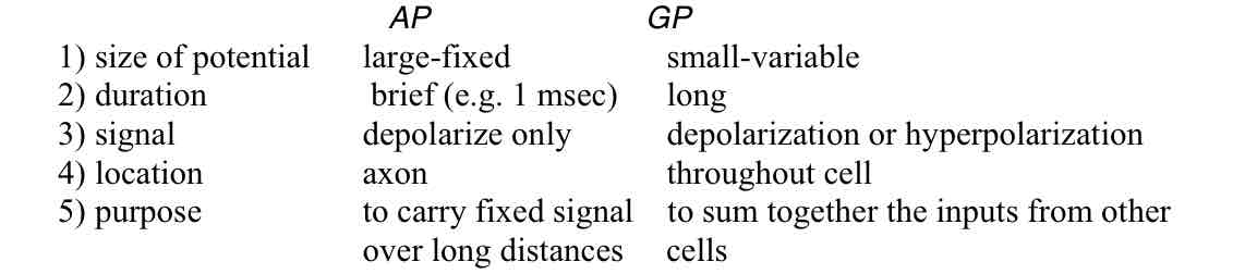
Differences between action potentials (red) and graded potentials (blue) : Size of potential
Acton potential : large-fixed
Graded potential : small-variable
Differences between action potentials (red) and graded potentials (blue) : Duration
Action : brief (e.g. 1 msec)
Graded : long
Differences between action potentials (red) and graded potentials (blue) : Signal
Action : depolarize only
Graded : depolarization or hyperpolarization
Differences between action potentials (red) and graded potentials (blue) : location
Action : axon
Graded : throughout cell
Differences between action potentials (red) and graded potentials (blue) : purpose
Action : to carry fixed signal over long distances
Graded : to sum together the inputs from other cells
Synapse
connection between neurons mediated by chemical neurotransmitters
Synapses can have excitatory vs. inhibitory effects on what type of cell
the post-synaptic cell
Receptive fields depend on the pattern of what
synaptic connections to the cell
Neurons can carry information by (3)
1. Size of response (graded, or number of action potentials)
2. Pattern of responses (e.g. temporal coding of sound frequencies)
3. Pattern of connections (labelled lines)
The problem of perception (3)
Stimuli are inherently ambiguous
eg: many different objects could give rise to the same retinal image
The brain must therefore make guesses or “unconscious inferences” in order to interpret the image
Perception is therefore “indirect” because it requires “information processing”
Levels of understanding perception (3)
ecological or computational
psychological
physiological
ecological or computational level
what is the goal of the perception, and whatproperties of the stimulus or the situation allow the problem to be solved?
Example: optic flow: as we move through the environment the retinal image changes in characteristic ways that provide cues about our motion
psychological Level
what are the processes underlying our perception and what sort of mental representations do they lead to?
physiological level
how are these processes and representations realized by the nervous system?
Informtation processing in sensory systems (3)
encoding, representation, and decoding
Serial vs parallel processing in sensory systems
Multiple-channel models and population codes
Serial processing in sensory systems
different stages along the visual pathway represent increasingly more abstract properties of the stimulus
parallel processing in sensory systems
different visual subsystems encode different properties
(e.g. obects vs. motion)
Coding efficiency and information theory (2)
Limited channel capacity
Creating more efficient representations by removing redundant signals
Examples of Limited channel capacity
neurons have a small range of response levels
Examples of Creating more efficient representations by removing redundant signals
redundancy
nearby locations typically have the same light level
Predictive coding
representing how stimuli deviate from expectations
Predictive coding Examples
coding a color by how it differs from gray or a face by how it differs from an average face
Plasticity in sensory representations example
developmental changes and perceptual learning
Bayesian inference in perception
decode the the sensory signals to estimate the stimulus by combining evidence from the senses with prior expectations
example of a prior: assume lighting is from above to interpret shading cues
Cornea and lens function
for focusing light to form image on the retina
Retina
thin layer of neurons along back of eye that absorb (receptors), process and transmit information about the light image
Fovea
small region of retina specialized for finest vision, corresponding to the center of gaze
Optic disk or blind spot
receptor-free “hole” in retina where ganglion cell axons leave the eye
Photopigments and transduction
conversion of physical energy to neural signal
Rods vs. cones
2 branches of dark adaptation curve reveal two mechanisms
Rods (purple) v Cones (yellow) : sensitivity to light
Rods : High
Cones : low
Rods (purple) v Cones (yellow) : Operating Range
Cones : moderate- bright
Rods : dim light
Rods (purple) v Cones (yellow) : Spatial Acuity (detail )
Rods: low
Cones: high
Rods (purple) v Cones (yellow) : Retinal Distribution
Rods : peak around 20 deg
Cones: peak in fovea
Rods (purple) v Cones (yellow) : # of receptor types
Rods : 1
Cones : 3
Rods (purple) v Cones (yellow) : Color Vision
Rods : no
Cones : yes
Receptive field (RF)
area on the retina to which a cell responds
Bipolar cell receptive fields
Center - surrounded
Different Bipolar cell receptive fields
1. Direct receptor input vs. indirect input through horizontal cells
2. on-center vs. off-center
3. Lateral inhibition: inhibition between spatially adjacent cells
Direct receptor input vs. indirect input through horizontal cells
1. forward – Receptors to bipolar cells to ganglion cells
2. Lateral – Horizontal cells and amacrine cells
on-center vs. off-center
on-center : responds to a bright spot of light that just covers the center
off-center : responds to a dark spot of light that just covers the center
Lateral inhibition
inhibition between spatially adjacent cells
Ganglion cell receptive fields similar to which cells
Bipolar cells
Information flow in the retina:
1. Straight through: receptor to bipolar to ganglion cell
2. Sideways: via horizontal cells and amacrine cells
Center-surround RF's emphasize contrast, de-emphasize absolute light level - why?
on-center cell responds best to a bright spot of light that just covers the center
off-center cell responds best to a dark spot that just covers the center
Examples of contrast effects
Simultaneous Contrast
Hermann Grid
Mach bands
Most ganglion cells send their axons to
Lateral geniculate nucleus (LGN)
Why do Nasal fibers cross over
so each LGN represents contralateral visual field
(ie opposite side of the world)
layers Of the LGN
magnocellular and parvocellular
These come from both eyes to line up signals
Cells have similar RF's to Which cells
Ganglion cells
Examples : center-surround and monocular
Monocular
Responds to signals only from one eye
Primary visual cortex also is known as
striate cortex or V1
What happens in the primary visual cortex
where most LGN cells send their axons
some RF properties first seen in cortex:
1. Orientation selectivity
2. Binocular
3. Increased selectivity for size
4. Sensitivity to direction of movement
5. simple cells v complex cells
Binocular
cells receive inputs from both eyes
Simple cells
spatially separated on and off subregions in receptive field, like center-surround cells
Complex cells
same stimulus selectivity as simple cells but no clear subregions within receptive field
Organization of different cell types in striate cortex
Retinotopic organization
Columnar organization
The hypercolumn
Retinotopic organization
spatial layout of retina is preserved by layout of cortex, but
Cortical magnification factor: much more of cortex is devoted to the fovea, much less to peripheral retina
Cortical magnification factor
much more of cortex is devoted to the fovea, much less to peripheral retina
Columnar organization
at each location cells in different layers have similar properties (e.g. preferred stimulus orientation), while RF’s change systematically as move from one point on the cortex to the next (e.g. preferred orientation changes smoothly from one column of cells to the next)
The hypercolumn
a functional module (~1mm square of cortex) that processes all orientations, eye combinations, colors, sizes, motion direction for a given location in space
Parallel pathways
Different areas are specialized for processing different visual tasks at the same time
Output of striate
about 30 distinct visual areas have been identified in primate cortex, suggesting a great deal of visual perception happens after the striate
Example of Different areas are specialized for processing different visual tasks at the same time (in parallel)
where pathway (parietal cortex) vs. what pathway (temporal)
Where do Parallel pathways begin with different cell types
In the retina
Two major pathways named for the LGN layers through which they pass:
magnocellular
parvocellular