BIOL 2160_Chapter 7_B
 Call Kai
 Learn
Learn Practice Test
 Spaced Repetition
 Match
Match Flashcards
 Knowt Play
1/59

There's no tags or description
Looks like no tags are added yet.
Name | Mastery | Learn | Test | Matching | Spaced | Call with Kai | Chat |
|---|
No analytics yet
Send a link to your students to track their progress
60 Terms
How do the biosynthetic differences between prototrophic and auxotrophic organisms connect cellular ATP consumption to their growth rates in nutrient-poor versus nutrient-rich habitats?
Prototrophs
Synthesize all essential growth factors from simple nutrients.
Can grow on minimal medium.
De novo biosynthesis consumes substantial ATP, carbon and reducing power.
In nutrient-poor habitats, this energy cost may slow growth, but biosynthetic independence allows continued growth.
In nutrient-rich habitats, they may suppress biosynthetic pathways and use available nutrients.
Auxotrophs
Lack one or more functional biosynthetic pathways.
Require specific preformed nutrients from the environment.
Importing available nutrients is generally less expensive than synthesizing them from simple precursors.
In nutrient-rich habitats, saved biosynthetic energy may support faster growth.
In nutrient-poor habitats, absence of the required nutrient can completely prevent growth.
Master Relationship
Nutrient-poor habitat:
Prototroph → high ATP cost → slower but possible growth
Auxotroph → required nutrient absent → little or no growth
Nutrient-rich habitat:
Auxotroph → nutrient import → biosynthetic ATP savings → potentially faster growth
Prototroph → pathway regulation and nutrient import → reduced biosynthetic cost
How do environmental temperature variations alter microbial plasma membrane lipid composition, and how does this relate to the evolutionary adaptation of hyperthermophilic archaea versus psychrophilic algae?
Effect of temperature
Low temperature → lipid tails pack tightly → membrane becomes rigid
High temperature → lipid movement increases → membrane becomes too fluid and permeable
Psychrophilic algae
Increase unsaturated fatty acids
Double bonds bend the lipid tails
Bent tails cannot pack tightly
Result → membrane remains fluid in cold environments
Hyperthermophilic archaea
Use heat-resistant ether-linked isoprenoid lipids
Often form tetraether monolayers that span the entire membrane
Result → membrane remains stable and prevents ion leakage at extreme temperatures
Evolutionary significance
Psychrophiles evolved flexible membranes for cold conditions
Hyperthermophiles evolved rigid, heat-stable membranes
Both adaptations maintain functional membrane fluidity and electrochemical gradients
Master relationship:
Cold → more unsaturated lipids → increased fluidity
Extreme heat → tetraether monolayer → increased stability and reduced leakage
How do the chemical defenses against reactive oxygen species (ROS) explain why obligate aerobes thrive in oxic habitats while obligate anaerobes are restricted to the human distal gut?
ROS: Reactive oxygen compounds produced when O₂ is incompletely reduced.
Main ROS: O₂•⁻, H₂O₂ and OH•
Aerobe defense:
SOD converts O₂•⁻ into H₂O₂.
Catalase or peroxidase removes H₂O₂.
ROS accumulation and cellular damage are prevented.
Obligate anaerobes:
Have insufficient ROS-defense capacity
May possess oxygen-sensitive enzymes
Accumulate oxidative damage when exposed to O₂
Master relationship:
Strong ROS defenses → greater oxygen tolerance
Weak ROS defenses → ROS accumulation → oxygen toxicity
Low intestinal oxygen → survival of obligate anaerobes
Common confusion:
Oxygen does not automatically kill every anaerobe. Toxicity depends on the organism’s ROS-defense capacity and oxygen-sensitive metabolism.
How does the human colon function as a natural continuous culture system, and how do nutrient availability and anoxia constrain the growth rate of resident Escherichia coli compared to its laboratory batch culture performance?
Natural continuous culture: Food residues continuously supply nutrients, while intestinal transit continuously removes waste and microbial cells.
Population stability: E. coli must reproduce fast enough to replace cells lost in feces.
Nutrient limitation: Host cells and competing microbes consume accessible nutrients, leaving little carbon for E. coli.
Anoxia: Limited oxygen forces E. coli to use anaerobic respiration or fermentation, generally producing less ATP.
Result: Resident E. coli may divide approximately once per day, compared with approximately every 20 minutes in rich, well-aerated laboratory culture.
Exam takeaway: The organism has the same genetic growth potential, but the colon’s environmental conditions prevent it from reaching its maximum growth rate.
4. Colon vs. Laboratory Batch CultureCondition
Human colon
Rich laboratory culture
Nutrient availability
Limited and strongly competed for
Abundant
Oxygen
Mostly anoxic
Often well aerated
Environment
Complex and spatially variable
Carefully controlled
E. coli generation time
Approximately 24 hours in the example
As short as approximately 20 minutes
Major growth constraint
Nutrient and energy limitation
Eventually nutrient depletion and waste accumulation
How does the metabolic activity of acidophilic lithotrophs like Acidithiobacillus ferrooxidans link inorganic compound oxidation to environmental acid mine drainage and severe pH stress on neutrophilic organisms?
Definition:
A. ferrooxidans is an acidophilic lithotroph that obtains energy by oxidizing Fe²⁺ and reduced sulfur compounds.
Mechanism:
Mining exposes pyrite to oxygen and water.
Pyrite oxidation releases Fe²⁺, sulfate, and H⁺.
A. ferrooxidans oxidizes Fe²⁺ to Fe³⁺ for energy.
Fe³⁺ oxidizes additional pyrite.
More H⁺ is produced, lowering the environmental pH and generating acid mine drainage.
Effect on neutrophiles:
High external H⁺ causes cytoplasmic acidification.
Protein function and membrane gradients are disrupted.
Cells consume energy pumping out H⁺.
Acid-solubilized metals add further toxicity.
Master relationship:
Increased pyrite oxidation → increased H⁺ production → decreased pH.
Decreased pH → increased proton and metal stress on neutrophiles.
Common confusion:
The bacterium does not simply secrete concentrated sulfuric acid. It accelerates iron and sulfur oxidation reactions that produce environmental acidity.
Exam takeaway:
Acidithiobacillus ferrooxidans gains energy by oxidizing ferrous iron and reduced sulfur compounds. Its production of ferric iron accelerates further pyrite oxidation, releasing sulfate and hydrogen ions that cause acid mine drainage. The resulting low pH disrupts cytoplasmic pH, proteins, membranes, and energy production in neutrophilic organisms.
How do the physiological adaptations expressed during the stationary phase of a batch culture connect nutrient exhaustion to bacterial survival mechanisms like endospore formation and horizontal gene transfer?
Definition: Stationary phase occurs when nutrient limitation and waste accumulation prevent continued population growth.
Main physiological change: Bacteria reduce growth-related activity and redirect energy toward stress resistance, maintenance, and nutrient scavenging.
Mechanism:
Nutrients become depleted and toxic products accumulate.
Stress-response pathways activate survival genes.
Some bacteria form resistant endospores.
Other bacteria become competent and take up extracellular DNA.
DNA may provide nutrients or useful genes through transformation.
Master relationship:
Greater nutrient limitation → stronger stress and survival responses
Sporulation → long-term physical survival
Competence → nutrient acquisition and possible genetic adaptation
Common confusion: Stationary phase does not mean that every cell has stopped dividing, and neither sporulation nor competence occurs in every bacterial species.
Exam takeaway: Nutrient exhaustion connects stationary phase to survival by shifting cellular resources from reproduction to protection, dormancy, scavenging, and genetic adaptation.

How do the structural features of bacterial endospores dictate the necessity of high-pressure autoclaving over standard boiling for achieving complete biological sterilization?
1. Endospore
Definition: A dormant, non-reproductive survival structure formed by certain bacteria under unfavorable conditions.
Main function: Protects the bacterial genome until environmental conditions improve.
Key structural defenses:
Dehydrated core: Very low water content reduces heat-induced damage.
Ca-DPA: Calcium–dipicolinic acid stabilizes the spore core and increases heat resistance.
SASPs: Small acid-soluble spore proteins bind to and protect DNA.
Cortex: A specialized peptidoglycan layer that helps maintain core dehydration.
Spore coat: Protective protein layers that resist chemicals and enzymes.
2. Why Boiling Is Insufficient
Boiling: Heating water to approximately 100°C at normal atmospheric pressure.
Kills most vegetative cells—actively growing and metabolizing microbial cells.
Some endospores can survive because their dehydrated, protected cores resist heat.
Therefore:
Boiling = disinfection, not reliable sterilization
3. How Autoclaving Works
Autoclaving: Sterilization using pressurized, saturated steam.
Typical conditions:
Temperature: 121°C
Pressure: approximately 15 psi above atmospheric pressure
Exposure time: commonly at least 15 minutes after the load reaches 121°C
Mechanism:
Pressure raises the boiling point of water.
Steam reaches a temperature above 100°C.
Steam condenses on the material and releases thermal energy.
Moist heat penetrates the endospore.
Essential proteins and cellular structures are irreversibly damaged.
The endospore loses its ability to germinate.
4. Key Clarification
Pressure does not directly kill endospores.
Pressure allows steam to reach approximately 121°C.
The actual killing agent is high-temperature moist heat.
5. Core Comparison
Method | Conditions | Effect |
|---|---|---|
Boiling | 100°C, atmospheric pressure | Kills vegetative cells but may not kill endospores |
Autoclaving | 121°C, pressurized steam | Destroys endospores and achieves sterilization |
Master Concept
Endospore protective structures → strong heat resistance → boiling may fail
Pressure ↑ → boiling point ↑ → steam temperature ↑ → endospore damage → sterilization
How does the presence of an obligate microbial consortium in biofilms connect metabolic interdependencies to the phenomenon known as the Great Plate Count Anomaly?
Definition: The Great Plate Count Anomaly is the large difference between the number of cells observed directly and the smaller number forming colonies on laboratory media.
Mechanism:
Biofilm microorganisms develop metabolic dependencies.
Isolation removes partner-produced nutrients, detoxification, pH control, and signals.
Some living cells cannot grow sufficiently on standard media.
Direct cell counts therefore exceed colony counts.
Master relationship:
Greater dependence on community interactions → lower recovery in pure culture
Restoration of partner species or required metabolites → increased culturability
Common confusion: Plate counts measure colony-forming cells under specific conditions, not every living cell.
Exam takeaway: Obligate microbial consortia help explain the Great Plate Count Anomaly because separating dependent microorganisms removes the interactions required for colony formation.
How do solar water disinfection (SODIS) mechanisms integrate electromagnetic radiation damage with photochemically generated reactive oxygen species to purify drinking water?
. Definition
SODIS: Solar water disinfection
Uses sunlight in transparent containers to inactivate waterborne pathogens.
Combines:
UV radiation
Photochemical ROS production
Solar heating
2. Radiation Damage
UV-A: 315–400 nm
Main solar UV component involved in SODIS
Excites cellular and waterborne photosensitizers
Produces mainly indirect oxidative damage
UV-B: 280–315 nm
Reaches Earth in smaller amounts
Can produce more direct nucleic-acid damage
Inhibits DNA replication and transcription
3. ROS Formation
Photosensitizer: A molecule that absorbs light and transfers energy or electrons to oxygen.
Mechanism:
Photosensitizer absorbs UV-A.
Photosensitizer enters an excited state.
Energy or electrons are transferred to dissolved O₂.
Reactive oxygen species are produced.
Major ROS:
Singlet oxygen: ¹O₂
Superoxide: O₂•⁻
Hydrogen peroxide: H₂O₂
Hydroxyl radical: •OH
4. Cellular Damage
ROS attack:
Membrane lipids → membrane leakage
Proteins → enzyme inactivation
DNA and RNA → replication and transcription failure
Respiratory systems → reduced energy production
5. Thermal Contribution
Solar radiation heats the water.
Higher temperature destabilizes microbial proteins and membranes.
At approximately 45–50°C or above, heat and UV produce a stronger combined effect.
Heat also reduces the microorganism’s ability to repair UV and oxidative damage.
6. Integrated Mechanism
Sunlight exposure
→ UV-induced molecular damage
→ photosensitizer activation
→ ROS formation
→ membrane, protein, and nucleic-acid oxidation
→ heat-enhanced cellular damage
→ pathogen inactivation
Key Clarification
Dissolved oxygen does not simply absorb UV-A and directly produce all ROS.
UV-A usually activates photosensitizers, which then transfer energy or electrons to O₂.
SODIS disinfects water but does not reliably remove toxic chemicals, heavy metals, or suspended particles.
How do the selective and differential properties of MacConkey agar connect mammal gastrointestinal physiology with the diagnostic identification of enteric pathogens?
Selective components: Bile salts and crystal violet inhibit most Gram-positive bacteria while permitting many bile-tolerant Gram-negative enteric bacteria to grow.
Differential components: Lactose and neutral red distinguish bacteria by lactose fermentation.
Mechanism:
Lactose fermenters produce organic acids.
The acids lower the medium’s pH.
Neutral red turns colonies pink or red.
Non-lactose fermenters remain pale or colorless.
Physiological connection: Enteric bacteria are adapted to the bile-rich mammalian intestine, so bile tolerance helps select them on MacConkey agar.
Diagnostic connection: Pink colonies suggest lactose-fermenting enterics, while colorless colonies raise suspicion for non-lactose-fermenting pathogens such as Salmonella or Shigella.
Exam takeaway: Selective = who grows; differential = how the growing colonies appear.
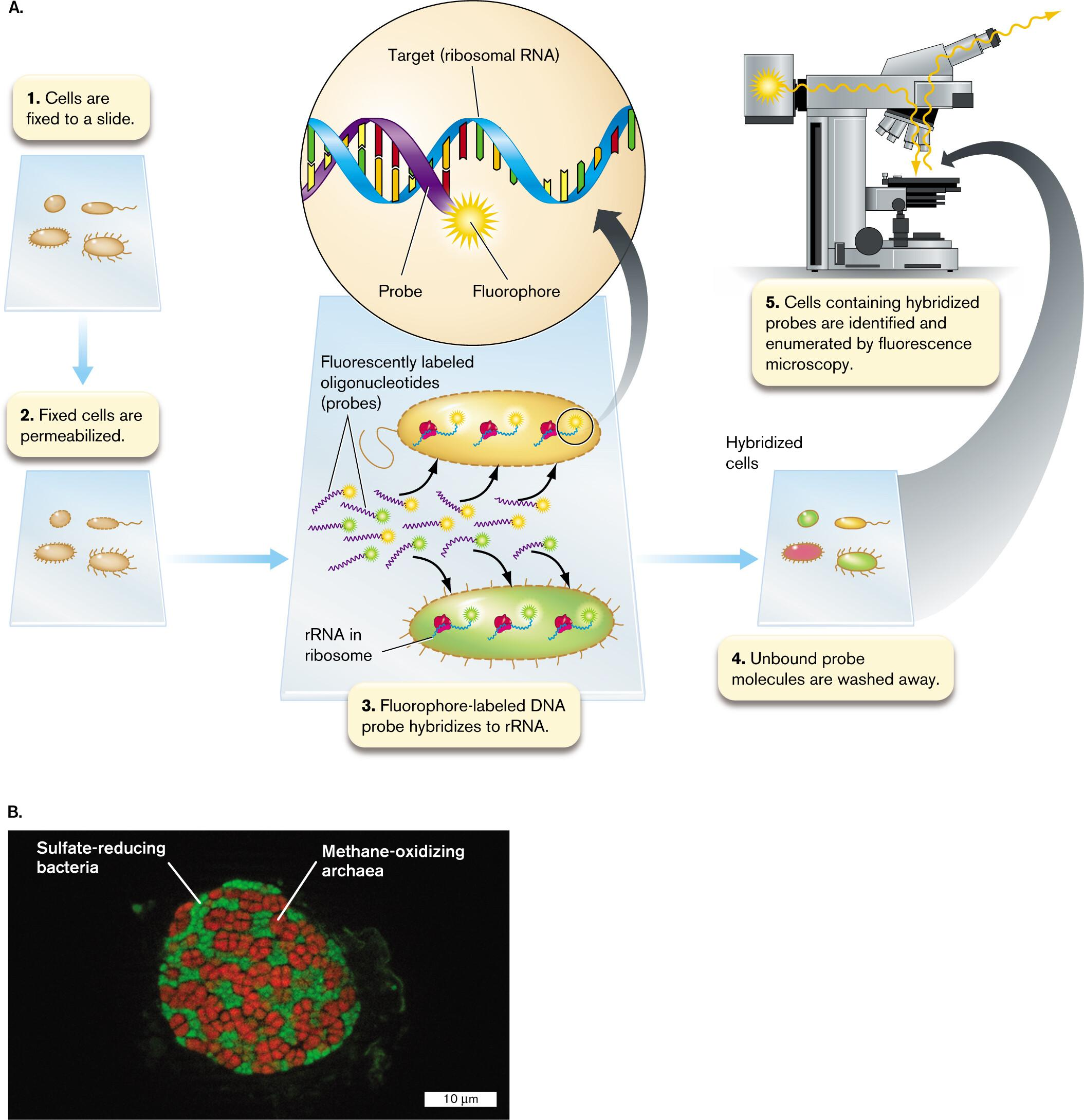
How do culture-independent 16S rRNA gene sequencing and Fluorescence In Situ Hybridization (FISH) integrate to uncover both the taxonomic identity and spatial organization of uncultivated microbes?
16S rRNA gene sequencing
Environmental DNA is extracted without culturing the microbes.
Conserved regions are used for PCR amplification.
Variable regions distinguish different microbial taxa.
Reveals which bacteria and archaea are present.
Does not preserve spatial information.
FISH
Fluorescently labeled DNA probes are designed from known 16S rRNA sequences.
Probes hybridize to complementary 16S rRNA molecules inside fixed cells.
Target cells fluoresce under a microscope.
Reveals location, physical arrangement, and relative abundance.
Integration
16S sequencing identifies an uncultivated microbe and provides the sequence needed for probe design.
FISH then locates that microbe within the intact community.
Together, they reveal who is present and where they are located.
Master relationship
16S sequencing → taxonomic identity
FISH → spatial organization
Combined approach → identity + location
Common confusion
16S sequencing usually analyzes the 16S rRNA gene in DNA.
FISH usually targets abundant 16S rRNA molecules inside cells.
Neither method alone directly proves the organism’s metabolic function.
How do the physical pore size parameters of membrane filtration versus ultrafiltration dictate their distinct applications in clearing bacterial cells versus viral particles from pharmaceutical solutions?
Comparison
Feature | Standard membrane filtration | Ultrafiltration |
|---|---|---|
Typical scale | 0.22–0.45 µm | Approximately 1–100 nm |
Main target | Cells and larger particles | Viruses and macromolecules |
Removes most bacteria? | Yes | Yes |
Reliably removes small viruses? | No | With a suitable membrane |
Pressure requirement | Lower | Generally higher |
Common pharmaceutical use | Sterile filtration and clarification | Virus removal or protein concentration |
Standard membrane filtration: Uses approximately 0.22–0.45 µm pores to remove bacterial cells while allowing the pharmaceutical solution to pass.
Ultrafiltration: Uses nanometre-scale pores or a defined MWCO (Molecular-weight-cutoff) to retain viruses and large macromolecules.
Mechanism: Particle larger than the effective pore → retained; particle smaller than the pore → may pass.
Master relationship: Smaller pore size → smaller particles removed, but fluid resistance and pressure requirements increase.
Common confusion: A 0.22-µm filter removes most bacteria but does not reliably remove all viruses or the smallest bacteria.
Exam takeaway: Use 0.22-µm filtration for most bacterial removal; use an appropriately validated ultrafilter for viral clearance.
How does the concept of Decimal Reduction Time (D-value) integrate microbial thermal death kinetics with food safety protocols to prevent endospore-borne botulism in commercial canning?
Definition: D-value is the time required at a specific temperature to reduce a microbial population by 90%, or one log.
Mechanism:
Heat kills a constant proportion of spores during each D-value interval.
Every additional D-value reduces survivors by another factor of 10.
Canning protocols multiply the D-value by the required log reduction.
Commercial application: Low-acid canned foods commonly use a 12D process against proteolytic C. botulinum spores.
Formula: Heating time = D-value × number of log reductions
Common confusion: Five D-values do not necessarily eliminate all organisms. Reducing 10⁵ to 10⁰ still predicts one survivor.
Exam takeaway: D-value connects microbial death kinetics to the heating time needed to achieve commercial sterility and prevent botulism.
How does the Soil Substrate Membrane System (SSMS) integrate low-nutrient environmental fluxes and natural chemical signaling to cultivate previously uncultured soil lineages like TM7?
The Soil Substrate Membrane System (SSMS) places soil bacteria on a polycarbonate membrane above a natural soil slurry.
The membrane retains the bacterial cells but allows dissolved nutrients, minerals, and natural signaling molecules from the soil to diffuse toward them.
This creates a low-nutrient environment similar to natural soil instead of using nutrient-rich laboratory media.
Slow-growing oligotrophic bacteria can therefore form microcolonies without being outcompeted by fast-growing species.
The resulting microcolonies can be detected and identified using FISH and 16S rRNA analysis.
This method enabled researchers to grow microcolonies belonging to previously uncultivated lineages such as TM7.
Core mechanism:
Natural soil compounds diffuse across the membrane → environmental conditions are reproduced → slow-growing soil bacteria form microcolonies. (Ferrari et al., 2005)
How do the chemical mechanisms of alkylating agents like glutaraldehyde compare to oxidizing agents like chlorine gas in achieving microbial control, and how do their residual properties dictate their use in water treatment versus medical fixatives?
Glutaraldehyde: Cross-Linking Agent
Reacts mainly with functional groups in proteins and other cellular molecules.
Forms stable covalent cross-links.
Immobilizes proteins and inactivates enzymes.
Kills microorganisms while preserving cellular morphology.
Stable cross-links make it useful as a medical and laboratory fixative.
Toxic chemical residues make it unsuitable for drinking-water treatment.
Chlorine: Oxidizing Agent
Reacts with water to produce hypochlorous acid, HOCl.
HOCl removes electrons from cellular molecules.
Oxidizes membranes, proteins, enzymes and nucleic acids.
Causes destructive cellular damage rather than structural fixation.
A low chlorine residual remains active in distribution pipes.
This residual provides continued protection against recontamination.
Master Relationship
Glutaraldehyde cross-linking → stable structural preservation → medical fixation
Chlorine oxidation + active residual → continued microbial killing → water treatment
Common Confusion
Both agents kill microorganisms, but their mechanisms and residual benefits differ:
Glutaraldehyde locks cellular structures together.
Chlorine chemically damages cellular structures through oxidation.
A clinical microbiologist receives a throat swab from a patient suspected of having strep throat. How can Blood Agar be applied to diagnose Streptococcus pyogenes infection?
Blood Agar for Detecting Streptococcus pyogenes
Blood agar is both an enriched medium and a differential medium.
A throat swab is streaked onto a blood agar plate.
The plate is incubated to allow bacterial colonies to grow.
S. pyogenes produces streptolysins that completely lyse nearby red blood cells.
This produces a clear zone around the colonies called β-hemolysis.
β-hemolysis provides presumptive evidence of S. pyogenes, but additional identification tests are required for confirmation.
Key relationship:
S. pyogenes growth → streptolysin production → complete RBC lysis → clear β-hemolytic zone
Common confusion: β-hemolysis suggests S. pyogenes, but it is not specific enough to confirm the species by itself.
A physician needs to isolate Neisseria meningitidis from a nasopharyngeal swab contaminated with abundant normal throat microbiota. How is Thayer Martin medium designed to achieve selective isolation?
Medium: Thayer–Martin medium
Enriched component: Chocolate agar supports the growth of fastidious Neisseria.
Selective components:
Vancomycin → inhibits Gram-positive bacteria
Colistin → inhibits most other Gram-negative bacteria
Nystatin → inhibits fungi
Trimethoprim → inhibits Proteus swarming
Final result: Normal throat microbiota are suppressed, allowing N. meningitidis to grow as isolated colonies.
An environmental engineer is tasked with isolating rare oil-degrading bacteria from a contaminated marine spill. How can an enrichment medium protocol be applied to accomplish this task?
Definition: Enrichment culture provides conditions that favor the growth of a desired microorganism over competing organisms.
Medium: Mineral salts and nutrients are supplied, but crude oil is used as the sole carbon and energy source.
Protocol:
Add the contaminated marine sample to the oil-containing enrichment broth.
Incubate under suitable marine and oxygen conditions.
Repeatedly transfer growing cells into fresh oil-containing medium.
Dilute and plate the enriched culture to obtain isolated colonies.
Test each isolate to confirm oil degradation.
Master relationship: Repeated growth on oil increases the relative abundance of bacteria capable of using hydrocarbons.
Common confusion: Enrichment does not produce a pure culture; plating and verification are still required.
Exam takeaway: The sole carbon source determines which metabolic type is enriched.
Enrichment protocol
Sample collection
오염된 바닷물이나 침전물을 채취합니다.Prepare enrichment broth
해양 환경과 비슷한 염도와 무기염류를 제공하되, crude oil을 sole carbon and energy source로 첨가합니다.⚠ “oil-only medium”은 정말 기름만 넣는다는 뜻이 아닙니다. 세균 성장에 필요한 물, 질소, 인, 무기염류는 제공하고 탄소원만 oil로 제한합니다.
Inoculation and incubation
환경 시료를 배지에 넣고 산소가 공급되도록 배양합니다. 많은 석유 분해 반응은 산소를 필요로 합니다.Successive transfer
성장한 배양액의 일부를 새로운 oil-containing medium으로 옮기는 과정을 반복합니다.
A food safety technician tests bottled drinking water for low-density bacterial contamination. Why is membrane filtration applied instead of standard direct plating, and how is CFU/mL calculated?
Why membrane filtration is used: Bottled water contains too few bacteria for reliable detection by plating a small sample volume.
Principle: A large volume of water is passed through a membrane, concentrating bacterial cells on the filter.
Mechanism:
Filter a known volume of water.
Bacteria are retained on the membrane.
Place the membrane on agar.
Incubate and count the resulting colonies.
Calculation:
CFU/mL = colonies counted ÷ volume filtered in mL
If the sample was diluted:
CFU/mL = colonies counted ÷ (volume filtered × dilution)
Example: 8 colonies from 100 mL = 0.08 CFU/mL = 8 CFU/100 mL.
Exam takeaway: Membrane filtration increases detection sensitivity by concentrating rare bacteria from a large sample volume.
희석한 시료를 여과한 경우CFU/mL = Number of colonies ÷ (Volume filtered × dilution)
예시:
Colony count = 25
Filtered volume = 10 mL
Dilution = 10⁻²
CFU/mL = 25 ÷ (10 × 10⁻²) = 250 CFU/mL
A research lab needs to measure the real-time growth of an Escherichia coli batch culture without taking overnight plating delays. How can spectrophotometric turbidity be applied, and what are its concentration limits?
A spectrophotometer measures culture growth in real time by directing a light beam at a wavelength of 550 to 600 nanometers through the liquid culture tube.
Definition: Spectrophotometric turbidity rapidly estimates bacterial density by measuring light scattering.
Mechanism:
Light passes through the E. coli culture.
Cells scatter light away from the detector.
More cells → less transmitted light → higher OD600.
Useful range: Approximately 1 × 10⁷–1 × 10⁹ cells/mL.
Below the range: Turbidity is too weak to detect reliably.
Above the range: Multiple scattering makes OD nonlinear; dilute the sample.
Common confusion: OD measures total biomass, not viable cells specifically.
Exam takeaway: OD600 provides fast, real-time growth monitoring, whereas plate counting is slower but measures viable cells.
An industrial facility uses a chemostat to produce a valuable bacterial enzyme continuously. How can the operator manipulate the dilution rate to maximize enzyme yield without causing culture washout?
Optimizing Dilution Rate in a Chemostat
The dilution rate is:
D = medium flow rate / culture volume
At steady state, the specific growth rate of the cells equals the dilution rate:
μ = D
Increasing D supplies nutrients and removes product more rapidly, which may increase enzyme productivity. However, as D approaches the critical dilution rate, cell concentration decreases. If cells are removed faster than they can reproduce, washout occurs.
The operator should:
Increase D gradually.
Allow the culture to reach steady state after each adjustment.
Measure biomass and enzyme concentration.
Calculate volumetric enzyme productivity as D × enzyme concentration.
Select the D that gives maximum productivity while remaining safely below the washout threshold.
Master relationship:
D increases moderately → growth and product-removal rate may increase
D approaches Dcritical → biomass begins to decrease
D exceeds Dcritical → washout occurs
Maximum D does not necessarily produce maximum enzyme yield
Common confusion: At steady state, μ equals D, but Dcritical is not always exactly equal to μmax. It also depends on nutrient concentration and growth kinetics.
A surgical center needs to sterilize heat-sensitive, plastic-packaged disposable scalpels. Why is gamma irradiation applied instead of autoclaving or UV light?
Gamma Irradiation of Disposable Medical Devices
Gamma irradiation is used for heat-sensitive, pre-packaged medical devices because it combines strong penetration with low-temperature processing.
Cobalt-60 releases high-energy gamma photons.
Gamma rays penetrate the plastic packaging and medical device.
The radiation ionizes cellular molecules and generates reactive oxygen species.
These effects damage microbial DNA, proteins, and membranes.
Microorganisms, including resistant endospores, lose their ability to survive or reproduce.
Why not autoclaving?
High-temperature pressurized steam may deform heat-sensitive plastics or packaging.
Why not UV?
UV radiation has poor penetration and mainly disinfects exposed surfaces.
Main advantage:
The device can be sterilized after sealing, reducing the risk of recontamination.
Following a bioterrorism incident involving Bacillus anthracis spore contamination inside an office building, public health officials selected chlorine dioxide gas for decontamination. Why was this chemical agent applied?
Anthrax spores are highly resistant + building contamination is widespread → use a penetrating, sporicidal gas
Clean Answer
Problem: Bacillus anthracis endospores are highly resistant to desiccation and many ordinary disinfectants.
Why chlorine dioxide gas was used:
It disperses throughout a sealed building.
It penetrates cracks, crevices, and ventilation systems.
It acts as a powerful oxidizing agent.
It damages essential spore proteins and other cellular components.
It provides whole-building sporicidal decontamination.
Master relationship:
Resistant spores + inaccessible contamination → penetrating sporicidal gas is required.Common confusion: Chlorine dioxide was selected not merely because it is a general disinfectant, but because its gaseous form can reach hidden areas and kill resistant endospores.
Exam takeaway: Chlorine dioxide gas combines sporicidal activity with whole-building penetration, making it suitable for anthrax decontamination.
A public health worker in a rural, resource-poor region needs a low-cost method to purify clear drinking water contaminated with enteric bacteria. How can Solar Water Disinfection (SODIS) be applied?
Definition: SODIS is a low-cost method that uses sunlight to disinfect clear water in transparent PET bottles.
Application:
Filter the water if it is cloudy or contains sediment.
Fill clean, transparent PET bottles of 2 L or smaller.
Lay the bottles horizontally in direct sunlight.
Expose them for at least 6 hours on sunny days or 2 consecutive days under cloudy conditions.
Mechanism: Solar UV-A, reactive oxygen species, and heat work together to damage and inactivate enteric bacteria.
Limitation: SODIS is less effective in turbid water, weak sunlight, continuous rain, or damaged and colored bottles.
Exam takeaway: Clear water + transparent PET bottle + sufficient sunlight → microbial inactivation without electricity or chemical disinfectants.
A daycare center wants to reduce gastrointestinal disease transmission among children using hand sanitizers. What active ingredients and concentrations must be specified for alcohol-based hand rubs (ABHR) to be effective?
Required formulation: At least 60% ethanol or 70% isopropanol.
Mechanism: Alcohol denatures microbial proteins and disrupts lipid membranes.
Why not 100% alcohol?: Water improves protein denaturation and slows alcohol evaporation.
Limitation: ABHR is less reliable against some gastrointestinal pathogens, particularly norovirus and bacterial spores.
Exam takeaway: Use correctly concentrated ABHR when hands are not visibly dirty, but use soap and water for visible contamination and pathogens resistant to alcohol.
A diagnostic lab uses Phenotype MicroArrays to characterize a newly isolated bacterial pathogen. How does this technology evaluate hundreds of metabolic traits simultaneously?
Definition: Phenotype MicroArrays measure bacterial metabolic activity across hundreds of different conditions simultaneously.
Components:
Multiwell plates
A different substrate or stress condition in each well
The same bacterial suspension in every well
Tetrazolium redox dye
Mechanism:
The bacterium metabolizes a usable substrate.
Cellular respiration generates electron flow.
The electrons reduce the tetrazolium dye.
Purple formazan develops.
A spectrophotometric system measures the color intensity.
Master relationship:
More metabolic activity → stronger purple color
No usable substrate or intolerable condition → little or no color
Common confusion: The technology measures phenotypic metabolic activity, not DNA sequences.
Exam takeaway: One well = one metabolic question; hundreds of wells = hundreds of traits tested in parallel.
A gastroenterologist treats a patient with chronic peptic ulcers. Why is antibiotic therapy targeting Helicobacter pylori applied instead of solely prescribing acid-suppressing proton pump inhibitors?
H. pylori: Causes chronic gastric inflammation and damages mucosal defenses.
PPI: Reduces acid and promotes ulcer healing but does not eliminate the infection.
Antibiotics: Eradicate H. pylori, removing the underlying cause of the ulcer.
Treatment logic: Antibiotics remove the cause; the PPI creates conditions for healing.
Exam takeaway: PPI treatment alone may temporarily heal an H. pylori-associated ulcer, but eradication therapy reduces recurrence.
A municipal water treatment plant uses chlorination to disinfect drinking water before distribution. Why is chlorine added at a late treatment stage, and what is the purpose of maintaining a chlorine residual?
Why chlorine is added late: Organic matter and suspended particles are removed first because they consume chlorine and can promote disinfection by-product formation.
Active disinfectants: Chlorine produces hypochlorous acid, HOCl, and hypochlorite ion, OCl⁻. Chloride ion, Cl⁻, is not the disinfecting species.
Mechanism:
Coagulation, sedimentation, and filtration remove particles and organic matter.
Chlorine is added to the clarified water.
HOCl and OCl⁻ oxidize microbial membranes, proteins, and enzymes.
A small amount of active chlorine remains in the finished water.
Purpose of chlorine residual: It prevents microbial regrowth and recontamination while water travels through storage tanks and distribution pipes.
Master relationship:
More organic matter → greater chlorine demand
Greater chlorine demand → less chlorine available for disinfection
Adequate residual → continued protection in the distribution system
Exam takeaway: Chlorine is added late to reduce chlorine demand and by-product formation; residual chlorine provides ongoing protection after the water leaves the treatment plant.
A molecular biologist needs to detect a non-culturable TM7 bacterial species in a complex dental plaque sample. How can a species-specific FISH probe be designed and applied to accomplish this?
Species-Specific FISH Detection of TM7
A fluorescently labelled, single-stranded DNA probe is designed to complement a variable region of TM7 16S rRNA that differs from the sequences of other oral bacteria.
Align the TM7 16S rRNA gene sequence with related bacterial sequences.
Select a region unique to the target TM7 species.
Synthesize a complementary oligonucleotide probe and attach a fluorophore.
Fix and permeabilize the dental-plaque cells on a slide.
Incubate the cells with the probe under optimized hybridization conditions.
Wash away unbound and mismatched probes.
Detect fluorescent TM7 cells using epifluorescence or confocal microscopy.
Master relationship:
Unique TM7 rRNA sequence → complementary probe binding → fluorescent target cell
Common confusion: The probe is designed using the 16S rRNA gene sequence, but inside the cell it usually binds the abundant 16S rRNA molecules—not the chromosomal gene itself.
Limitation: Fluorescence identifies and localizes the target cells, but standard FISH does not necessarily prove that the cells are alive.
A hospital central supply department needs to disinfect delicate endoscopes that cannot tolerate autoclaving or harsh corrosive bleaches. Why is glutaraldehyde applied as a high-level cold disinfectant?
Definition: Glutaraldehyde is a liquid dialdehyde alkylating agent used as a cold chemical sterilant for heat-sensitive and corrosion-sensitive medical equipment.
Main components: Five-carbon aliphatic chain with two terminal aldehyde groups (-CHO).
Mechanism:
Penetrates microbial cells and endospores at room temperature.
Forms covalent cross-links with amino, carboxyl, and hydroxyl groups on proteins and nucleic acids.
Permanently immobilizes essential enzymes, structural proteins, and DNA, resulting in microbial death without heat.
Master relationship:
If instrument is heat-sensitive -> Use glutaraldehyde to avoid melting optical fibers or damaging synthetic seals.
If contact time is short -> High-level disinfection (vegetative cells/viruses destroyed).
If contact time is extended -> Sporicidal cold chemical sterilization.
Common confusion: Confusing glutaraldehyde's mechanism (alkylation and covalent cross-linking) with oxidizers like hydrogen peroxide or bleach, or assuming that high-level disinfection instantly achieves sporicidal sterilization without extended soak times.
Exam takeaway: Glutaraldehyde is the standard chemical choice for flexible endoscopes because it achieves sporicidal sterilization via alkylating cross-links at ambient temperatures without thermal damage or metal corrosion.
Compare and contrast defined (synthetic) media with complex (chemically undefined) media regarding their chemical composition, typical ingredients, and research applications.
Defined media are formulated using precise, known quantities of pure inorganic salts and organic chemicals such that every molecular constituent is identified. Complex media incorporate digested biological extracts—such as peptone, yeast extract, or blood—whose exact chemical identities and concentrations are undefined and variable. Defined media like M9 minimal salts are used in precise physiological and metabolic research to test specific nutrient requirements. In contrast, complex media like Lysogeny Broth (LB) are easier to prepare and supply abundant amino acids and vitamins to support rapid microbial growth in general laboratory and clinical settings.
Compare and contrast selective media and differential media in terms of their primary purpose, mechanisms of action, and clinical diagnostic utility, using MacConkey agar as an example.
Definition: Selective media suppress non-target organisms to allow target microbes to grow; differential media make co-growing organisms look visually distinct through metabolic reactions.
Main components (MacConkey Agar):
Selective: Bile salts + crystal violet (inhibit Gram-positive bacteria).
Differential: Lactose (metabolic substrate) + neutral red (pH indicator dye).
Mechanism:
Bile salts and crystal violet block Gram-positive cell division; only Gram-negative enterics survive.
Surviving bacteria either ferment lactose or utilize alternative peptides.
Lactose fermentation generates acid end-products, dropping pH below 6.8.
Neutral red turns dark pink/red in acidic environments, staining lactose fermenters red and leaving non-fermenters colorless.
Master relationship:
If Gram-positive -> No growth (selective barrier).
If Gram-negative + lactose fermenter (E. coli) -> Growth + red colonies.
If Gram-negative + non-fermenter (Salmonella) -> Growth + colorless/pale colonies.
Common confusion: Believing differential media inhibit non-target organisms; differential media allow multiple species to grow, but visually distinguishes between them.
Exam takeaway: MacConkey agar is both selective (inhibits Gram-positives via bile salts and crystal violet) and differential (distinguishes lactose fermenters from non-fermenters via neutral red)[cite: 3].
Compare and contrast the streak plate method with the spread plate method regarding their mechanics, primary objectives, and suitability for quantifying viable cell density.
Clean Answer Streak Plate Method
Mechanism: An inoculating loop progressively spreads fewer cells across successive agar sectors.
Type of dilution: Mechanical dilution on the agar surface.
Primary objective: To obtain isolated colonies for pure-culture isolation.
Sample volume: Not accurately measured.
Quantification: Not suitable for calculating CFU/mL because the transferred volume and number of cells are unknown.
Spread Plate Method
Mechanism: A known volume of a liquid dilution is distributed uniformly across the agar surface.
Type of dilution: Measured serial dilution followed by surface spreading.
Primary objective: To quantify viable microorganisms.
Sample volume: Accurately measured with a pipette.
Quantification: Suitable for calculating CFU/mL using colony count, dilution factor and plated volume.
Master Relationship
Streak plate → progressive cell separation → isolated colony → pure culture
Spread plate → known dilution + known volume → countable colonies → CFU/mL
Compare and contrast batch culture systems with continuous culture systems (chemostats) regarding nutrient availability, growth phases, and environmental stability.
Batch culture:
Closed system
Fixed initial nutrients
Nutrients decrease and waste accumulates
Passes through lag, exponential, stationary and death phases
Environmental conditions continuously change
Chemostat:
Open system
Fresh medium continuously enters
Equal culture volume continuously exits
Maintains a relatively stable, nutrient-limited environment
Cells remain metabolically active at steady state
Cell growth rate = cell removal rate
Common confusion:
In a chemostat, cells continue dividing, but the population inside the vessel does not keep increasing because cells are continuously removed.
Compare and contrast direct microscopic cell counting using a Petroff-Hausser chamber with viable cell counting via serial dilution plating.
Petroff–Hausser count:
Counts cells directly in a chamber of known volume.
Reports total cells/mL.
Rapid, but normally includes both living and dead cells.
Serial dilution plate count:
Dilutes the sample, plates it on agar, and counts colonies after incubation.
Reports CFU/mL.
Measures cells capable of reproduction under the selected culture conditions.
Main relationship:
Direct count is usually higher than or equal to viable plate count.
One colony does not always equal one cell because a cell cluster may produce one CFU.
Exam takeaway:
Need a rapid total count → Petroff–Hausser chamber.
Need the concentration of culturable cells → serial dilution plating.
Compare and contrast disinfectants and antiseptics regarding their site of application, toxicity levels, and typical chemical formulations.
Disinfectants vs. Antiseptics
Both are chemical agents used to reduce or destroy microorganisms, but they differ mainly in their site of application.
Disinfectants are formulated for inanimate objects and environmental surfaces. They may contain stronger or more irritating concentrations because direct tissue compatibility is not their primary requirement.
Antiseptics are formulated for living tissues, such as skin. Their concentrations and ingredients must balance antimicrobial activity with acceptable tissue toxicity.
Typical formulations:
Disinfectants: sodium hypochlorite, hydrogen peroxide, phenolics, and quaternary ammonium compounds
Antiseptics: alcohol, chlorhexidine, and povidone-iodine
Master relationship:
Same chemical + different concentration/formulation/application → different classification
Common confusion: Antiseptics are not completely non-toxic. They are only safe enough when used at the approved concentration and application site.
Compare and contrast physical removal by 0.2 μm membrane filtration with thermal destruction by autoclaving when sterilizing laboratory liquids.
0.2 μm filtration:
Physically removes most bacteria and larger microorganisms.
Does not kill the trapped organisms.
Suitable for heat-sensitive solutions.
Most viruses and dissolved toxins may pass through.
Autoclaving:
Uses pressurized saturated steam, commonly at 121°C.
Heat destroys proteins, membranes, nucleic acids, and endospores.
Suitable for heat-stable liquids.
May damage vitamins, antibiotics, enzymes, or serum.
Common confusion:
Pressure does not directly kill microorganisms; it allows steam to reach a temperature above 100°C.
Neither method guarantees removal of every biological hazard—for example, filtration may not remove viruses, and some heat-stable toxins or prions may resist standard autoclaving.
Exam answer:
A 0.2 μm membrane sterilizes heat-sensitive liquids by physically removing most bacterial cells without heating the solution, but viruses and some very small organisms may pass through. Autoclaving uses pressurized saturated steam, commonly at 121°C, to destroy vegetative cells, viruses, and bacterial endospores, but it is suitable only for heat-stable liquids.
Compare and contrast the antimicrobial mechanisms and physical limitations of ultraviolet (UV) radiation versus ionizing gamma radiation.
UV radiation:
Non-ionizing
Produces pyrimidine dimers
Blocks DNA replication and transcription
Has poor penetration
Mainly disinfects exposed surfaces, air and clear water
Gamma radiation:
Ionizing
Directly damages DNA and indirectly produces free radicals
Causes single- and double-strand DNA breaks
Penetrates packaging and solid materials
Can sterilize prepackaged medical supplies and food
Master relationship:
Longer wavelength and lower energy → lower penetration
Shorter wavelength and higher energy → ionization and deeper penetration
Common confusion:
UV mainly produces pyrimidine dimers; gamma radiation mainly produces ionization, free radicals and DNA strand breaks.Exam takeaway:
UV = surface disinfection; gamma = deep, packaged-product sterilization.
Compare and contrast pasteurization with autoclaving in terms of temperature parameters, targeted microbial populations, and final sterility outcomes.
Pasteurization exposes liquids like milk to controlled sub-boiling heat for brief periods, whereas autoclaving applies superheated steam at 121°C under 15 psi pressure for at least 15 minutes. Pasteurization targets pathogenic vegetative bacteria and spoilage microbes to extend product shelf life while preserving taste and nutrition. Autoclaving achieves complete sterilization, destroying all living vegetative cells, viruses, and heat-resistant endospores. Consequently, pasteurized products are not sterile and require refrigeration, whereas autoclaved materials are entirely sterile.
Compare and contrast obligate aerobes, facultative anaerobes, and obligate anaerobes with respect to their oxygen requirements, terminal electron acceptors, and protective antioxidant enzymes.
Clean Answer
Obligate aerobes
Require O₂ for growth
Use O₂ as the terminal electron acceptor
Produce superoxide dismutase (SOD) and catalase to remove reactive oxygen species (ROS)
Cannot grow without O₂
Facultative anaerobes
Can grow with or without O₂
With O₂ → aerobic respiration → higher ATP yield
Without O₂ → anaerobic respiration or fermentation
Produce SOD and catalase
Usually grow better when O₂ is available
Obligate anaerobes
Do not use O₂; O₂ may be toxic
Use non-oxygen terminal electron acceptors, such as NO₃⁻ or SO₄²⁻, or perform fermentation
Lack sufficient protective antioxidant enzymes
O₂ exposure → ROS accumulation → cellular damage or death
Master relationship
Adequate SOD and catalase → survival in oxygen
Insufficient antioxidant enzymes → ROS accumulation → cellular damage
Common confusion
Facultative anaerobes do not require O₂; they use it when available because aerobic respiration produces more ATP.
Fermentation does not use an electron transport chain or an external terminal electron acceptor.
Compare and contrast psychrophiles and hyperthermophiles regarding their optimal growth temperature ranges, protein structural adaptations, and plasma membrane lipid compositions.
Clean Answer 1. Growth Temperature
Psychrophiles
Cold-adapted organisms
Grow near 0°C
Optimal growth temperature: ≤15°C
Maximum growth temperature: usually ≤20°C
Hyperthermophiles
Extreme heat-adapted organisms
Optimal growth temperature: ≥80°C
Most are Archaea
2. Protein Adaptations
Psychrophiles
Produce flexible enzymes
Fewer salt bridges and stabilizing interactions
Active at low thermal energy
Easily denatured at warmer temperatures
Hyperthermophiles
Produce compact, heat-stable proteins
More salt bridges, ionic networks, and hydrophobic interactions
Resist heat-induced denaturation
May use chaperones to maintain protein folding
3. Membrane Adaptations
Psychrophiles
More unsaturated fatty acids
Double bonds create bends in lipid tails
Prevent tight lipid packing
Maintain membrane fluidity in the cold
Hyperthermophilic Archaea
Ether-linked isoprenoid lipids
Often contain membrane-spanning tetraether lipids
Form stable monolayers
Prevent excessive fluidity and ion leakage at high temperatures
Master Comparison
Feature | Psychrophiles | Hyperthermophiles |
|---|---|---|
Main environmental problem | Low molecular movement | Heat-induced structural damage |
Protein strategy | Increased flexibility | Increased stability |
Membrane strategy | Increased fluidity | Increased rigidity and stability |
Overall result | Function maintained in cold | Function maintained in extreme heat |
Cold → flexible proteins + unsaturated lipids
Extreme heat → stable proteins + tetraether membranes
Compare and contrast 16S rRNA gene profiling with shotgun metagenomics for analyzing uncultivated environmental microbial communities.
Both methods analyze microbial communities directly from environmental DNA without culturing the organisms.
16S rRNA gene profiling: Uses PCR and universal primers to amplify the 16S rRNA gene. It mainly identifies which bacteria and archaea are present and estimates their relative abundance. However, it provides limited information about their functions.
Shotgun metagenomics: Randomly sequences all extracted DNA. It can identify a wider range of organisms and reveal genes involved in metabolism, transport, antibiotic resistance, and other cellular processes.
Main difference:
16S profiling → Who is present?
Shotgun metagenomics → Who is present, and what could they do?
Shotgun metagenomics provides greater taxonomic and functional detail but is more expensive and computationally demanding. Detected genes indicate functional potential, not necessarily active gene expression.
Compare and contrast the spread plate method with the pour plate method regarding sample preparation, colony distribution, and suitability for heat-sensitive microbes.
Feature | Spread Plate | Pour Plate |
|---|
Agar condition | Pre-solidified agar | Molten agar cooled to about 45°C |
Sample preparation | A small diluted sample, usually 0.1 mL, is placed on the agar | A diluted sample, usually up to 1 mL, is placed in an empty Petri dish |
Procedure | Sample is spread across the agar surface | Sample is mixed with molten agar and allowed to solidify |
Colony distribution | Colonies grow only on the surface | Colonies grow both on the surface and within the agar |
Colony appearance | Larger and easier to observe or collect | Embedded colonies are smaller and harder to observe |
Oxygen exposure | High; suitable for aerobic microbes | Lower inside the agar; can support organisms preferring reduced oxygen |
Heat-sensitive microbes | More suitable because cells are not exposed to heat | Less suitable because molten agar may injure or kill sensitive cells |
Process
Spread plate
Diluted sample → placed on solid agar → spread across surface → surface colonies form
Pour plate
Diluted sample → mixed with molten agar at about 45°C → agar solidifies → colonies form throughout the agar
Key Contrast
Spread plate: no heat exposure + surface colonies + easier colony collection
Pour plate: brief heat exposure + 3D colony distribution + smaller embedded colonies
Therefore, spread plating is better for heat-sensitive microbes.
Compare and contrast the antimicrobial mechanisms and applications of alcohols (ethanol/isopropanol) versus phenolic compounds (triclosan).
Feature | Alcohols: Ethanol / Isopropanol | Phenolic compound: Triclosan |
|---|---|---|
Main target | Cell membranes and proteins | Cell membrane and fatty-acid synthesis |
Mechanism | Dissolve membrane lipids + denature proteins | Binds enoyl-ACP reductase → inhibits bacterial fatty-acid synthesis |
Effect | Rapid, broad antimicrobial action | Slower but more persistent antibacterial action |
Residue | Evaporates quickly; no residue | Remains on the treated surface |
Applications | Hand sanitizers, skin antisepsis, small-surface disinfection | Formerly common in antibacterial soaps and consumer products |
Limitation | Ineffective against bacterial endospores; reduced activity on dirty surfaces | Overuse can select resistant bacteria with altered target enzymes |
Main mechanism
Alcohol: membrane disruption + protein denaturation → rapid cell death
Triclosan: inhibition of fatty-acid synthesis → defective membrane production and inhibited bacterial growth
Key contrast
Alcohols act quickly and broadly but do not persist.
Triclosan acts on a more specific bacterial target and can persist, but repeated use may promote resistance.
Compare and contrast chlorine treatment with ozone treatment for large-scale municipal drinking water and wastewater disinfection.
Similarity: Both kill microorganisms through oxidation.
Chlorine: Cheaper, slower and leaves a protective residual, but may produce taste, toxicity and chlorinated disinfection by-products.
Exam takeaway: Choose chlorine when continued protection is essential; choose ozone when rapid oxidation and minimal lasting disinfectant are preferred.
For drinking water, ozone may be used first for strong primary disinfection, followed by a small chlorine or chloramine dose for residual protection:Ozone treatment → rapid initial disinfection → chlorine residual → protection inside pipes
For wastewater, ozone avoids releasing residual chlorine, while chlorinated wastewater may require dechlorination before discharge to protect aquatic organisms.
ComparisonFeature
Chlorine
Ozone
Disinfection speed
Moderate
Very rapid
Oxidizing strength
Strong
Stronger
Residual protection
Yes
No
Storage
Can be stored or supplied chemically
Must be generated on-site
Operating cost
Usually lower
Higher equipment and electricity cost
Taste and odor
May create chlorine taste or odor
Often improves taste and odor
Important by-products
Trihalomethanes and haloacetic acids
Bromate when bromide is present
Wastewater concern
Usually requires dechlorination before discharge
No lasting disinfectant residual
Main advantage
Protects water throughout distribution
Rapid, powerful treatment without chlorine residual
Main disadvantage
Residual toxicity and chlorinated by-products
High cost and no continued protection
A technician prepares nutrient agar plates, autoclaves them, but forgets to allow the molten agar to cool to \45°C before pouring into Petri dishes. What physical and operational problems will occur?
Clean Answer
Problem: Agar is poured while still too hot (>60°C).
Physical effects
Hot agar releases large amounts of water vapour.
The vapour cools on the Petri dish lid.
Heavy condensation forms on the underside of the lid.
Plastic Petri dishes may warp from the heat.
Operational effects
Condensation drips onto the agar surface.
The droplets spread the streaked bacterial cells.
Colonies run together instead of remaining separated.
Isolated single colonies cannot be obtained reliably.
Additional risks
Increased risk of burns and spills
Longer cooling and solidification time
Possible destruction of heat-sensitive supplements
Main sequence
Agar poured too hot → excessive evaporation → lid condensation → droplets fall onto agar → bacterial cells spread → poor colony isolation
Correct procedure
Cool molten agar to about 45–50°C before pouring it into Petri dishes.
A student inoculates a fastidious respiratory pathogen onto basic M9 minimal salts agar, but no bacterial growth occurs after 48 hours. What nutritional deficiency explains this culture failure, and how should the media be modified?
Deficiency: M9 lacks preformed amino acids, vitamins, hemin, NAD, and other growth factors required by fastidious bacteria.
Mechanism:
The pathogen cannot synthesize all essential cellular components.
M9 does not provide the missing components.
Biosynthesis and cell division stop, so no colonies form.
Media modification: Replace M9 with an enriched medium such as chocolate agar, or supplement the medium with the organism’s required growth factors.
Exam takeaway: Fastidious organism + minimal medium + no growth → suspect missing organic growth factors and use enriched media.
A researcher sets up an anaerobic jar to culture obligate anaerobes but forgets to add the palladium catalyst pellet to the jar lid. Why will the culture fail to create an anoxic environment?
Definition: A palladium catalyst is an inorganic agent in an anaerobic jar that drives the reaction between ambient O2 and generated H2 to establish an oxygen-free growth chamber.
Main components: Palladium pellet (catalyst), gas generator packet (produces H2 and CO2), ambient O2 (target contaminant).
Mechanism:
Gas packet generates H2 gas inside the sealed vessel.
Palladium lowers the activation energy for the reaction: 2 H2 + O2 -> 2 H2O.
Without the catalyst, H2 and O2 cannot react at room temperature; O2 remains intact as a gas.
Master relationship:
If palladium is absent -> Reaction does not occur -> Atmospheric O2 remains -> Obligate anaerobes are killed by oxidative stress.
If palladium is active -> O2 converts to liquid water condensation -> Anoxic state achieved.
Common confusion: Confusing the catalyst (which removes oxygen) with the redox indicator strip (which only reports the presence or absence of oxygen).
Exam takeaway: Generation of hydrogen gas is useless without the palladium catalyst because the activation energy to form water from H2 and O2 is too high at room temperature.
A student performs a 1:10 serial dilution of an E. coli culture and plates 0.1 mL of the (1 \times 10^{-1}) dilution, obtaining over 500 overlapping colonies. Why is this plate excluded from CFU/mL calculations, and which dilution should be evaluated?
A 10⁻¹ dilution means that the original culture was diluted tenfold. The student plated 0.1 mL of this diluted sample and obtained more than 500 overlapping colonies.
This plate cannot be used for an accurate CFU/mL calculation because it is TNTC (too numerous to count).
When too many bacterial cells are placed close together:
Colonies grow into one another.
Separate colonies become difficult to identify.
Several bacterial cells may appear as one merged colony.
The colony count can therefore underestimate the actual number of viable bacteria.
A reliable plate should generally contain 30–300 separate colonies.
Because each step in a 1:10 serial dilution reduces the expected colony number by a factor of 10:
10⁻¹: >500 colonies → 10⁻²: >50 colonies → 10⁻³: >5 colonies
Therefore, the 10⁻² dilution should be evaluated first, because it is most likely to produce a countable plate. The 10⁻³ dilution may contain fewer than 30 colonies.
The provided answer is therefore partly incorrect: it is correct that the 10⁻¹ plate must be excluded, but it should not automatically skip to 10⁻³.
A laboratory technician monitors an E. coli culture using a spectrophotometer. The culture reaches an optical density (OD) reading of 1.8, but cell counts calculated from OD significantly underestimate actual cell numbers. What spectrophotometric limitation caused this error?
Clean Answer
Limitation: Loss of linearity at high cell density
Cause: Multiple light scattering in an overly dense culture
Result: OD readings flatten out → cell concentration is underestimated
Solution: Dilute the culture, measure OD again, and multiply by the dilution factor
Exam answer:
The OD of 1.8 exceeded the spectrophotometer’s linear measurement range. Multiple scattering caused the OD reading to plateau, resulting in an underestimate of the actual cell concentration.
An industrial fermentation batch culture halts exponential growth earlier than expected despite abundant glucose. Analysis reveals the media pH dropped from 7.0 to 4.2. What metabolic byproduct caused this disruption, and how could it be prevented?
Clean Answer
Problem:
The culture stops growing even though glucose is still abundant.
Cause:
The microorganisms produce organic acids, such as lactic acid or acetic acid, while metabolizing glucose.
Mechanism:
Cells rapidly consume glucose.
Acidic metabolic byproducts accumulate in the medium.
The medium pH decreases from 7.0 to 4.2.
The acidic environment inhibits enzymes and membrane functions.
Exponential growth stops even though glucose remains available.
Prevention:
Add a buffer to resist sudden pH changes.
Use automatic pH control to add a base when the pH drops.
Improve aeration to reduce excessive fermentation.
Supply glucose gradually using a fed-batch system.
Master Relationship:
More organic acid accumulation → lower pH → stronger growth inhibition
Common Confusion:
The exact acid cannot be identified without knowing the microorganism. The safest general answer is organic acid byproducts.
Exam Takeaway:
Growth may stop before nutrients are depleted because toxic metabolic waste products can accumulate and create an unfavorable pH.
A research team isolates a novel bacterium from a deep-sea hydrothermal vent at 95°C. When the bacterium is transferred to a standard laboratory incubator at 37°C, it fails to divide. What changes in its enzymes and cell membrane explain this growth failure?
The organism is a hyperthermophile—a microorganism whose cellular machinery is adapted to extremely high temperatures, commonly above 80°C.
At 95°C:
High thermal motion → enzymes remain flexible enough to change shape → substrates bind and reactions proceed
At 37°C:
Low thermal motion → enzymes become too rigid → conformational changes slow → metabolism becomes extremely slow
The proteins do not necessarily misfold at 37°C. The more accurate explanation is that many become insufficiently flexible and catalytically inefficient.
Growth Stops
Enzyme activity decreases + membrane function declines
→ nutrient uptake and ATP production decrease
→ DNA, protein and cell-wall synthesis cannot proceed sufficiently
→ cell mass does not double
→ binary fission stops
Definition: A hyperthermophile is adapted to grow optimally at extremely high temperatures.
Mechanism:
At 37°C, its heat-stable enzymes become too rigid and catalytically inefficient.
Its high-temperature-adapted membrane loses fluidity.
Nutrient transport, ATP production and biosynthesis decline.
The cell cannot accumulate enough biomass or complete binary fission.
Master relationship: Temperature far below the optimum → enzyme flexibility and membrane fluidity decrease → growth rate decreases.
Common confusion: Low temperature generally does not denature the proteins; it makes their activity too slow.
Exam takeaway: The growth failure results from impaired enzyme kinetics and excessive membrane rigidity at a temperature far below the organism’s optimum.
A hospital central supply department attempts to sterilize surgical instruments by boiling them in water at (100^\circ\text{C}) for 30 minutes. Several patients subsequently develop post-operative Clostridium infections. Why did boiling fail to prevent these infections?
Problem: Boiling at 100°C does not reliably destroy bacterial endospores.
Organism: Clostridium species produce heat-resistant endospores.
Mechanism:
Boiling killed most vegetative cells.
Clostridium endospores survived on the instruments.
The spores entered the patients’ tissues.
They germinated into pathogenic vegetative cells and caused infection.
Required method: Steam sterilization in an autoclave, commonly at 121°C and about 15 psi gauge pressure for an appropriate exposure time.
Common confusion: Boiling provides disinfection, not reliable sterilization.
A student prepares hand sanitizer using pure 100% absolute ethanol, but tests show it performs worse at killing surface bacteria than a 70% ethanol solution. What biochemical mechanism explains this unexpected result?
Why 70% ethanol works better than 100% ethanol
100% Ethanol
Evaporates very rapidly → short contact time
Rapidly coagulates surface proteins
This coagulated protein layer can reduce deeper penetration
Contains too little water for efficient protein denaturation
Result → incomplete microbial killing
70% Ethanol
Contains 30% water
Water slows evaporation → longer contact time
Water helps ethanol penetrate the cell
Water supports complete protein denaturation
Ethanol dissolves membrane lipids and denatures cellular proteins
Result → more effective microbial killing
Main Mechanism
70% ethanol
→ slower evaporation
→ better cellular penetration
→ stronger protein denaturation
→ membrane and enzyme destruction
→ cell death
Key Point
Water is not merely a diluent. It increases ethanol’s antimicrobial effectiveness by improving penetration, prolonging contact time, and supporting protein denaturation.
A quality control lab evaluates a new disinfectant against a bacterial population of (1 \times 10^6) cells. If the disinfectant has a (D)-value of 2 minutes under test conditions, how many minutes of treatment are required to reduce the surviving population to 1 cell?
Definition: The D-value is the time required to reduce a viable microbial population by 90%, or one log₁₀.
Given:
Initial population = 10⁶ cells
Target population = 10⁰ cell
D-value = 2 minutes
Calculation:
Reducing 10⁶ cells to 10⁰ requires 6 decimal reductions.
Treatment time = 6 × 2 minutes
Treatment time = 12 minutes
Answer: 12 minutes
Common confusion: One expected surviving cell does not mean that the sample is sterile.
A pharmaceutical company attempts to sterilize a heat-sensitive liquid antibiotic using a 0.45 (\mu)m membrane filter, but the filtered drug product causes viral infections in trial patients. What design flaw caused this sterilization failure?
Design flaw: The company incorrectly relied on a 0.45 µm membrane filter to achieve viral sterilization.
Mechanism: Many viruses are much smaller than the filter’s 450 nm pores and therefore passed into the final product.
Additional issue: A 0.45 µm membrane is not normally considered a sterilizing-grade filter; 0.20–0.22 µm filters are commonly used for bacterial removal.
Exam takeaway: Standard membrane filtration may remove bacteria, but it does not reliably remove viruses. Viral clearance requires a separately validated removal or inactivation process.
A researcher uses a chemostat to grow E. coli at a steady-state cell density. When the operator increases the fresh medium pumping rate from 100 mL/hr to 800 mL/hr in a 1-liter vessel, the culture turns completely clear. What phenomenon occurred?
Phenomenon: Culture washout
Dilution rate:
D = 800 mL/hr ÷ 1,000 mL = 0.8 hr⁻¹Required doubling time:
0.693 ÷ 0.8 hr⁻¹ ≈ 0.866 hr ≈ 52 minMechanism:
Pumping rate increases.
Dilution rate increases to 0.8 hr⁻¹.
D exceeds the maximum growth rate of E. coli under those conditions.
Cells leave faster than they reproduce.
Cell density approaches zero, making the culture clear.
Master relationship:
μ = D → stable cell population
μ > D → cell population can increase
D > μmax → washout
Exam takeaway:
A chemostat becoming clear after the flow rate increases indicates washout caused by an excessively high dilution rate.
0.693의 정체: ln(2)왜 2인가?: Doubling은 세포 수가 2배가 되는 것이기 때문
공식: Doubling time = 0.693 ÷ specific growth rate
이 문제: 0.693 ÷ 0.8 hr⁻¹ ≈ 0.866 hr ≈ 52 min
Exam takeaway: 0.693이 보이면 보통 지수성장에서의 doubling과 관련 있다.
A microbiology lab attempts to isolate pure cultures of a TM7 bacterium from an SSMS microcolony by transferring cells onto rich Nutrient Agar, but no growth occurs. Why did subculturing on rich media fail, and what media should be used?
Reason for failure:
TM7 bacteria often have reduced metabolic capabilities.
They may depend on neighboring or host bacteria for essential metabolites and growth signals.
Transfer to ordinary Nutrient Agar removes these natural interactions.
Rich medium may also be unsuitable for organisms adapted to low-nutrient soil conditions.
Appropriate cultivation:
Maintain the organism in an SSMS or diffusion-based soil system.
Alternatively, use a low-nutrient soil-extract medium.
If the strain is host-dependent, culture it together with its required bacterial host.
Master relationship:
More nutrients do not necessarily produce more growth.
Removing the ecological partner → removal of essential metabolites/signals → no growth.
Exam answer:
Subculturing failed because TM7 has limited biosynthetic capacity and may depend on natural soil conditions or a bacterial host for essential metabolites and growth signals. It should be maintained in an SSMS or cultured on a low-nutrient soil-based medium, with an appropriate host organism if required.
A wastewater treatment plant switches from chlorine gas to ozone ((O_3)) treatment. While effluent discharged into the river meets bacterial safety standards, water stored in distribution tanks develops bacterial re-growth hours later. What chemical property of ozone caused this re-growth?
Chemical property: Ozone is highly unstable and rapidly decomposes into O₂.
Immediate effect: It kills bacteria effectively during direct contact.
Later effect: It leaves no persistent disinfectant residual.
Result: Surviving or newly introduced bacteria can regrow in storage tanks.
Exam takeaway: Rapid ozone decomposition → no residual protection → bacterial re-growth.
An investigator tests a new disinfectant against a mixed culture containing Escherichia coli vegetative cells, Staphylococcus aureus, influenza virus, and Bacillus subtilis endospores. After 5 minutes, only B. subtilis survives. What structural feature protected B. subtilis?
Protective structure: Endospore
Main components:
Thick protein spore coat
Peptidoglycan cortex
Low-permeability inner membrane
Dehydrated core
Ca-DPA and DNA-protective SASPs
Mechanism:
The protective layers restrict disinfectant penetration.
The dehydrated core reduces damaging chemical reactions.
Ca-DPA and SASPs stabilize cellular components and DNA.
The dormant endospore survives while vegetative cells and influenza virus are inactivated.
Common confusion:
B. subtilis survived because it formed endospores, not simply because it is Gram-positive.Exam answer:
B. subtilis survived because its endospores contain a thick protective coat, cortex, low-permeability inner membrane, and dehydrated core that provide strong resistance to chemical disinfectants.