Central Nervous System (CNS) and the Brain
1/59
Earn XP
Description and Tags
4.2 Biology- Anatomy & Physiology
Name | Mastery | Learn | Test | Matching | Spaced |
|---|
No study sessions yet.
60 Terms
Central and Peripheral Nervous Systems (CNS and PNS)
The 2 Nervous System Structures
The Brain
A Complex Mass of Nerve, Connective, & Epithelial Tissue
Cerebrum, Diencephalon, Cerebellum, & Brainstem
The 4 Anatomical and Functional Regions
Cerebrum
Largest region & occupies most of the space in the cranial cavity; split into 2 cerebral hemispheres (left & right) & each hemisphere are made up of lobes
Diencephalon
Pair of oval-shaped structures deep into the cerebrum; protruding laterally in the lateral ventricles, then meets medially encapsulating the 3rd ventricle; it contains multiple nuclei and is dubbed as the gateway to the cerebral cortex
Cerebellum
Second largest region of the brain; located inferior to the cerebrum but posterior to the brainstem. It consists of a left & right cerebellar hemispheres
Brainstem
A long structure that extends inferiorly from the diencephalon; 10 of the 12 cranial nerves originate from here; it is made up 3 sections/parts/regions: the superior midbrain, the middle round-shaped pons, & inferior medulla oblongata
Cerebrum
Structure A

Diencephalon
Structure B

Cerebellum
Structure D
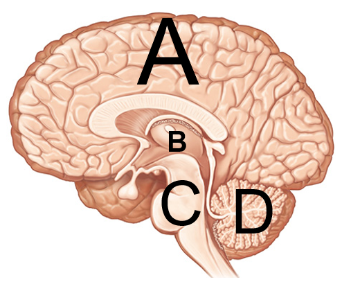
Brainstem
Structure C

Fissures
Deep grooves/furrows, usually separating bigger parts of the brain.
Sulci
Shallow grooves found in the cerebrum
Gyri
Folds found in the cerebrum; think of a gyro sandwich which is folded
Gyri; Sulcus; 2 Sulci; Gyrus
It takes two _ to form a _ & _ are found on each side of a _.
Cranial Gray Matter
Located superficial in the cerebrum & cerebellum; they are mainly composed of neuron cell bodies, dendrites & synapses- surrounds white matter
Cerebellar & Cerebral Cortexes
Superficial layer on both the cerebrum & cerebellum
Nuclei
Deep masses of gray matter; surrounded by white matter
White Matter
Lies deep to the gray matter & is mainly composed of bundles of axons/tracts.
Cranial Meninges
A set of 3 connective tissue membranes surrounding the brain and spinal cord
Dura Mater
Tough layer protecting the brain; made up of 2 layers
Periosteal Layer
Outer layer surrounding the cranial bones, equivalent to periosteum
Function of the Meningeal Layer
Function: Surrounds and Supports Dural senses that drain deoxygenated blood from the brain
Meningeal Layer
inner layer facing other meninges, extends from the vertebral canal and has 3 structures
Falx Cerebri
In Meningeal Layer; Separates cerebral hemisphere in longitudinal fissure
Tentorium Cerebelli
Separates each Cerebellar Hemisphere; located in the transverse fissure
Falx Cerebelli
Separates each cerebellar hemisphere; located inferiorly to the cerebellum
Subdural Space
A potential space found below the dura mater and above the arachnoid mater; can be opened if needed for brain bleeds etc.
Dural Sinuses
Formed by the dura mater and collects deoxygenated blood and CSF
Arachnoid Mater
Second Layer resembling a spider web; contains the Arachnoid Granulations
Arachnoid Granulations
tree-like protrusions piercing through the dura mater that reabsorbs CSF into the Dural Sinuses
Pia Mater
very thin and delicate layer that closely follows every contour of the brain
Ventricles
A system of fluid filled cavities; lined with a spongy mass of capillaries
Choroid Flexus
Spongy Mass of Capillaries found in ventricles
Cerebrospinal Fluid
Clear and Colorless fluid following in the ventricles; derived from plasma and serves to protect the brain by allowing it to float in the insulating fluid of the CSF
500mL per day
How much CSF is produced by the ependymal cells lining the choroid plexus found in each ventricle and pathways
Functions of the CSF
Buoyancy; Protection; Chemical Stability
Buoyancy in the CSF
allows the brain to attain considerable size without being impaired by its own weight
Protection in the CSF
protects the brain from striking the cranium when the head is jolted in CSF
Chemical Stability in the CSF
rinses metabolic wastes from the nervous tissue and regulates its chemical environment
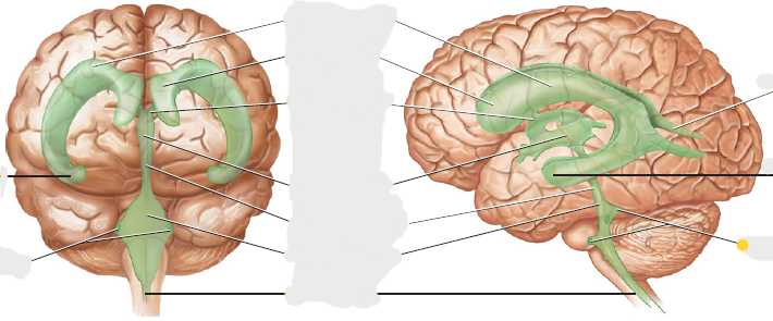
Lateral Ventricles
forms an arch with anterior, posterior, and inferior horns in the right and left hemispheres
Interventricular Foramen
Inferior to the anterior horn; connects lateral ventricles to the 3rd ventricle
3rd Ventricle
Below the corpus callosum and within the medial part of the thalamus
Cerebral Aqueduct
In midbrain; connects the 3rd and 4th ventricles
4th Ventricle
between the pons and cerebellum
Central Canal and Apertures
near the medulla oblongata; drains CSF into the subarachnoid space
Subarachnoid Space
Space that surrounds the brain and spinal cord, located below the arachnoid mater
Arachnoid Granulations
Reabsorbs CSF into the Dural Sinuses
Lateral Ventricle (image)

Anterior Horn (image)

Interventricular Foramen (image)

Third Ventricle (image)

Cerebral Aqueduct (image)

Fourth Ventricle (image)

Central Canal (image)
Apertures Anterior View (image)

Inferior Horn Anterior View (image)

Posterior Horn (image)

Inferior Horn Lateral View (image)

Apertures Later View (image)