signs, symptoms, complications and risk factors for cardiac disease
1/168
There's no tags or description
Looks like no tags are added yet.
Name | Mastery | Learn | Test | Matching | Spaced | Call with Kai | Chat |
|---|
No analytics yet
Send a link to your students to track their progress
169 Terms
what is a sign?
objective evidence of disease
something that can be seen
what is a symptom?
subjective evidence of disease
it is a feeling
headache : sign or symptom
symptom
cyanosis : sign or symptom
sign
SOB : sign or symptom
symptom
edema : sign or symptom
sign
what is angina?
chest pain caused by myocardial ischemia
stable vs unstable angina
stable
chest pain controlled by nitroglycerin
or by reducing/eliminating activity at the time of pain
unstable
chest pain at rest
why is echo used for angina symptom?
to identify WMA
reduced EF%,
rule out ao stenosis, HCM, etc
why would a stress echo be used for angina?
induce symptoms and visualize changes on echo
what can be administered to treat angina?
propanolol (beta blocker)
nitroglycerin
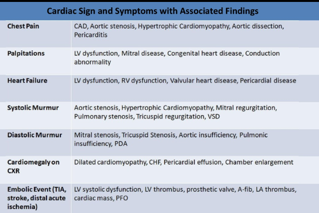
SOB is associated with
valvular stenosis
pulmonary HTN
significant regurg
CHF
decreased EF%
what is dyspnea on exertion?
difficulty breathing with exercise
dyspnea on exertion is associated with
valvular stenosis
pulmonary HTN
significant regurg
CHF
decreased EF%
what is hemoptysis?
bloody sputum
hemoptysis is caused by
pulmonary edema
hemorrhage
hemoptysis is associated with
significant MS
significant acute MR
what is cyanosis?
bluish discoloration of skin and mucous membranes
cyanosis is associated with
pulmonary disease
eisenmenger syndrome
TOF
other r to l shunts
what can lead to clubbing of fingers and toes?
cyanosis
what is edema
accumulation of fluid in tissues/cells
what is pitting edema
ability to indent the skin and it takes time for the skin contour to return to normal
what can lead to edema?
LV/RV failure
systemic HTN
how is pitting edema measured?
scale of +1 - +4 with +4 being very slow normalization of skin contour
what is brawny edema
occurs when chronic tissue edema leads to tissue fibrosis (excessive buildup of fibrous connective tissue)
edema and skin discoloration occurs without pitting
what kind of edema is commonly seen with left heart failure? right heart failure?
left : pulmonary edema
weaked LV fails to pump oxygenated blood so blood backs up into LA and pulm veins
right : bilateral pedal edema
RV fails to pump blood forward into the lungs and blood backs up into the systemic venous circuation
compare venous insufficiency swelling vs heart failure swelling
venous insufficiency : swelling in affected leg(s)
heart failure : swelling of legs AND feet
what is jugular vein distention
increased right heart pressure causes back log of blood into the vena cava and their tributaries
jugular vein distention can be due to
tricuspid stenosis
pulm htn
severe tricupid regurg
constrictive pericarditis
cardiac tamponade
what is pulsus paradoxus
>10mmHg drop in systolic BP with inspiration
pulsus paradoxus is associated with
cardiac tamponade (rapid fluid accumulation in pericardial sac)
constrictive pericarditis (pericardium becomes rigid, scarred and thick)
pulm embolism
COPD (lung disease)
what are palpitations?
when pt is aware that heart is beating
palpitations can be caused by
exercise
anxiety
caffeine
smoking
what is an arrhythmia?
abnormal heart rate or rhythm
what is syncope?
fainting/passing out
syncope can be caused by
hypotension
arrhythmia
myocardial infarction
subao stenosis
severe ao valvular stenosis
what is a murmur
audible sound associated with turbulent flow
what aids in murmur diagnosis
timing and duration of murmur
murmurs are associated with
valvular disease/defects
septal defects
what is a thrill?
palpable vibration and loud harsh murmurs
thrills are associated with
VSD
ao/pulm stenosis
fever/malaise is seen with
endocarditis (bacteria/fungi entering bloodstream causing heart lining/valves to be inflamed)
what is cachexia?
overall state of ill health (malnutrition and wasting) due to chronic heart disease
elevated serum levels of these three can indicate acute myocardial infarction
troponin
creatine kinase MB
lactic dehydrogenase (LDH)
** usually increased during and for 24-48 hours after an acute MI
abnormal carnitine levels result in
impaired muscle metabolism which can lead to dilated cardiomyopathy
what are signs of infection?
presence of staphyloccocus
fever on unknown origin
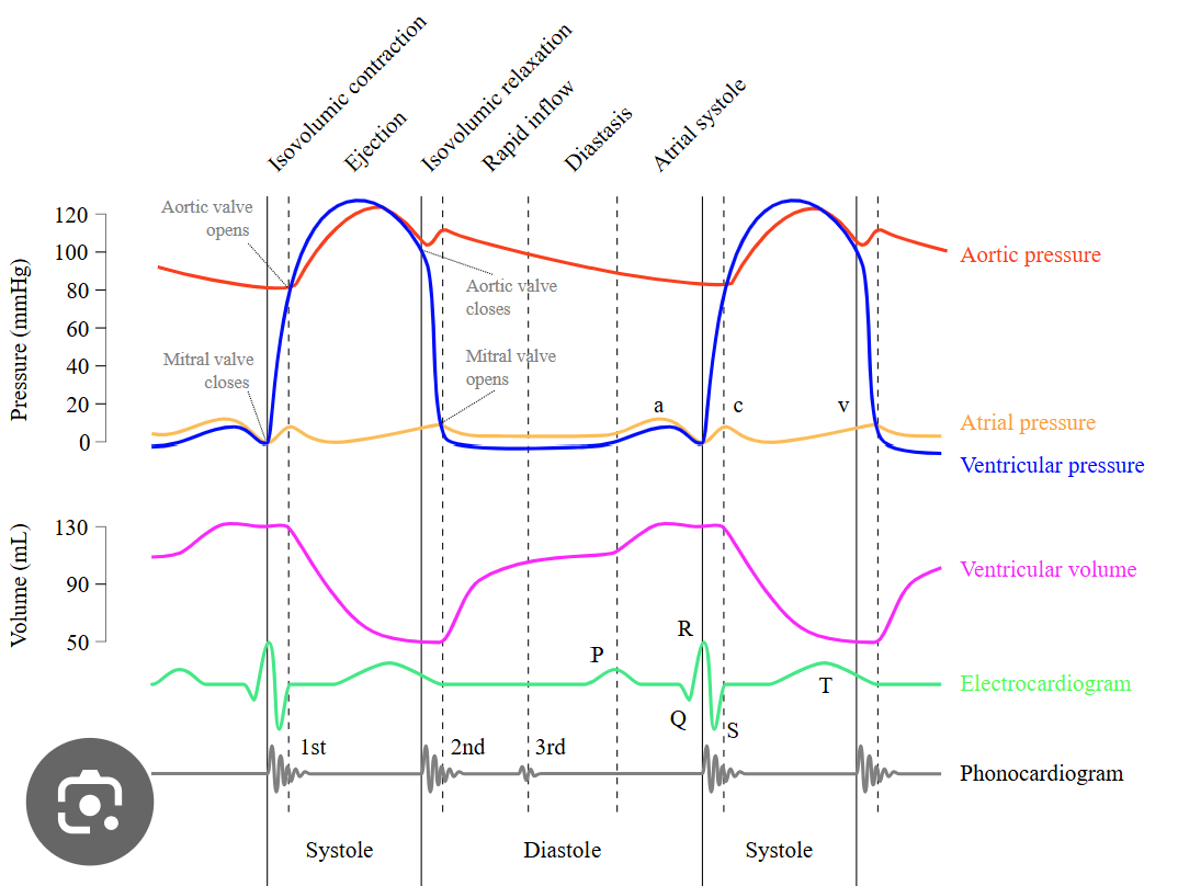
what causes the first heart sound (S1 lub)
mitral and tricuspid valve closing after atrial contraction
when does S1 occur on the EKG?
onset of QRS wave
what causes the second heart sound (S2 lub)
ao and pulm valve closing after ventricular contraction
when does S2 occur on the EKG?
end of T wave
which heart sound has a higher pitch?
s2
what closes first : mv or tv
mv
what closes first : ao or pulm
ao
what is s3? caused by?
abnormal heart sound : ventricular gallop
caused by oscillation of blood back and forth between the walls of the ventricals due to the inflow of blood from the atria
when does s3 occur?
in diastole
early diastole just after s2
s3 indicates
rapid ventricular filling due to
pregnancy
anemia
CHF
dilated cardiomyopathy
severe valvular regurg
l to r shunting
high cardiac output
what is s4? caused by?
abnormal : presystolic gallop or atrial gallop
caused by sound of blood being forced into a stiff/hypertrophic ventricle during atrial kick
when does s4 occur?
diastole
late right before s1
s4 is seen with
htn
severe ao/pulm stenosis
cad
cardiomopathy
which abn heart sound is normal is children/YA?
s3
after age 40 = abnormal & correlated with dysfunction or volume overload
when does the opening snap occur? associated with?
onset of diastole
mitral stenosis
what causes ejection click
abnormal tethering of cusps and it is the sounds they make when the valve tries to open under significant pressure
when does the ejection click occur?
right after S1 and occurs with ventricular contraction
when does the midsystolic click occur? associated wtih?
mid-late systole
associated with MVP (leaflets billow back into LA)
what is pericardial rub?
friction sounds caused by inflammation due to acute pericarditis
what is pericardial knock?
heart knocking against thickened rigid pericardial sac
pericardial knock is associated with
constrictive pericarditis
when is pericardial knock heard?
early diastole
during filling blood passively rushes from atria into ventricles
due to rigid pericardium the heart is restricted and unable to expand
causes sudden stopping or braking of blood creating a loud knocking sound
compare functional vs pathologic murmur
functional : caused by rapid inflow across normal cardiac valve
pathologic : caused by turbulence of flow across disease cardiac valves
murmurs are characterized by
location (apex, sternal border, arch)
timing (systole, diastole, continuous)
intensity (grade 1-6, 1 barely audible; 6 very loud with palpable thrill)
pitch (low to high)
quality (harsh, rubbing, blowing, rumbling)
sound profile (crescendo, descrescendo, holosystolic)
in terms of heart sounds, when do systolic murmurs occur?
between s1 and s2
what are the systolic murmurs
mid systolic
late systolc
holosystolic
systolic ejection
describe what causes a midsystolic murmur
ao or pulm stenosis
increased flow through normal semiluar valves
dilation of ao root
dilation of pulm trunk
describe what causes a late systolic murmur
mitral valve prolapse
tricuspid valve prolapse
pap muscle dysfunction
describe what causes a holosystolic murmur
mitral and tricuspid regurg
vsd
when does the holosystolic murmur occur
immediately after s1 and continues to end systole
occurs during isovolumic contraction period & when ao valve is opening
when does the systolic ejection murmur occur
when valve is opening BUT NOT during isovulimc contraction period
slowly after s1 and increases in intensity relative to severity of valve obstruction
what causes systolic ejection murmur
semilunar valve stenosis (as,ps)
hypertrophic obstructive cardiomyopathy
when do diastolic murmurs occur
during interval between s2 of one cardiac cycle and s1 of another cardiac cycle
what are the diastolic murmurs
regurg diastolic murmurs
diastolic ejection murmur
late diastolic
austin flint
dock murmur
when do regurg diastolic murmurs occur?
early diastole; immediately after S2
what causes regurgitant diastolic murmurs
semilunar valve regurg
when do diastolic ejection murmurs occur?
when av valves are opening BUT NOT during isovolumic relaxation period
mid diastolic murmur
what causes diastolic ejection murmurs
av valve stenosis
when does late diastolic mumur occur?
right before s1
how do large l to r shunt affect AV flow
increased av valve flow (PDA/ASD)
what is the austin flint murmur?
functional diastolic murmur caused by significant aortic regurg
sound from vibratory motion of MV caused by aortic insufficiency jet
what is a dock murmur? associated with?
early diastolic murmur
associated with stenosis of LAD artery
what causes a continuous murmur
continuous flow through systole and diastole
continuous murmurs are seen in patients with
PDA
AV fistula (abn direct connection between artery and vein)
anomalous coronary origin
what is amyl nitrate?
vasodilator
venous return increases and BP drops
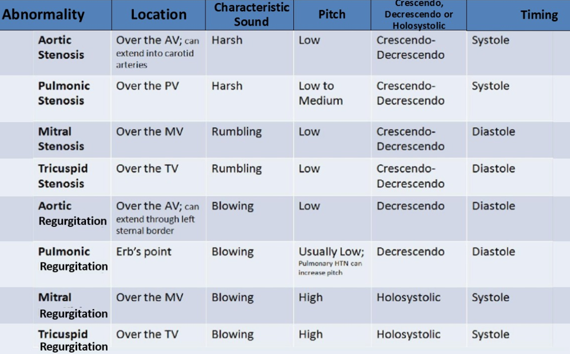
which valvular abnormalities are holosystolic?
mr and tr
which valvular abnormalities are crescendo-descrescendo?
ao/pulm stenosis
mitral/tricuspid stenosis
which valvular abnormalities are descrescendo?
ao regurg
pulm regurg
what is aids and what is the most common cardiac complication?
virus that damages the immune syste and inhibits the body’s ability to fight infection/disease
most common cardiac complication : pericardial effusion
what is acromegaly and what is it associated with?
when the pituitary gland produces too much growth hormone
associated with
LV hypertrophy
increased LV mass
impaired relaxation
what finding are normal with aging?
increased LV wall thickness and mass
sigmoid ventricular septum
increased LA size
reduced cardiac output and stroke volume
mitral annular calcification
mitral valve leaflet thickening
increased mitral A velocity (reduced E/A ratio)
prolonged IVRT
MR/TR
increased ao root size
ao valve calcification
alcohol abuse can cause
acute MI
reduced systolic ventricular function
dilated cardiomyopathy