303 Week 2 - Pulse Sequences
 Call Kai
 Learn
Learn Practice Test
 Spaced Repetition
 Match
Match Flashcards
 Knowt Play
1/9

There's no tags or description
Looks like no tags are added yet.
Name | Mastery | Learn | Test | Matching | Spaced | Call with Kai | Chat |
|---|
No analytics yet
Send a link to your students to track their progress
10 Terms
What is each axis?
X-Axis = Frequency Encoding Grading
Y-Axis = Phase Encoding Gradient
Z-Axis = Slice Select Gradient
Spin Echo
90 deg. → 180 deg. → Rest → Repeat
Takes too Long
Great SNR in T1 images
Fast Spin Echo
90 deg. → 180 deg. → Repeat
Signal does slowly and slowly get smaller so eventually a 90 degree pulse would be needed
FatSat
180 deg. (fat + water then recover at diff. times) → 90 deg. pulse (to saturate out the signal)
T2 makes fat bright so we use a T2 fat sat to keep fluid bright but Fat Dark
FLAIR
Fluid Attenuated Inversion Recovery
Good for : M.S. + Plaque around Lateral Ventricles
180 deg. → 90 deg.
Suppresses Fluid
Used for Brain Path to supress Lat Ventricles
STIR
Short Tau Inversion Recovery
Used for MSK to supress fat over Large FOV
Why is Fat still bright in T2?
J Coupling: Fat appears bright in T2 when it should be Dark
When are RF is sent, it is constantly excited and never relaxes and gives artefact of a signal
Gradient Echo
Alpha Pulse (<90 deg.) → We then Change Frequency Encoding Gradient (X-axis) [changing the Magnet, north to south rapidly]
INCLUDES: SWI (Susceptiblity Weighted Image)
USED FOR: Head Scans
Downside: Susceptible to artefacts (unlike spin echo)
SAFETY CONSIDERATION: Due to rapid changing of Magnetic Field --> Twitching -> Nerve Stimulation -> Faradays Law of Induction
FLOWING BLOOD: Bright
Stationary Blood: Dark (for SWI)
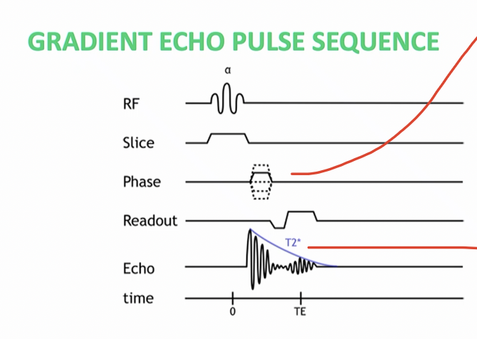
What is bottom red line pointing to
FID: Fourier Induction Decay
Signal to Noise Ratio:
Larger Voxel = Better SNR = Faster Scans (BUT WE LOSE Resolution)