hematology exam 1 - hemoglobin structure and fxn, iron metabolism, iron lab tests
 Call Kai
 Learn
Learn Practice Test
 Spaced Repetition
 Match
Match Flashcards
 Knowt Play
1/18

There's no tags or description
Looks like no tags are added yet.
Name | Mastery | Learn | Test | Matching | Spaced | Call with Kai | Chat |
|---|
No analytics yet
Send a link to your students to track their progress
19 Terms
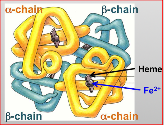
hemoglobin molecule
6composed of 4 heme grps and 2 pairs of dissimilar polypeptide chains
4 polypeptide chains: (two al6pha and two beta)
4 iron (ferrous state, Fe2+66) molecules (heme group)
what is the most functional iron a constituent of
hemoglobin and myglobin (heme proteins that carry oxygen)
25% is in a storage form
where is most non-heme iron stored
in ferritin or hemosiderin in hepatocytes or macrophages
how are body iron levels regulated
absorption → humans cannot actively excrete iron
how is iron absorbed and transported
by enterocytes in the duodenum and jejunum and carried to basolateral membrane where it passes to plasma via ferroportin
hephaestin converting ferrous to ferric iron
transferrin: carrier protein, binds to iron and transports it in the plasma
transferrin receptors: located on all cells (except mature RBCs)
provide cells with transferrin bound iron w/in the cell
in hemopoietic centers (mainly bone marrow), enters immature erythroid cells AFTER transferrin has attached to transferrin receptors on the cell membrane
hepcidin
antimicrobial peptide
regulates basolateral iron transport
when hepcidin is elevated in chronic inflammation and infection, can interfere with absorption = iron deficiency anemia
ferritin and hemosiderin
storage forms of iron
ferritin: water soluble complex
hemosiderin: insoluble complex
made up of ferritin aggregates found in liver and bone marrow
degradation product of ferritin produced by digestion of protein + release of iron
normally most stored iron is in ferritin form, but when iron stores are abundant, proportion of hemosiderin increases
where is iron stored
liver, bone marrow and spleen
when iron is needed from iron stores, returned to transferrin to be used by cells that need iron for metabolism
laboratory tests for iron
serum ferritin
serum iron
serum transferrin
transferrin saturation
bone marrow or liver biopsy
serum transferrin receptor analysis
erythrocyte protoporphyrin assay
serum ferritin
adult reference range: 12-300 mcg/L
indicates: iron stores
serum iron
adult reference range: 10-30 mmol/L
indicates: tissue iron supply
serum transferrin (TIBC)
adult reference range: 47-70 mmol/L
indicates: tissue iron supply
Because an indirect measure of total circulating transferrin, it indicates the body's iron-carrying capacity. The liver increases transferrin (raising this analyte) when tissue iron levels are low, as an adaptive mechanism to scavenge more iron.
transferrin saturation
adult reference range: 16-60%
indicates: tissue iron supply
bone marrow or liver biopsy
adult reference range: normal stores seen in tissue
indicates: direct observation of functional iron
relationship b/w hemolysis and clinical events
decrease in RBC, HgB, Hct = symptoms of anemia, pallor, fatigue, tachycardia
increase of bilirubin = jaundice
releases massive amounts of hemoglobin into the bloodstream, which is broken down into heme.
The heme is rapidly converted into excess unconjugated (indirect) bilirubin, temporarily exceeding the liver's capacity to process and clear it
hemoglobinemia = blood-tinged plasma
excess free Hb in plasma
hemoglobinuria = blood-tinged urine
excess fre Hb in urine
hyperplasia: increase in RBC precursors = M:E of 1:2
normally 3:1 or 4:1
peripheral blood smear shows polychromasia = a laboratory finding where red blood cells appear larger and have a bluish-gray tint under a microscope
rapidly releasing reticulocytes (immature RBCs) into bloodstream to compensate for loss of RBCs
spherocytosis may appear (if Abs present) = when RBCs go through spleen the Abs are sheared off and spherocytes form
extravascular hemolysis
90% of hemolysis
occurs in spleen, liver, lymph nodes, bone marrow, and RES
RBC destroyed and contents phagocytized by macrophages
leads to release of Heme and globin groups
globin AA are recycled into AA pool and the heme components go diff pathways
iron is transported by transferrin (protein carrier) to bone marrow for erythropoiesis
intravascular hemolysis
10% is intravascular and it occurs when RBCs are destroyed directly in the blood vessel
Hb released into plasma
can be observed in the red-tinged plasma after blood is centrifuged
hemoglobinuria (blood in urine)
ex of potential causes: transfusion incompatibility, complement mediated hemolysis
how to distinguish extravascular from intravascular hemolysis in lab
hemoglobin released from ruptured RBCs binds haptoglobin (transport protein) in plasma
Hgb - Haptoglobin complex is too large to be filtered by kidneys
not excreted, but transported to liver to be destroyed
Undetectable Haptoglobin = Intravascular: Because cells break directly in the blood, a massive flood of hemoglobin is released. Haptoglobin quickly binds to this hemoglobin and is rapidly cleared by the liver, leaving almost zero haptoglobin remaining.
Low or Normal Haptoglobin = Extravascular: Because the destruction happens in the spleen or liver, the hemoglobin is safely processed inside macrophages. It leaks into the blood at much lower levels. Therefore, haptoglobin is moderately decreased but rarely fully consumed.
idek results to differentiate b/w extravascular and intravascular hemolysis
Useful Lab tests:
1. Plasma Haptoglobin is low during hemolysis (no place for transporting HB)
intravascular
2. Hemoglobin conc., Hematocrit, and RBC count decreased
extra and intra
3. Reticulocyte count elevated
extra and intra
4. Plasma LDH enzyme elevated (RBC enzyme)
lactate dehydrogenase enzyme, normally present in high lvls inside RBC
5. Hemoglobinemia (hemoglobin in plasma elevated)
6. Hemoglobinuria (hemoglobin in urine).