Ch 6: Abdominal Vascular Structures
 Call Kai
 Learn
Learn Practice Test
 Spaced Repetition
 Match
Match Flashcards
 Knowt Play
1/96

There's no tags or description
Looks like no tags are added yet.
Name | Mastery | Learn | Test | Matching | Spaced | Call with Kai | Chat |
|---|
No analytics yet
Send a link to your students to track their progress
97 Terms
The layers of vessel walls
Tunica Intima: inner endothelial lining
Tunica Media: middle smooth muscle layer
Tunica Adventitia: outer layer consisting of collagen fibers
Segments of the Aorta
Ascending: comes out of the heart
Arch: where a lot of cerebral vascular vessels come from
Descending: heads down after arch
Thoracic: extension of descending through thoracic cavity
Abdominal: after it enters hiatal opening in diaphragm and feeds extremities/trunk of body

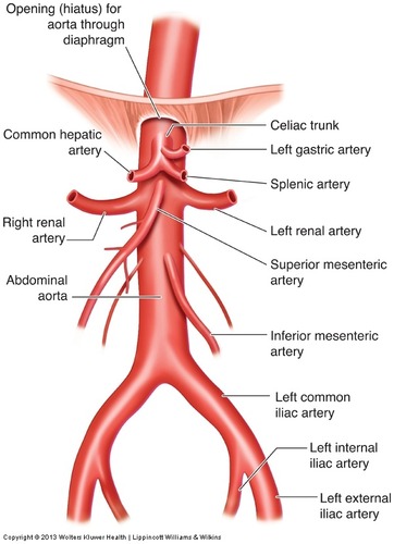
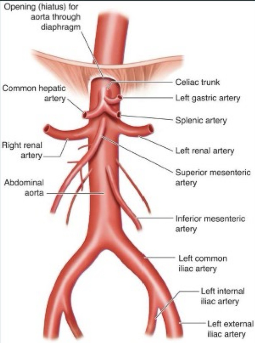
What are the major abdominal aortic branches that should be recognized from superior to inferior?
Celiac trunk, superior mesenteric artery, renal arteries, inferior mesenteric artery, and common iliac arteries
Artery wall Characteristics
Thicker/more elastic, tend to maintain constant shape, do not readily collapse, pulsatile in nature, and stiff (will not collapse if you apply pressure)
Vein wall characteristics
Less smooth muscle and less elastic tissue, unable to contract to force blood flow, requires pressure gradient (which comes from breathing, skeletal muscle contractions, and valves in extremities)

Which veins form the main portal vein
Splenic vein and superior mesenteric vein (SMV)

Where do hepatic veins drain
Into the IVC

What is atherosclerosis
It is a form of arteriosclerosis where intimal lining of arteries is altered by the presence of focal accumulation of lipids, blood and blood products, or calcium deposits.
media of arterial wall is also changed/hardened (harder to contract)
Atherosclerosis risk factors/signs and symptoms
Risk factors are increased lipids, high cholesterol, hypertension, smoking, or diabetes.
Symptoms are rare until significant stenosis (narrowing of a vessel) develops, but may have pain with walking or back pain.
Atherosclerosis Sonographic appearance
Luminal irregularities (low level echoes (soft gray indicating soft plaque/not calcified yet)along internal walls of aorta), tortuosity (vessel gets wavy in appearance) which is easier to see in transverse, and wall calcification (soft plaque begins to harden/becomes more echogenic and often produces shadows). If there’s calcification on anterior wall, it can produce shadows inside the vessel, making it tricky to see inside

What is an aneurysm
Abnormal dilation of a blood vessel
True aneurysm
Involve all 3 layers of the arterial wall and has dilation greater than 3cm and lacks normal tapering
False aneurysm (pseudoaneurysm)
Extra-vascular hematoma communicating with the intravascular space, often post-procedure or from trauma. These have the yin-yang blood flow sign
What does the yin-yang sign represent in pseudoaneurysm?
Blood flowing in and out of the pseudoaneurysm sac/the swirling bidirectional flow
Aneurysm Etiology (cause/origin)
risk factors are smoking which cases hardening of the arteries, and atherosclerosis which is fatty plaque build up
Aneurysm Symptoms
Usually asymptomatic, and often suspected by a doctor during a physical exam when patient has a palpable pulsating mass in umbilicus area. patient may have vague unexplained low back/abdominal pain and doctor may hear “wooshing” sound when listening to abdominal vessels (blood flow velocity is different)
Why should you identify the origin of renal/iliac vessels
To determine AAA location or vessel involvement. If they are located near bifurcation, examination of iliacs is also necessary. Knowing the location of the AAA will help know if the patient could have a procedure done to fix it or if its too risky.
Where do most AAA occur
Below the level of the renal arteries
Most true aneurysms are what type?
fusiform and circumferential
What is a Fusiform Aneurysm
a gradual progressive dilation of the vessels circumference. they vary in diameter and length

What is a Saccular Aneurysm
A “neck” or connecting stalk in an aneurysm off the aorta. bulging in one area of the vessel
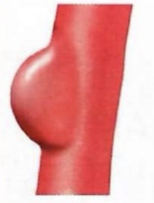
What is a Dissecting Aneurysm
Longitudinal tear of the intimal lining of the vessel, which allows bleeding into the wall. the lest common type and occur more often in the thoracic AO

Where do saccular and fusiform aneurysms often occur
In abdominal AO
Aneurysm Sonographic Appearance
Ectasia of AO, true aneurysm has dilation greater than 3cm and lacks normal tapering, thrombus is common finding within AAA (along anterior and posterior wall where vessel is dilated)
What is Ectasia
Diffuse, mild enlargement of AO without distal tapering
Normal AO measurement at diaphragm
2.5cm and tapers distally
Why most total aneurysm size be assessed rather than only patent lumen
Assessment of the total outer diameter of the blood vessel is needed because that determines the risk of rupture
What is Mural Thrombus
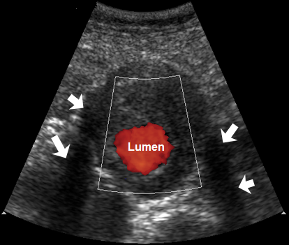
Thrombus that attaches/bulges out of the wall of the blood vessel

What is the crescent sign described in an AAA with mural thrombus
An anechoic crescent caused by liquification within the thrombus

How does color Doppler help when an AAA contains mural thrombus?
It demonstrates the residual flowing lumen and helps distinguish flowing blood from thrombus.
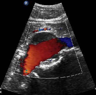
What is an aortic dissection
Separation of the layers of the arterial wall by blood or hemorrhage after a tear in the inner wall
Aortic dissection causes
Are not clear but could be hypertension (increase pressure in vascular structure), pregnancy (changes in the body/hormones), Marfan syndrome (genetic condition that affects the connective tissue and because of the tissue blood vessels are made from, it could cause problems), or general valve disease within vascular system
Aortic dissection signs/symptoms
intense chest pain (because they mostly occur in thoracic aorta), could be associated abdominal pain, headaches, syncope (fainting/passing out from temporary drop in blood flow to the brain), or transient blindness
What is the key B-mode sonographic sign of an aortic dissection?
A thin echogenic linear intimal flap within the lumen.
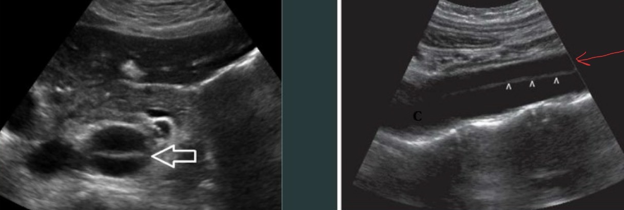
What happens to blood flow in an aortic dissection?
Blood may flow through the normal lumen and through a false channel created within the vessel wall.
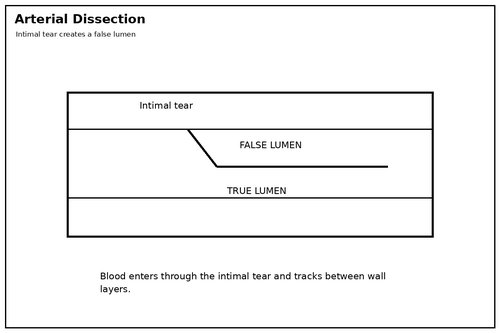
What is DeBakey type I aortic dissection?
A dissection involving the ascending aorta and extending into the arch and potentially beyond.

What is DeBakey type II aortic dissection?
A dissection involving the ascending aorta and aortic arch, and associated with Marfan syndrome. least common type

What is DeBakey type III aortic dissection?
A dissection beginning in the descending aorta below the left subclavian artery.

Which DeBakey types have high mortality risk and why
types I and II because of their location, it will often spread to pericardium
What is an aortic rupture
Disruption of aortic wall with bleeding outside the vessel, primarily from ruptured AAA
When can an AAA rupture
at any size, but the risk increases with aneurysm is larger than 7cm
Why is an aortic rupture a medical emergency
its mortality rate is 100% if left untreated because it can cause rapid internal bleeding, hemorrhage, hypotension, or syncope
Aortic rupture signs/symptoms
central back pain (mid/lower back) or very quick drop in blood pressure (may feel faint/pass out)
Aortic rupture sonographic appearance
disruption of aorta wall and bleeding in abdomen (intra-abdominal fluid collections). Could be hard to see the aneurysm because of the wall disruption/bleeding
Most common type of splanchnic artery aneurysm
Splenic artery aneurysm
Where do splenic artery aneurysms commonly occur?
In the main splenic arterial trunk. The image is near the splenic hilum.

Which sex has a higher prevalence of splenic artery aneurysm
Females
Are splenic artery aneurysms common?
No they are rare but can be life-threatening.
Splenic artery aneurysm etiology
Pancreatic inflammation, peptic ulcer disease, mycotic lesions (fungal rashes/growths), portal hypertension, and multiple pregnancies (changes in blood flow, connective tissue, and hormones may weaken vessel walls). Overall, anything that causes increased pressure along portal system/splenic vessels
Splenic artery aneurysm symptoms
LUQ pain, N/V (nausea/vomiting), or palpable mass
How can color Doppler help evaluate a suspected splenic artery aneurysm?
It demonstrates blood flow within the dilated vascular structure and helps distinguish it from a nonvascular cystic mass

2nd most common splanchnic vessel aneurysm
Hepatic artery aneurysm
What percentage of hepatic artery aneurysms are extrahepatic
75%
Which hepatic artery is more often affected
right hepatic artery
Which sex has a higher prevalence of hepatic artery aneurysm
males
Hepatic artery aneurysm etiology
Systemic infection (liver filters toxins within the body), arteriosclerosis (hardening of arteries), blunt abdominal trauma, and chronic cholecystitis (GB inflammation, close proximity to these vessels)
Hepatic artery aneurysm symptoms
Epigastric pain, GI bleeding, hemobilia (blood in bile), and jaundice (yellowing of skin/eyes)
What important complication of hepatic artery aneurysm is emphasized?
A propensity (strong tendency) to rupture
How may a hepatic artery aneurysm appear on color Doppler?
As a dilated vessel or mass with turbulent mixed red-and-blue swirling flow
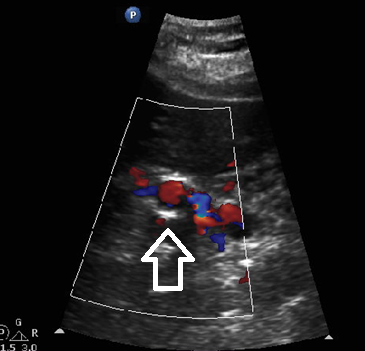
Splanchnic artery aneurysm sonographic appearance
distinguishing feature is location in abdomen, may appear as anechoic or complex abdominal mass, demonstrate continuity of mass with vessel, and doppler should always be used to assess for characteristic swirling blood flow pattern
What vessel abnormality is an iliac artery aneurysm most often associated with?
Abdominal aortic aneurysm (AAA)
What is the most common major complication of an untreated iliac artery aneurysm
rupture
Iliac artery aneurysm symptoms
Urologic, gastrointestinal, or neurologic symptoms because of proximity (such as hydronephrosis, uti, or kidney infection), pain, or a palpable mass
Iliac artery aneurysm etiology
most are arteriosclerosis in origin, external or surgical trauma, or pregnancy
Iliac artery aneurysm sonographic appearance
Primarily anechoic mass in pelvis, smaller aneurysms may be difficult to see due to bowel gas, thrombus along periphery, calcific wall changes, and may see doppler turbulent arterial signal (swirling blood flow pattern and PW doppler will show very turbulent waveform)
Why should both iliac arteries be evaluated when an iliac aneurysm is found?
Iliac aneurysms tend to be bilateral
What is an aortic graft, endograft, or prosthesis and how is it named
A man-made structure used to repair or bypass diseased vascular segments, including aortic aneurysms (taking part of one vessel and connecting it to another to to bypass damaged area). they are named in reference to the vessel they are attached to
Aortic graft sonographic appearance
A ribbed or railroad-track-like echogenic structure

What is aortofemoral bypass surgery
A graft connecting the aorta to the femoral artery to bypass diseased iliac arterial segments, placed anterior and adjacent to native vessel. The diseased segment is left intact and an end-to-end anastomotic technique is used
What is endovascular repair of an abdominal aortic aneurysm (EVAR)
a minimally invasive surgical procedure that involves deploying a stent graft into the AO with exclusion of the AAA (to direct blood through the graft and reduce pressure and flow within the aneurysm sac)

Why is surveillance important after graft or EVAR placement?
To confirm continued patency and detect complications such as leaks, thrombosis, or infection
Vascular graft complications
Pseudoaneurysm, hematoma, abscess or infection, endoleaks (persistent blood flow into aneurysm sac outside an aortic stent graft after EVAR), and graft occlusion (blood can become stagnant and clot, or the graft can become blocked because of procedural or other complications)
What happens to blood velocity at the site of a stenosis?
Velocity increases
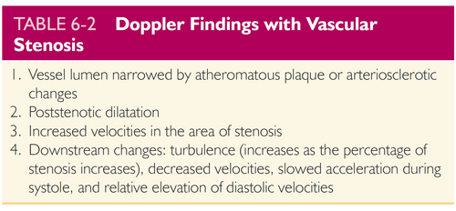
What happens to flow immediately downstream/after from a significant stenosis?
turbulence develops and velocity drastically drops

What is renal artery stenosis
Narrowing of a renal artery that reduces renal blood flow which can produce elevated blood pressure and is associated with uncontrolled hypertension
Renal artery stenosis complications
Decreased glomerular filtration rate (GFR) and ischemic renal damage
Renal artery stenosis sonographic appearance/findings
elevated velocity in the main renal artery and parvus/tardus pattern in intrarenal arteries. must evaluate prox, mid, and dist segments of all types of arteries in the kidney (segmental, interlobar, and arcuate arteries)
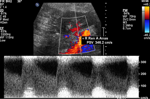
What does a parvus-tardus waveform look like conceptually?
A delayed, dampened systolic upstroke with a lower, rounded systolic peak, and increase in acceleration index

What happens to the IVC caliber below an obstruction?
It increases or becomes dilated below the point of obstruction
IVC obstruction causes
Most common cause is right-sided heart failure, but could also be hepatomegaly, enlarged lymph nodes around aorta, retroperitoneal mass/tumor, or anything that can apply pressure to the IVC
IVC obstruction signs/symptoms
depends on where it is/how severe it is, but you can experience abdominal pain, ascites, or lower extremity edema (if obstruction is really severe)
IVC obstruction appearance
A distended IVC containing intraluminal echoes with absent color Doppler signal.
dilation below the obstruction, respiratory changes decrease or absent below the obstruction, or in right sided heart failure, the proximal IVC and hepatic veins become congested

What are primary IVC tumors
tumors that form in the IVC, this includes leiomyosarcoma, and they are rare
Which sex do primary IVC tumors tend to occur
women
Malignant Invasion IVC tumors
Metastasis or extension of tumors that may occur from renal cell carcinoma (most common, especially the right side because RRV is shorter than left), adrenal tumors, or hepatocellular carcinomas
IVC tumor sonographic appearance
An enlarged vessel containing an intraluminal mass of varying echogenicity. larger tumors may be heterogeneous with areas of necrosis. doppler will show distinct blood flow changed around the tumor
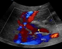
What is portal vein thrombosis
Thrombus within the portal venous system
Portal vein thrombosis etiology
Portal hypertension, abdominal inflammatory processes (cirrhosis), trauma, postsurgical complications, or hypercoagulable states (BCPs, pregnancy, polycythemia vera)
Portal vein thrombosis symptoms
Abdominal pain, abdominal rigidity (hardened feeling), and elevated liver function tests
Portal vein thrombosis appearance
Varies depending on stage (starts with echogenic thrombus and small collaterals).
normal caliber PV is 13mm or less but a thrombus could make it dilate because of improper flow

What is portal hypertension
Increased pressure within portal venous system
Advanced cases of Portal hypertension can cause what
ascites or GI bleeding
What is important to look for when diagnosing portal vein hypertension
secondary effects of increased pressure
What are some secondary findings of portal hypertension
Increased portal vein diameter, ascites, splenomegaly, recanalized umbilical vein (ligamentum teres/remnants of umbilical cord), and bidirectional flow (both red and blue flow on color doppler)
What is an AV (arteriovenous) Fistula
an abnormal connection between an artery and vein
What indications are there to have an AV fistula exam
Presence of bruit (buzzing/vibrations) or thrill (swishing or humming sound) associated with dilated pulsatile mass, low back and abdominal pain, or swelling of lower trunk and extremities
AV Fistula doppler findings
Pulsatile, arterialized venous waveform, increased systolic and diastolic arterial velocities proximal to fistula but reverts to normal diastolic flow distal to fistula