Anatomy 2 - Blood Vessels, Blood Pressure and the Lymphatic System
1/50
There's no tags or description
Looks like no tags are added yet.
Name | Mastery | Learn | Test | Matching | Spaced | Call with Kai |
|---|
No analytics yet
Send a link to your students to track their progress
51 Terms
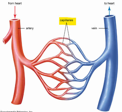
What are the 3 principal categories of blood vessels?
Arteries - carry oxygenated blood away from the heart; veins - carry deoxygenated blood back to the heart; capillaries - connect arteries to veins to create a circuit
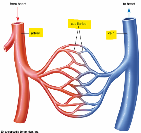
This is the simplest and most common route for blood
Heart > arteries > arterioles > capillaries > venules > veins
True or false; during one cycle, blood passes through only one network of capillaries
True
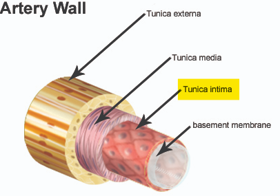
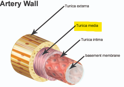
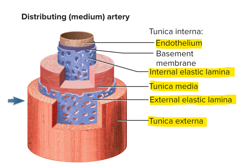
Walls of arteries and veins have 3 layers, what are they?
Tunica interna (deep), tunica media (middle) and tunica externa (superficial)
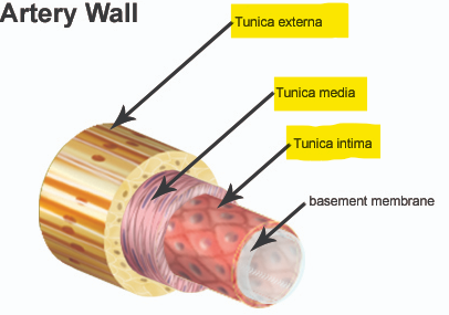
This layer of the blood vessel wall is simple squamous epithelium and faces the lumen
Tunica interna
This layer of the blood vessel supports endothelium from rupturing and is for contraction
Tunica media
Do veins or arteries have an elastic lamina layer?
Only in arteries, not in veins
These are the biggest arteries; what are some examples?
Conducting (elastic or large) arteries. Examples: aorta, pulmonary trunk - blood from heart to lungs, common carotid - blood to the brain, subclavian - blood to upper limbs and common iliac arteries - blood to lower limbs
This artery delivers blood to specific arteries; what are some examples?
Distributing (muscular or medium) arteries. Examples: brachial - arm, femoral - legs, renal - kidneys and splenic arteries - spleen
These are the smallest of resistance arteries; what is their function?
Arterioles. Function: Control the amount of blood to various organs; lumen is modulated with vasoconstriction and vasodilation
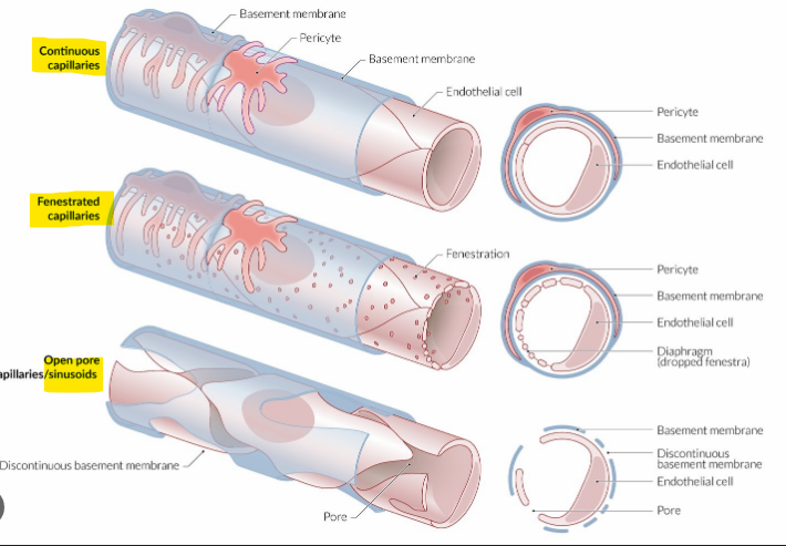
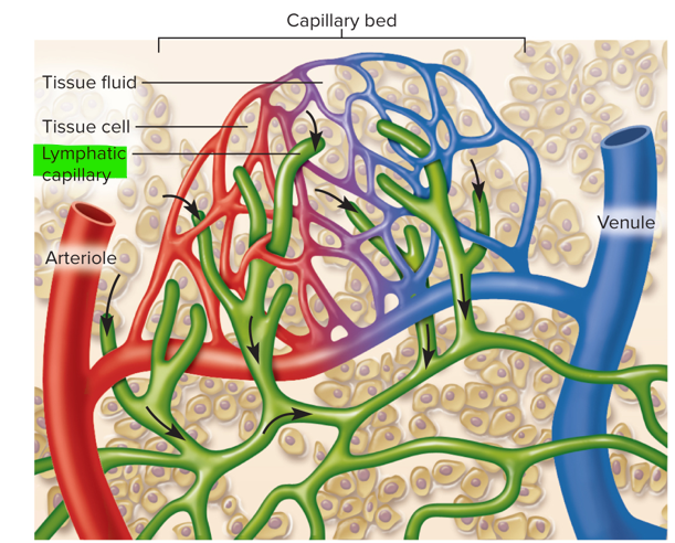
These are exchange vessels
Capillaries; where gasses, nutrients, wastes and hormones pass between the blood and tissue fluid
What are the 3 capillary types from least permeable to most permeable?
Continuous, fenestrated and sinusoids
These have a greater capacity for blood containment than arteries
Veins; have a steady blood flow and low blood pressure
Give an example of a large vein
Venae cavae, pulmonary veins, internal jugular veins and renal veins
What is blood pressue?
The force that blood exerts against a vessel wall
Where is blood pressure measured?
Brachial artery
These are the two pressures recorded when taking blood pressue
Systolic (ventricular contraction) and diastolic (ventricular relaxation) pressure
What is the normal blood pressure for a young adult?
120/75 mm Hg - systole/diastole
This is the difference between systolic and diastolic pressue
Pulse pressure (systolic pressure - diastolic pressure)
How does salt influence blood pressure?
Less salt = less volume so BP goes down; high salt = high volume so BP goes up
Albumin is produced by the _____
Liver
This is the most powerful influence on blood flow
Vessel radius; only significant way of controlling resistance
Define whether low and high viscosity speeds or slows the flow of blood
Low viscosity speeds the flow of blood; high viscosity slows the flow of blood
Where is the vasomotor center and what does it do?
Medulla oblongata; it exerts sympathetic control over blood vessels throughout the body
These are the 3 autonomic reflexes of the vasomotor center
Baroreflexes, chemoreflexes and medullary ischemic reflex
This autonomic reflex responds to changes in blood pressure; what is able to detect this change?
Baroreflex; carotid sinuses detect increases in BP
What does an increase in BP lead to?
Inhibition of sympathetic cardiac and vasomotor neurons - leads to vasodilation and lower cardiac output; activation of the parasympathetic NS - slow heart rate and thus reduces BP
What does a decrease in BP lead to?
Activation of sympathetic cardiac and vasomotor neurons - leads to vasoconstriction and higher cardiac output; inhibition of the parasympathetic NS - increases heart rate and thus raises BP
This autonomic reflex responds to changes in blood chemistry
Chemoreflex
What are the 2 chemoreceptors?
Aortic bodies located in the aortic arch and carotid bodies located in the carotid arteries
What is the primary role of chemoreflexes?
To adjust respiration to changes in blood chemistry; stimulate breathing to eliminate CO2 and get more O2
What does hypoxemia (low O2), hypercapnia (high CO2) and acidosis stimulate? What does this increase?
Chemoreceptors to cause widespread vasoconstriction; increases BP and lung perfusion, leading to an increase gas exchange
This is the automatic response to a drop in perfusion of the brain
Medullary ischemic reflex; the medulla oblongata monitors its own blood supply
What is ischemia and what does it trigger?
Insufficient perfusion; triggers the medullary ischemic reflexes; increases heart rate and contraction force, causes widespread vasoconstriction and increases BP so that more oxygen will go to the brain
True or false; hormones also influence BP
True
This hormone raises BP and is a potent vasoconstrictor
Angiotensin II
This hormone is the salt-retaining hormone and promotes Na+ retention by the kidneys and supports BP
Aldosterone
This hormone is secreted by the heart and lowers BP; how does it lower BP?
Natriuretic peptides; these peptides lower BP by inhibiting aldosterone which promotes vasodilation
This gland produces the antidiuretic hormone (prevents urination); what does this promote?
The posterior pituitary gland; promotes water retention and raises BP, it also acts as a vasoconstrictor
These are adrenal and sympathetic catecholamines
Epinephrine and norepinephrine; these cause vasoconstriction which raises BP
What are the general functions of the lymphatic system?
Fluid recovery - fluids leaked from capillaries, 85% are reabsorbed; immunity - excess filtered fluid picks up foreign cells and chemicals from the tissues; lipid absorption - lacteals in small intestine absorb dietary lipids that are not absorbed by the capillaries
This is the recovered fluids of the lymphatic system
Lymph
These transport the lymph
Lymphatic vessels
What are lymphatic tissues composed of?
Aggregates of lymphocytes and macrophages
This is the beginning of the lymphatic system
Lymphatic capillaries (terminal lymphatics)
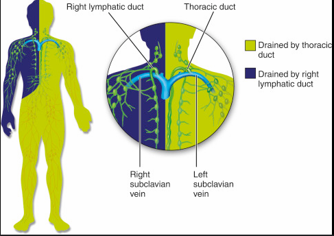
What are the 2 collecting ducts of the lymphatic system?
Right lymphatic duct - empties into right subclavian vein; thoracic duct - empties into the left subclavian vein
These are all lymphatic cells
Neutrophils - antibacterial; natural killer (NK) cells - large lymphocytes that kill anything foreign; T lymphocytes (T cells) - mature in the thymus; B lymphocytes (B cells) - antibodies that mature in bone; macrophages - antigen-presenting cells (APCs); dendritic cells - alert immune system; reticular cells - build lymphatic organs
These are the 2 primary lymphatic organs and where lymphocytes mature
Red bone marrow and thymus
These are the secondary lymphatic organs
Lymph nodes, tonsils and spleen; there is no maturation of lymphocytes here
What is red bone marrow involved in?
Hematopoiesis (blood formation) and immunity
This is a member of the endocrine, lymphatic and immune systems
The thymus - houses developing T lymphocytes (T cells)