Overview of the Sensory Inputs and Motor Outputs of the Brain
1/24
There's no tags or description
Looks like no tags are added yet.
Name | Mastery | Learn | Test | Matching | Spaced |
|---|
No study sessions yet.
25 Terms
Sensory Input (______) → to CNS to be ________, then exits via Motor Output (______) → to the ______ (____ or ____)
afferent, integration, efferent, effector, muscle, gland
What are the three main functions performed by neurons?
- Sensory input: Neurons that collect information about the external environment and internal conditions of the body
- Integration: Neurons that receive the sensory input, analyze it, and make decisions about the appropriate response to make
- Motor output: Neurons that carry out the instructions that result from integration by impulses sent to various effector organs that include muscles and glands
What is sensation?
A physical feeling or perception for something that happens to or comes into contact with the body
How can sensory inputs arrive to the CNS?
Spinal nerve: Nerve that carries signal between the spinal cord and the body (From the sensory receptor to dorsal ganglion to dorsal root)
Cranial nerve: Nerve that carries signal between the forebrain or brainstem and mainly the head and neck
Cranial nerves emerge from the brain, primarily innervating the head and neck, while spinal nerves branch from the spinal cord to serve the trunk and limbs.
How do sensory neurons appear? (Anatomy)
Are unipolar: Most sensory neurons are pseudounipolar, which means they only have one axon which is split into two branches.
What are the sensory receptors?
Receptors: Sensory endings of peripheral branches of sensory fibers
Different modalities:
- Touch
- Temperature
- Movement
- Chemicals
- Pressure
- Light
- Sound
How can the receptors be classified?
• Anatomical: Encapsulated and non- encapsulated
• Location:
- Exteroreceptors: Near the surface of the body. Sensitive to change in external environment
- Proprioceptors: Sensitive to movement of muscles, tendons and joints
- Interceptors: Located within the viscera. Sensitive to change in internal environment
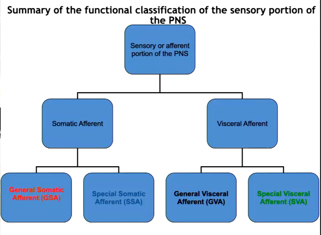
The sensory portion of the PNS can be classified by location of what?
Note the two main zones and their subsequent subdivisions.
Dendritic Zone = the receptive area of a neuron.
- Somatic afferent: the dendritic zone is on or near the surface of the body.
General somatic afferent (GSA):
Touch, temperature, proprioception and noxious stimuli
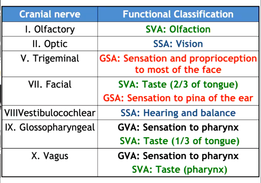
Cranial nerve V for the head
Spinal nerves for the rest of the body
Special somatic afferent (SSA):
Vision: Cranial nerve II
Sound: Cranial nerve VIII
- Visceral afferent: the dendritic zone in in the wall of the various viscera of the body
General visceral afferent (GVA):
Organ content, distention, chemicals
Cranial nerves VII, IX and X to visceral structures in the head
Cranial nerve X and spinal nerves to the viscera and blood vessels of the rest of the body
Special visceral afferent (SVA):
• Taste: Cranial nerves VII, IX and X
• Olfaction: Cranial nerve I
Summarize the functional classification of the sensory portion of the PNS.
Summarize the sensory cranial nerves and their functional classifications.
A majority of the sensory inputs travel to various tracts within the spinal cord on the ____ aspect.
dorsal
Describe the sensory spinal tracts and their main function.
• Fasciculus gracilis:
Proprioceptive information from pelvic limbs
• Fasciculus cuneatus:
Proprioceptive information from thoracic limbs
• Dorsal and ventral Spinocerebellar tracts:
Information to cerebellum for coordination
• Spinothalamic tracts:
Information to thalamus about pain, itch, touch and temperature
What are the motor functions of the brain?
Movement involving a muscular component
How does information output or exit the spinal cord?
Two ways
Exits via the Lower motor neuron (LMN): Neurons located in either the ventral horn of the spinal cord or the cranial nerve nuclei of the brainstem with motor function
Upper motor neuron (UMN): Motor neurons that originate in the motor region of the cerebral cortex or the brain stem and carry motor information down to the lower motor neurons
Activates UMN → Stimulates LMN
Upper motor neurons (UMNs): the "command neurons" in the brain that initiate and control movement.
Lower motor neurons (LMNs): the "final pathway" neurons that directly cause muscles to contract.
Describe the anatomy of the lower motor neuron.
Multipolar, large cell body, with many dendrites with a long axon
How can the motor portion of the PNS be classified?
• The motor portion of the PNS can be classified based on the location of the axon terminals in the body:
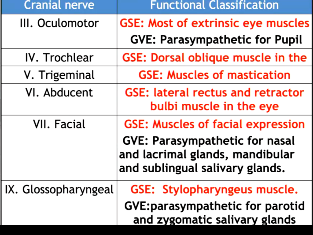
- General somatic efferent (GSE):
• Axon terminals in striated muscle throughout the body
Cranial nerves: all of them except from I, Il and VIlI
Ventral nerve roots and spinal nerves for the rest of the body
- General Visceral efferent (GVE):
• Axon terminals in involuntary smooth muscle of viscera
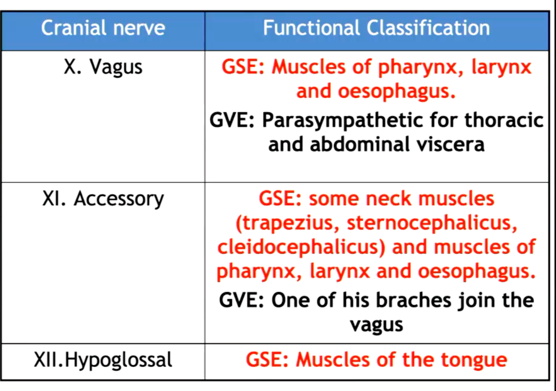
Cranial nerves: Ill, VII, IX, X and XI
Spinal nerves for the rest of the body
Summarize the motor cranial nerves.
Summary Pt. 2
Where are the motor spinal tracts mainly located?
Central + Lateral
Describe the motor spinal tracts in the spinal cord and their main functions.
Corticospinal tracts (pyramidal system):
Motor information directly from cerebral cortex to spinal cord for precise movement
Poorly developed in domestic animals
Rubrospinal tracts (extrapyramidal system):
Motor information from red nucleus in the brainstem to spinal cord
More developed in domestic animal, quick, non-precise movement
Reticulospinal tracts (extrapyramidal system):
motor information from reticular formation in the brainstem to spinal cord
• Vestibulospinal tracts (extrapyramidal system):
Motor information from vestibular nuclei in the brainstem to spinal cord
•Tectospinal tracts (extrapyramidal system):
Motor information from the tectal region in the brainstem to spinal cord
How do we assess proprioception?
Proprioceptive placing (knuckling response)
Placing dogs carpus down and watching for corrective action
Hopping Reaction
Support all legs, should properly use remaining leg to jump beneath self
Hemiwalking
Used to determine if one side is more affected than the other
Holding both limbs laterally and assessing the other side of the body and the hopping response
Wheelbarrowing
Want to determine if thoracic limbs are affected, must support neck to block vision and ensure vision cannot compensate
Extensor Postural Thrusting
Walking backwards and looking for same position while holding thoracic limbs
Hip Sway
Evaluating strength, pushing hips to side and looking for corrections
Visual and Tactile Placing
Movement toward table, evaluating body preparing to come closer to the table
Can cover eyes to evaluate proprioceptive pathways
How can we assess motor function?
Gait
Walking on non-slippery surface, observe from behind and the front, as well as the side
Assess Muscle Tone and Bulk
Compare legs and palpate muscles
Patellar Reflex
Can determine LMN and UMN
Withdrawal (Flexor) Reflex)
Pinching paw to see that foot retracts
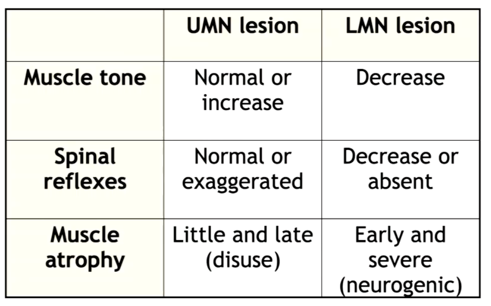
What are the clinical differences between UMN and LMN signs?
How do we assess severity of damage of spinal cord?
Different susceptibility to damage of the different tracts
Motor function:
Normal → back pain → paralysis weakness → total
• Sensory function:
Normal → loss of proprioception → loss of superficial pain perception → loss of deep pain perception
Describe the grading of spinal injuries:
0. Normal
1. Pain (no neurological deficits)
2. Ambulatory paresis (Able to walk but weak)
3. Non-ambulatory paresis (Not able to walk but able to move with support)
4. Plegia (No movement of legs)
5. Plegia with absent deep pain sensation (Cannot move, cannot feel)