Post-Lab Quiz 4 - 232 Lab
 Call Kai
 Learn
Learn Practice Test
 Spaced Repetition
 Match
Match Flashcards
 Knowt Play
1/51

There's no tags or description
Looks like no tags are added yet.
Name | Mastery | Learn | Test | Matching | Spaced | Call with Kai | Chat |
|---|
No analytics yet
Send a link to your students to track their progress
52 Terms
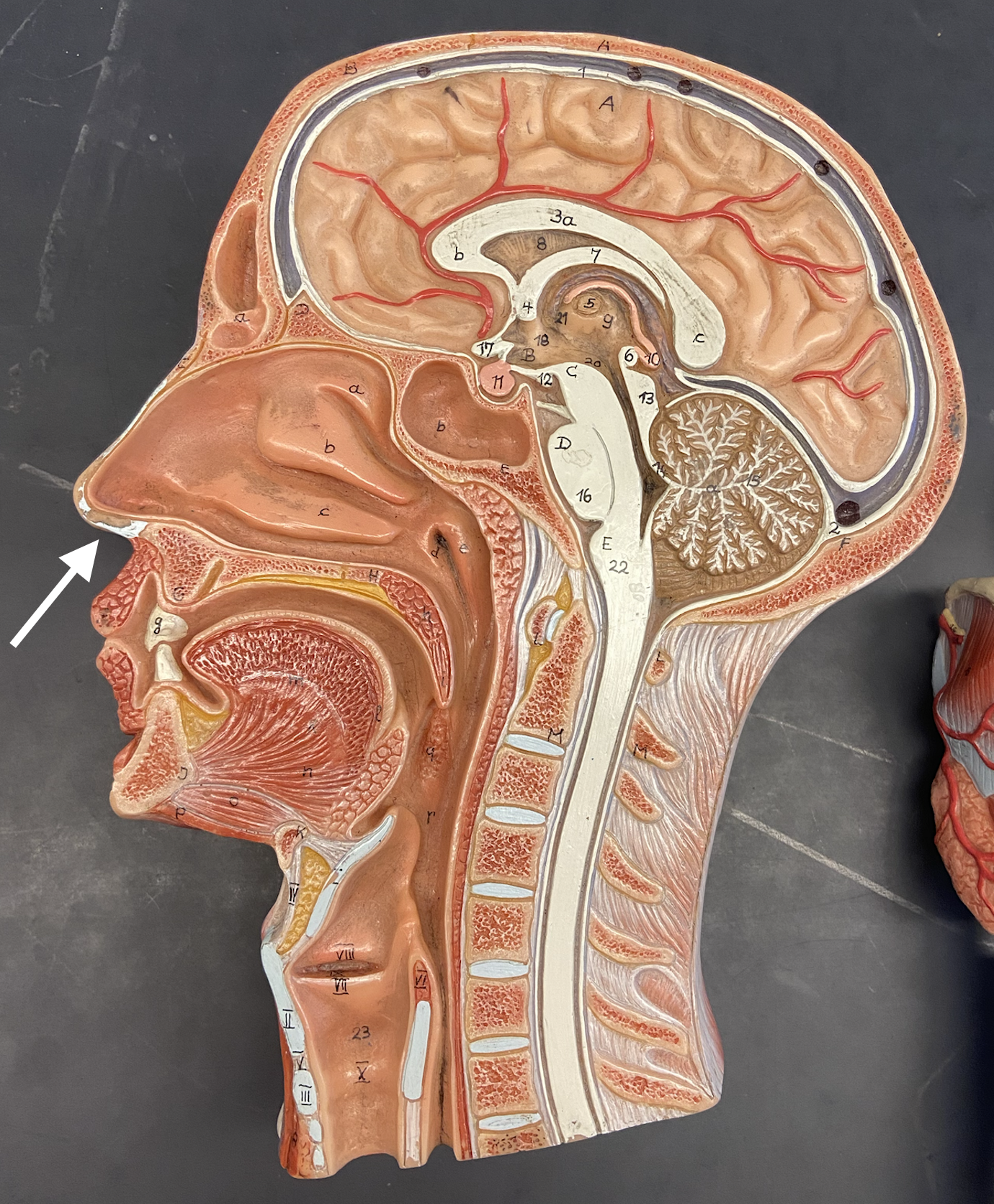
External nares
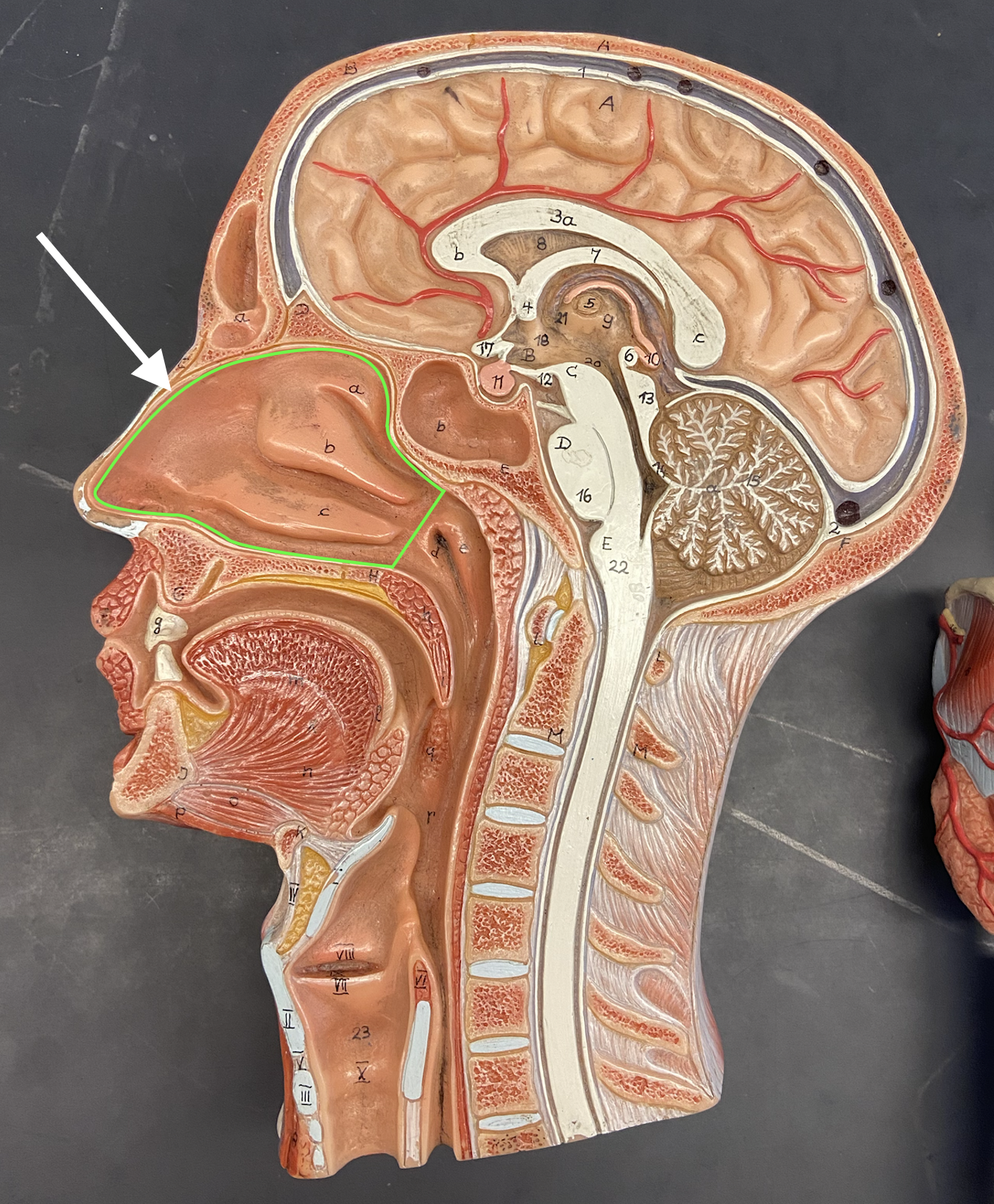
Nasal Cavity
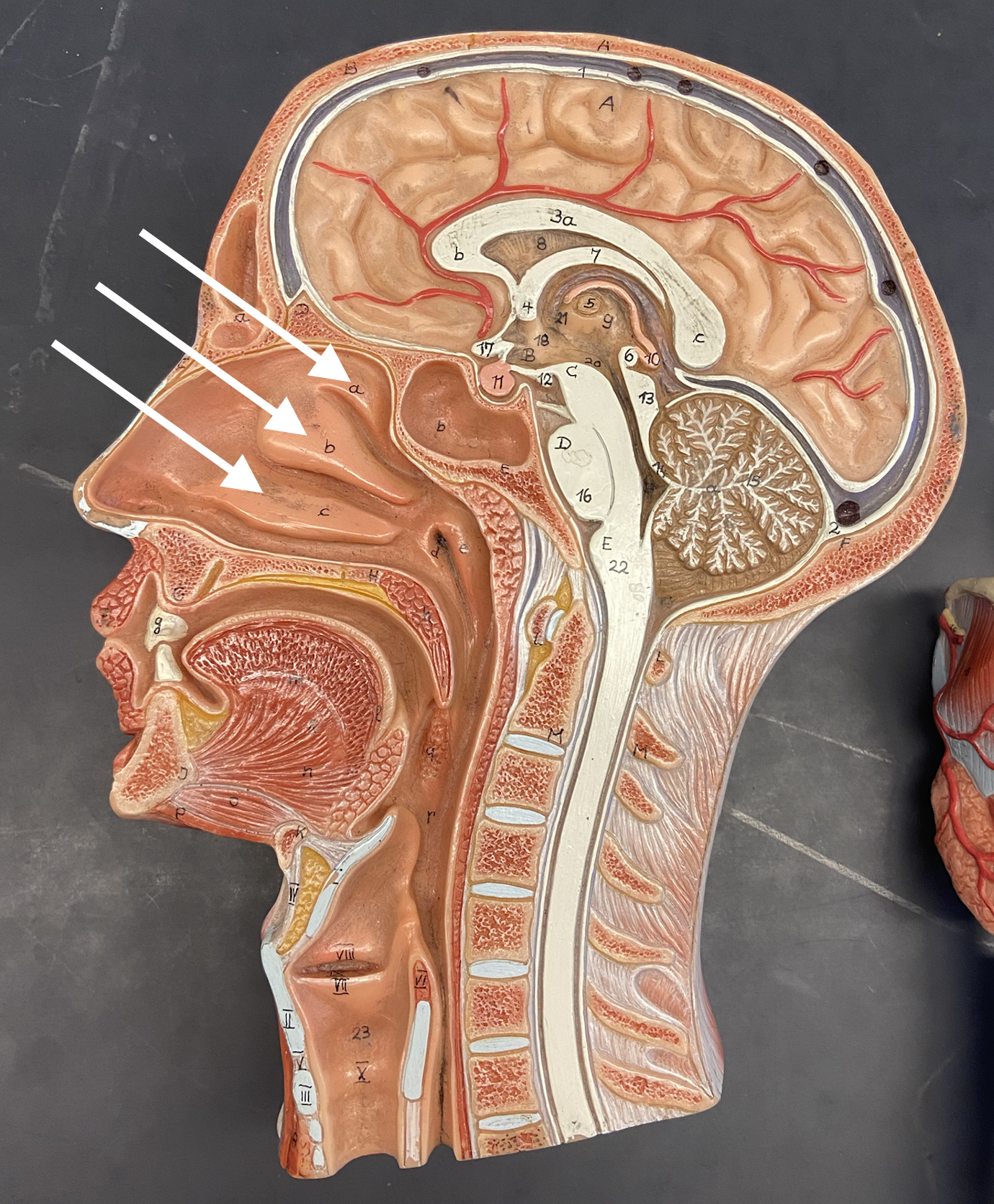
Concha
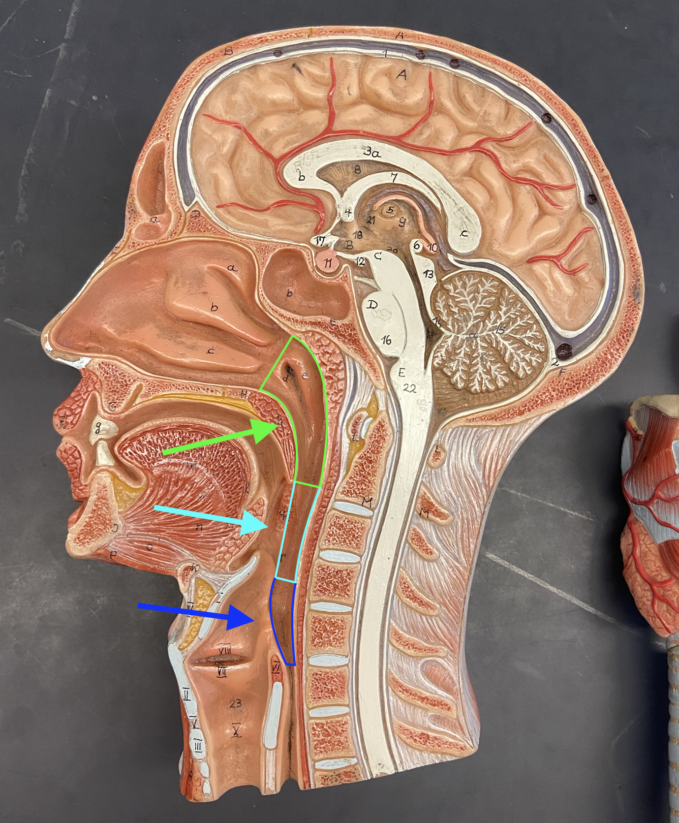
Pharynx: Nasopharynx, Oropharynx, Laryngopharynx
Larynx
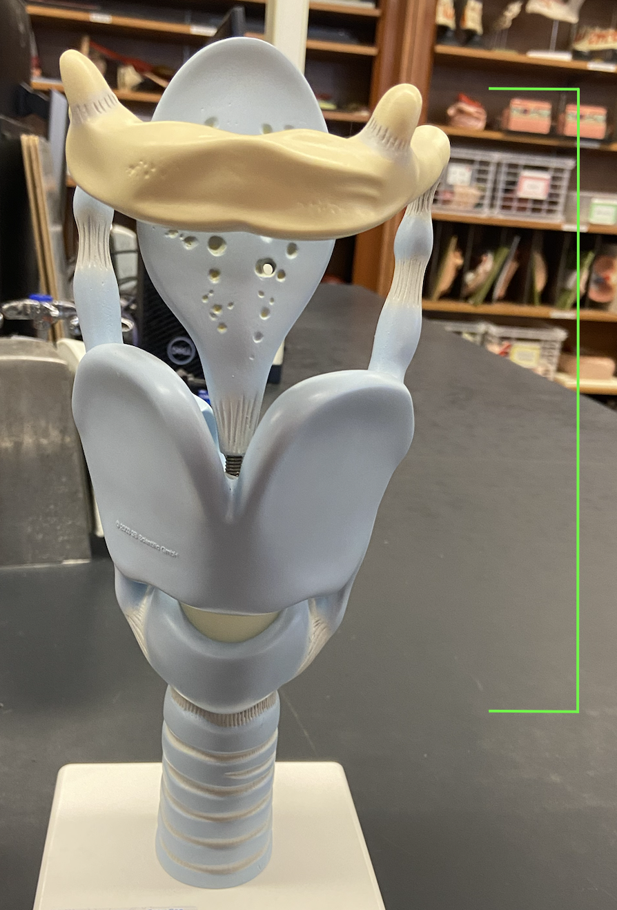
Epiglottis
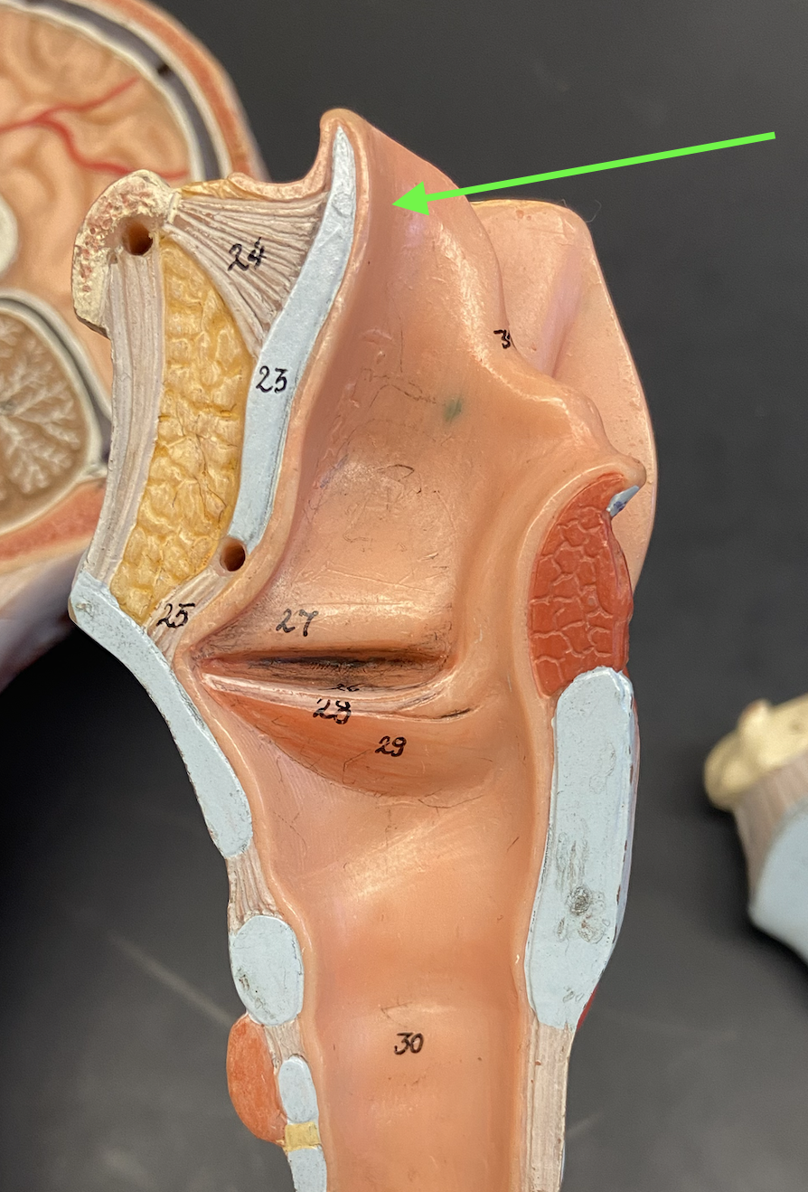
Glottis
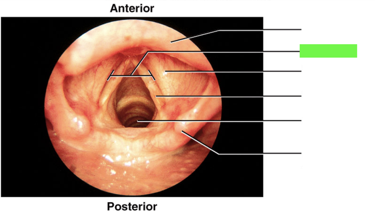
Vocal Cord / Vocal Folds
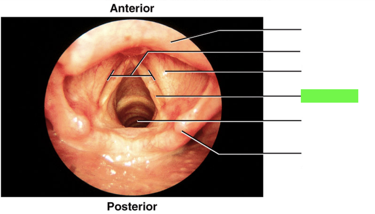
Thyroid Cartilage
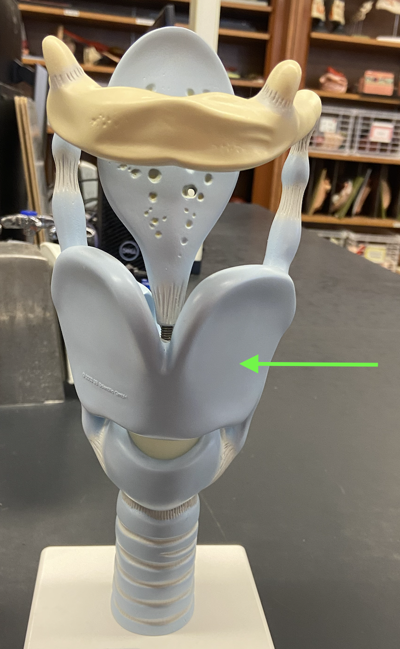
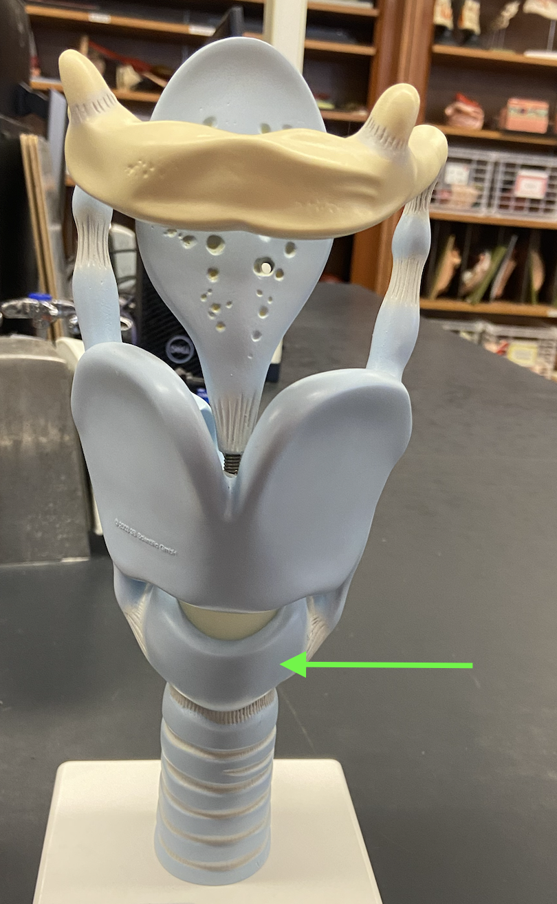
Cricoid Cartilage
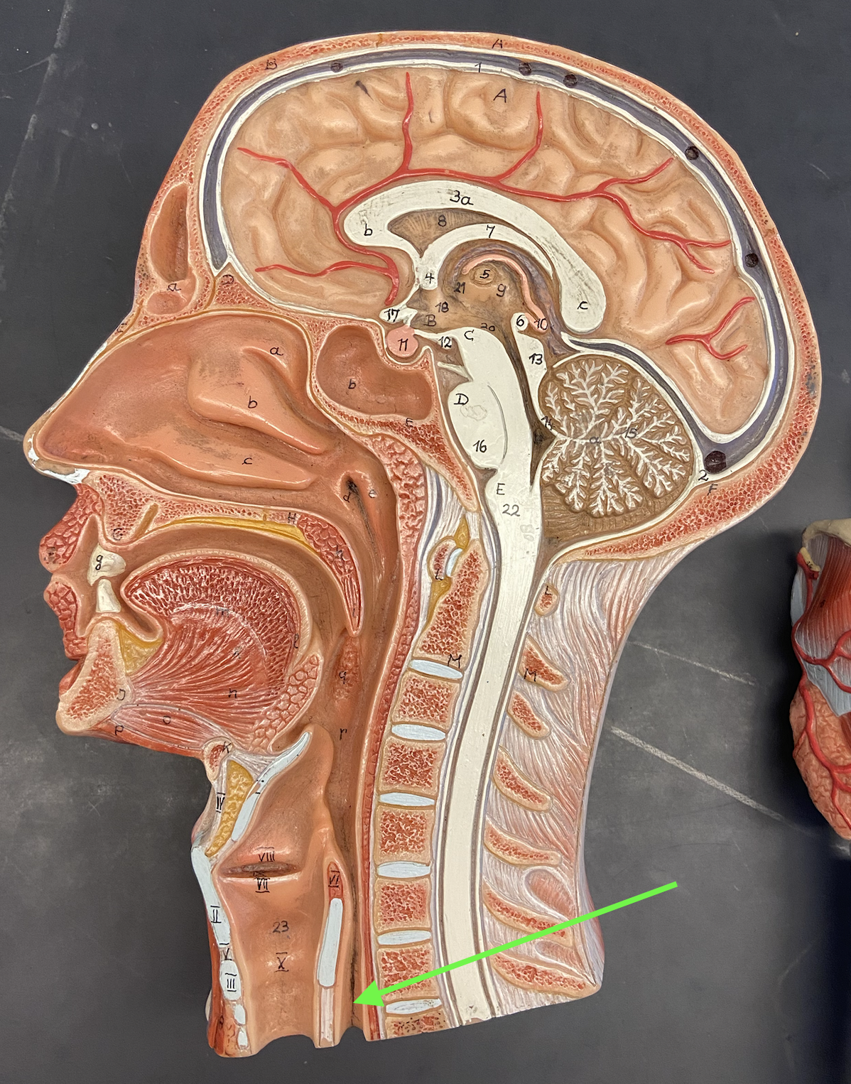
Esophagus (not respiratory)
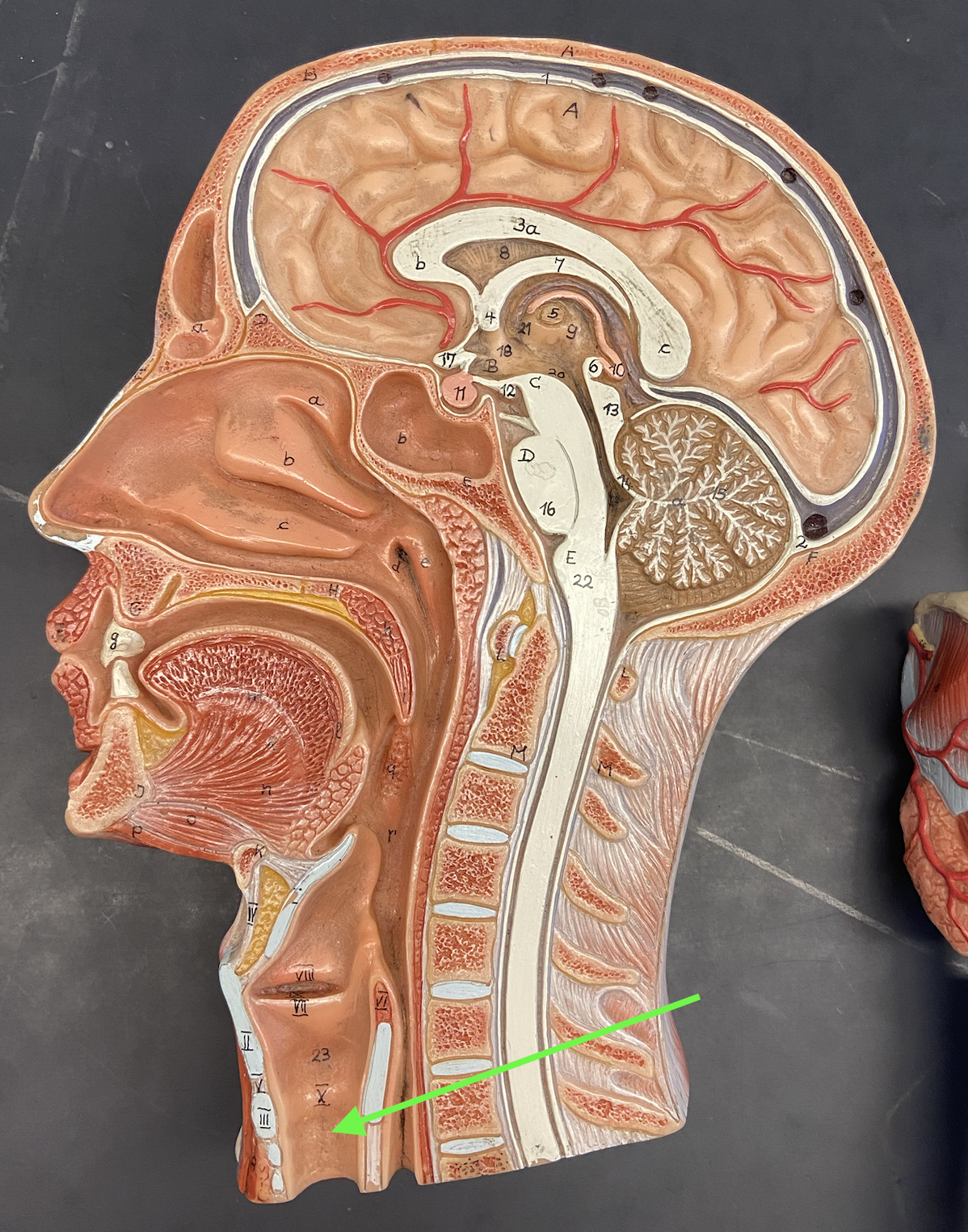
Trachea
1° Bronchi (main/primary)
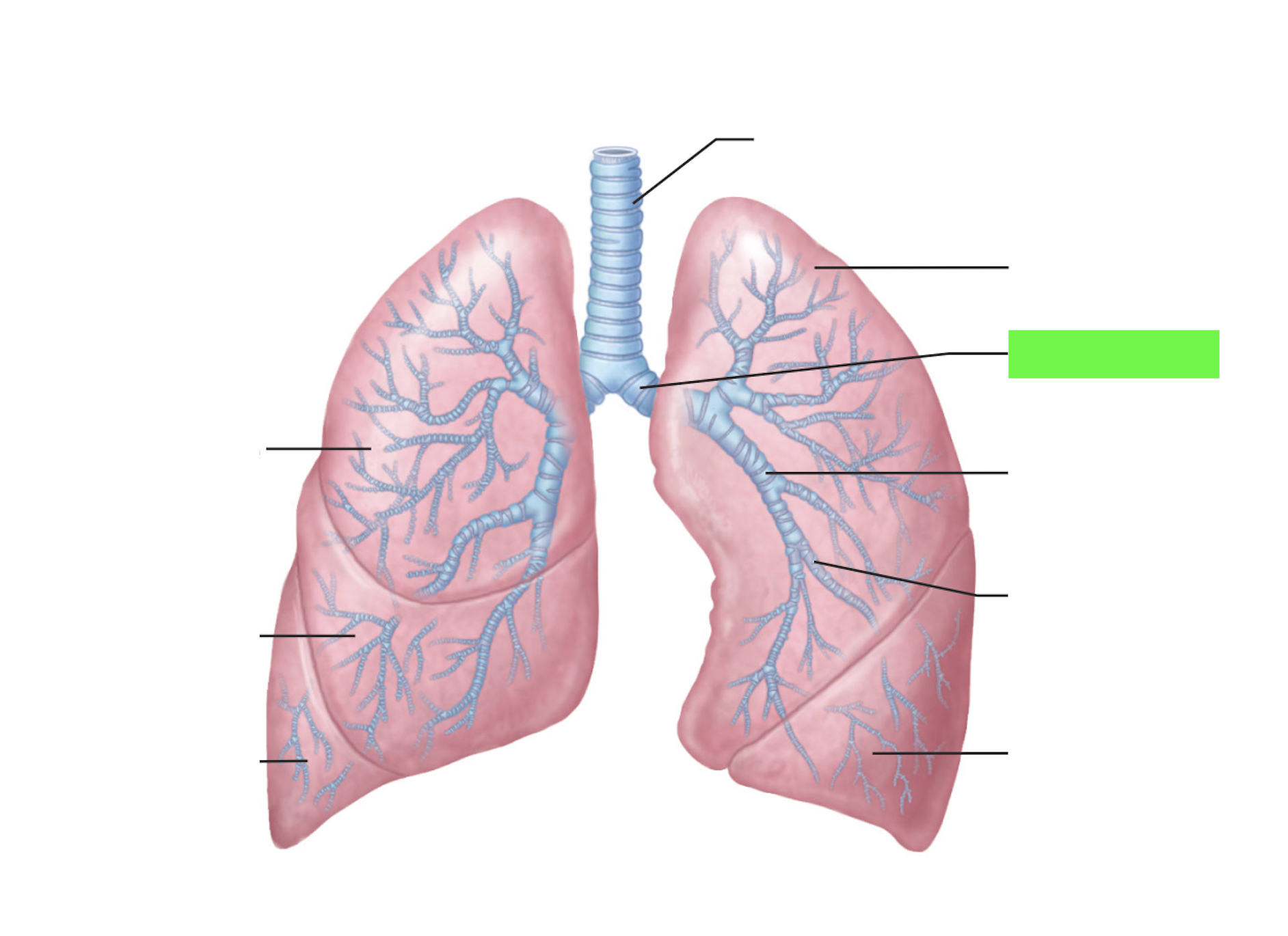
2° Bronchi (lobar/secondary)
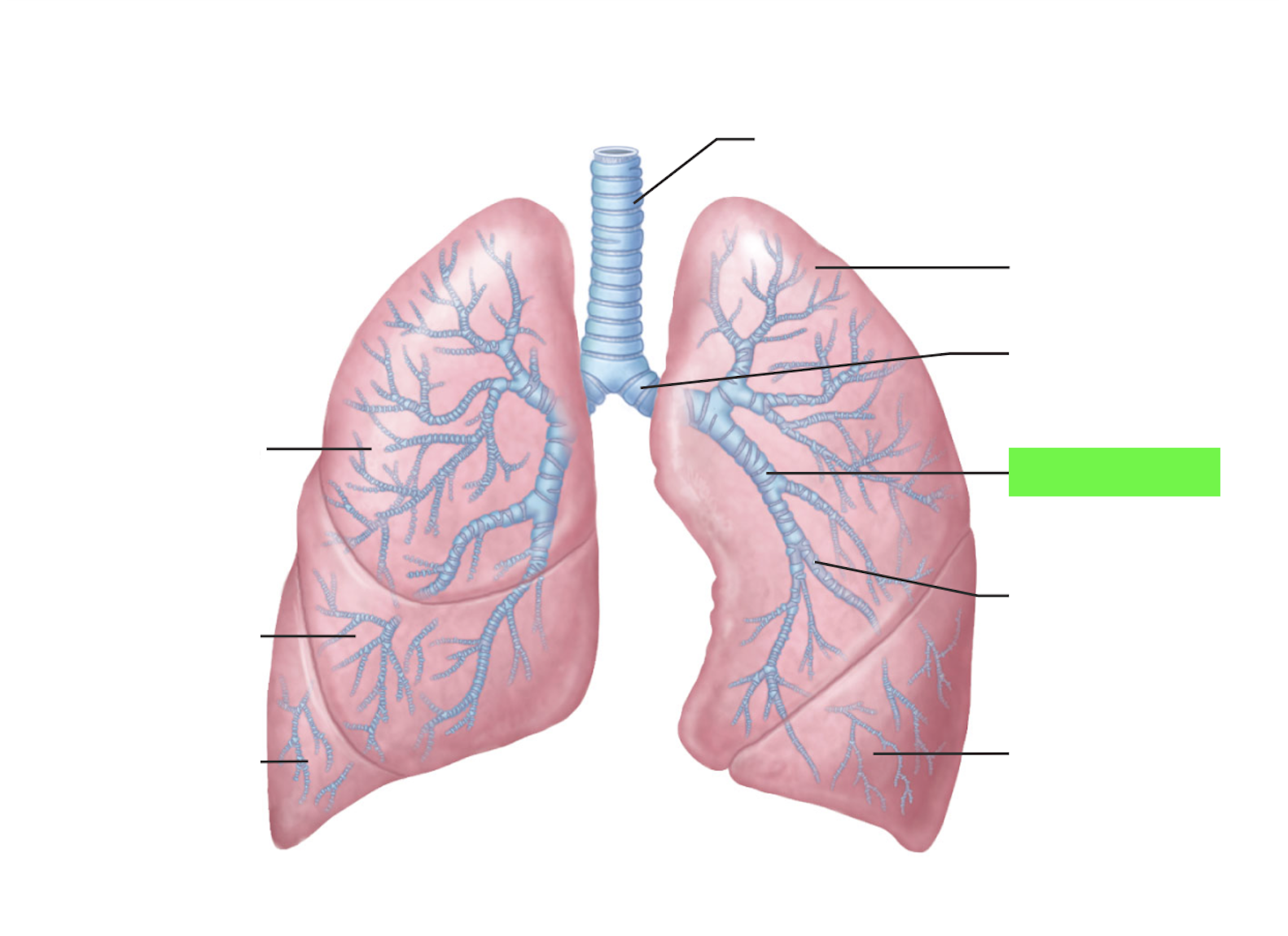
3° Bronchi (segmental/tertiary)
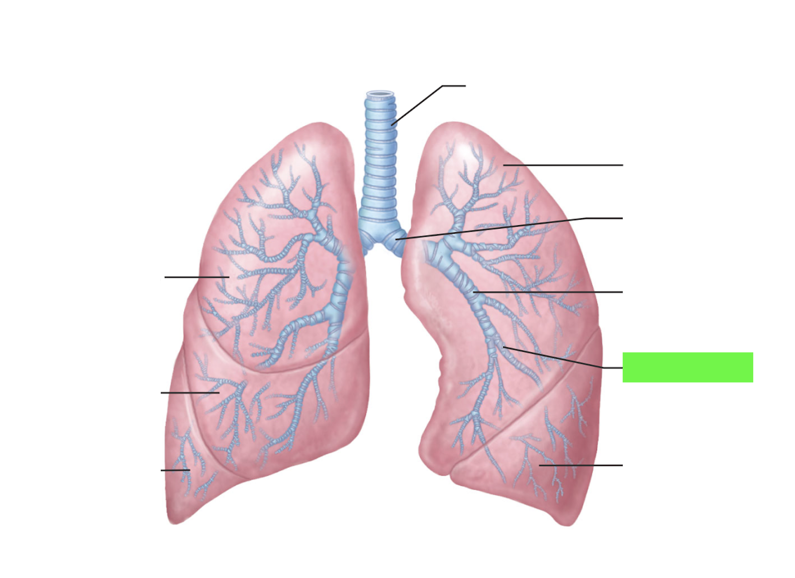
3° Bronchi (segmental/tertiary) - Histology slide
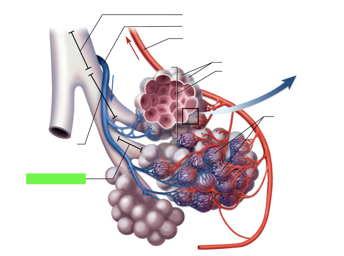
Bronchiole
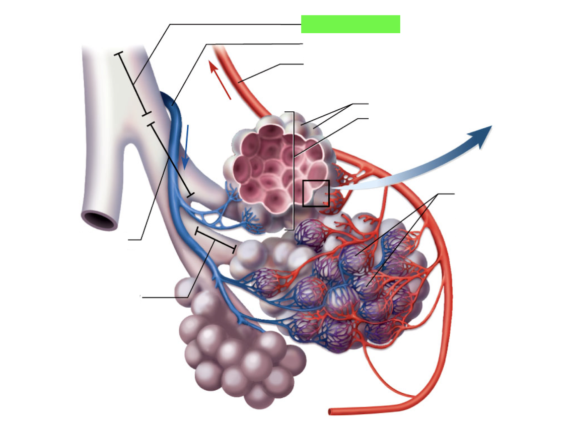
Terminal Bronchiole
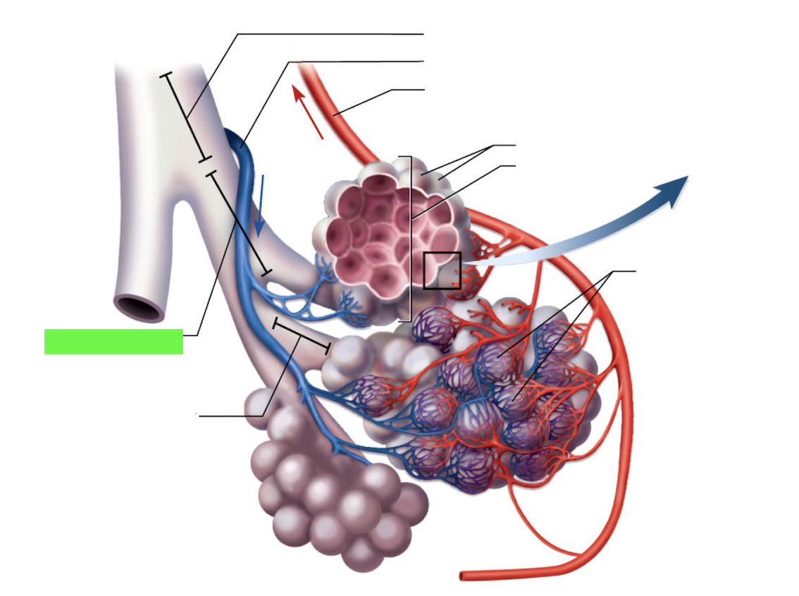
Respiratory Bronchiole
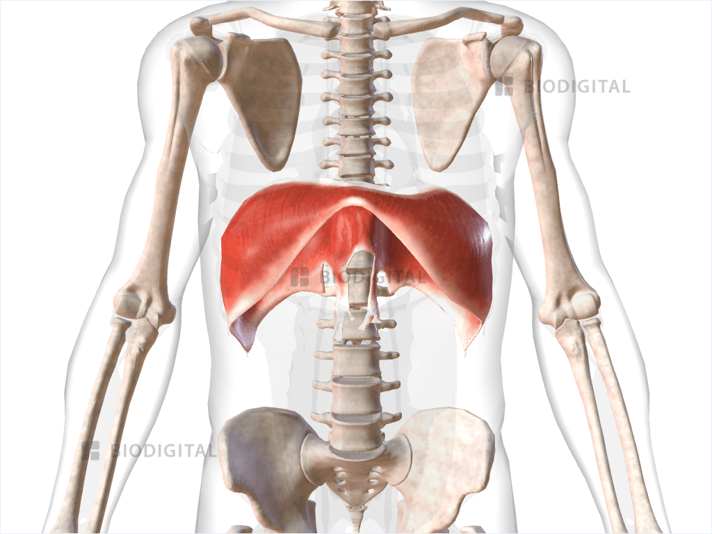
Diaphragm
Left Lung: Superior and Inferior Lobes
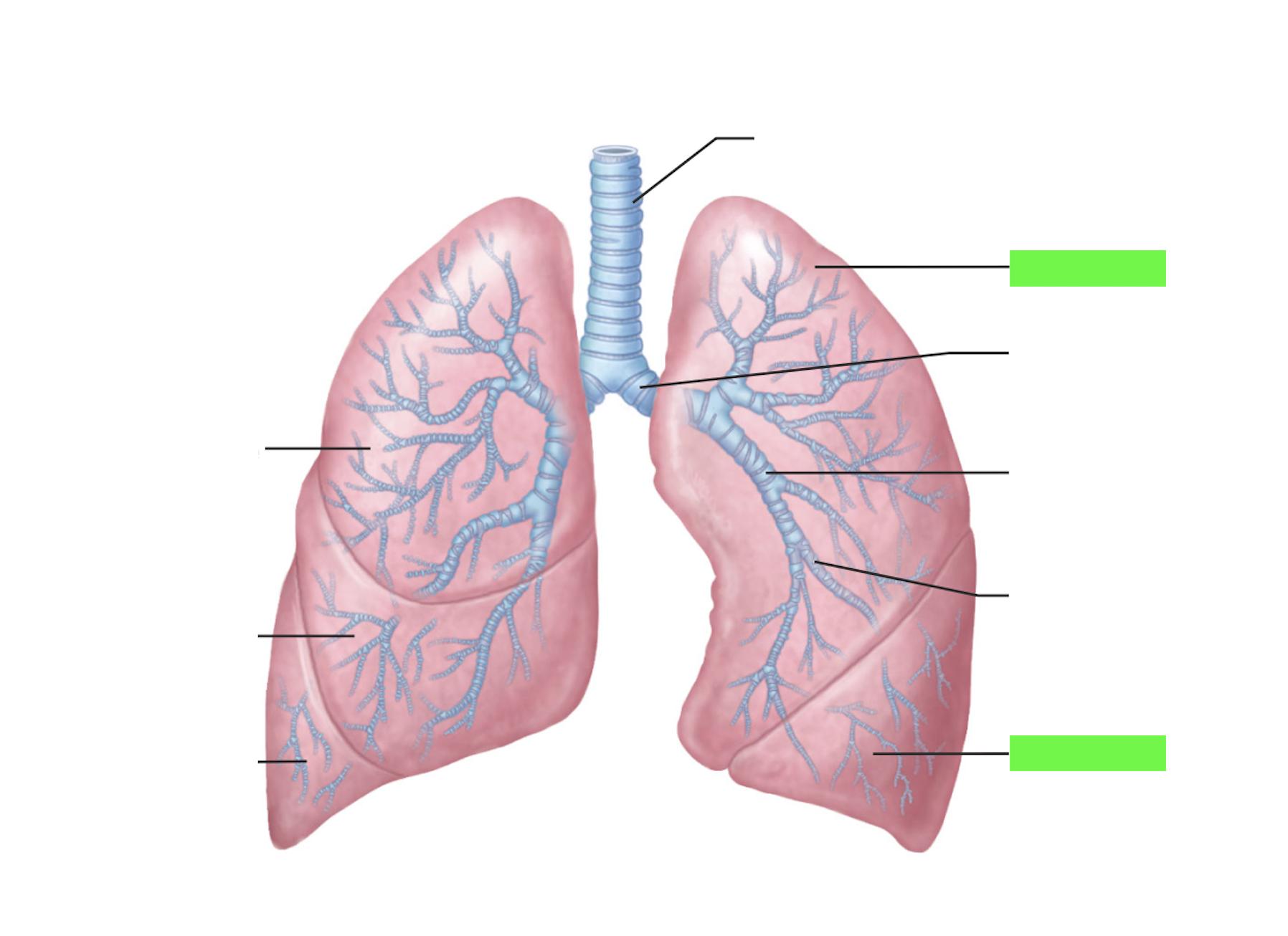
Right Lung: Superior, Middle, and Inferior Lobes
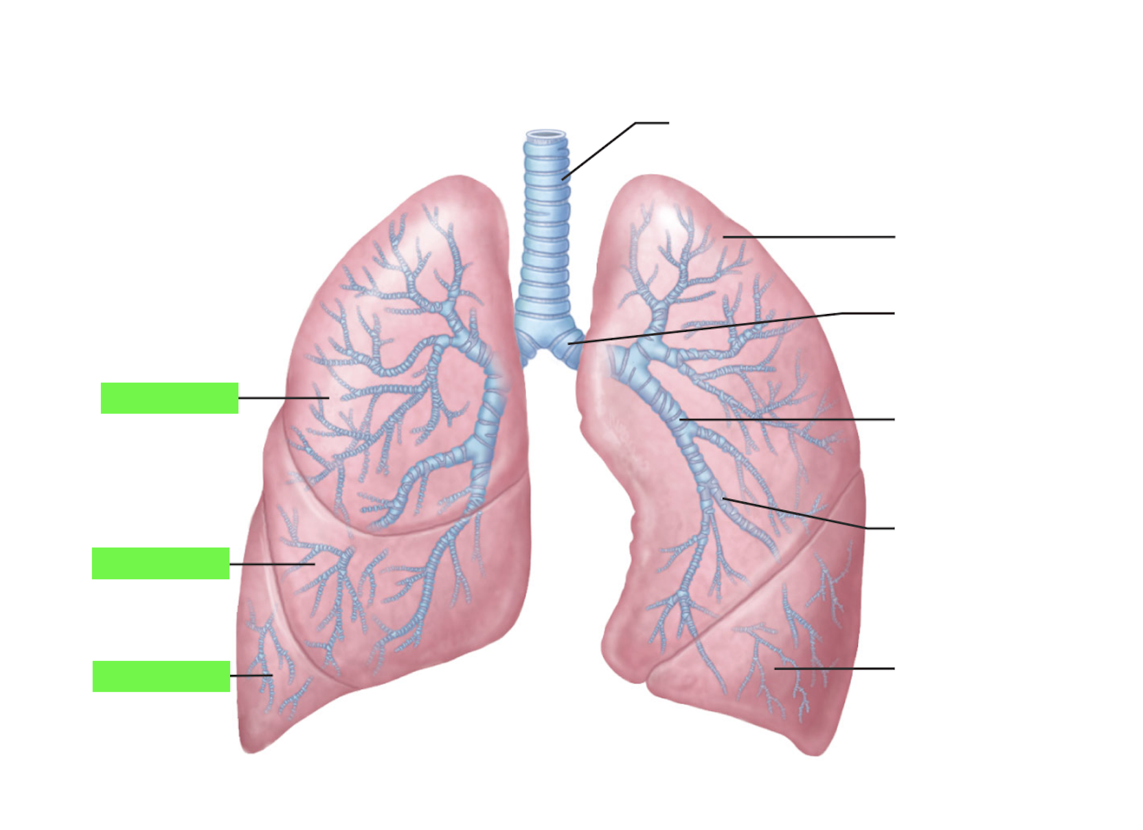
Histology: Terminal and Respiratory Bronchioles
Simple Cuboidal Epithelium
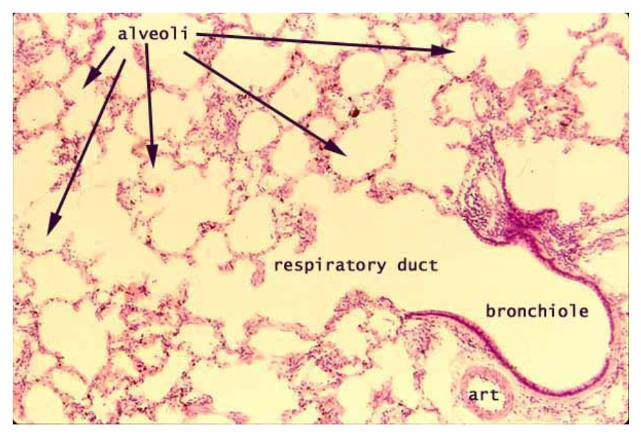
Histology: Alveolar ducts (No)
Histology: Alveolus (alveoli) (Yes)
Histology: Progressive Epithelial tissue - Stratified Squamous
Pseudosstratified Ciliated Columnar Epithelium (Yes)
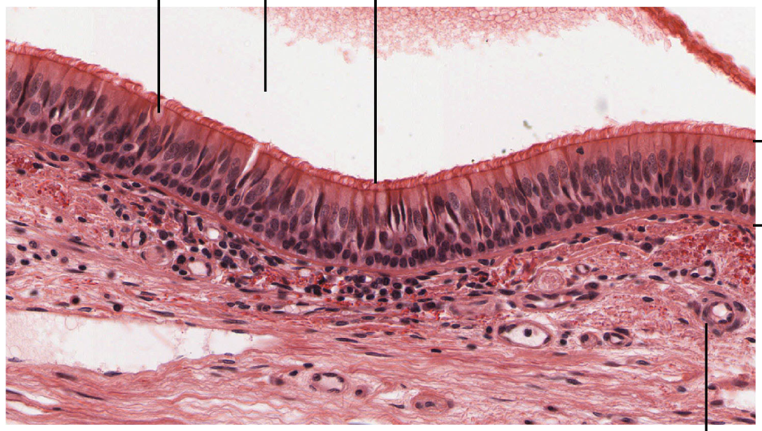
Simple cuboidal (No)
Simple Squamous Epithelium (Yes)
Alveolar ducts and alveoli
Learning Objectives
List 4 functions of the respiratory system
move air
gas exchange
smell
make sound
Compare and contrast the structural and functional organization of the respiratory system
Structural
Upper = superior to larynx
Lower = trachea and inferior
Functional
Conducting = transports air
Respiratory = gas exchange starts at the respiratory bronchioles
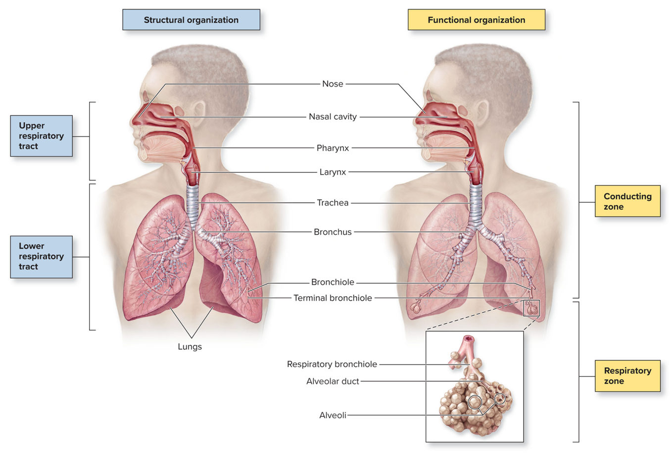
Identify the structures of the upper respiratory tract.
nose = nares, nasal cavity, concha
pharynx
nasopharynx
oropharynx
laryngopharynx
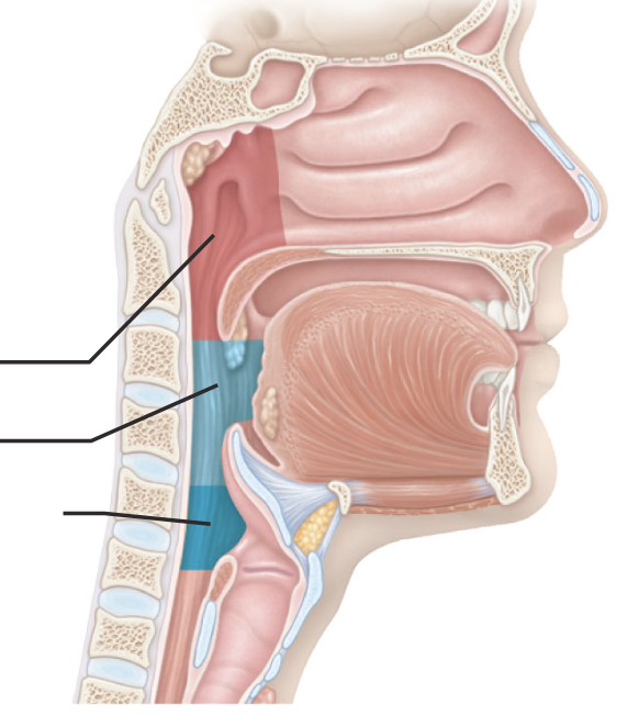
Explain how the nasal cavity conditions air
Identify and describe the structures of the larynx
Vocal cord (glottis)
Vestibular fold
Epiglottis
Thyroid cartilage
Cricoid cartilage
Identify and describe the 4 structures of the larynx
Identify, locate, and describe the function of the epiglottis
closes over trachea during swallowing
Identify the structures of the lower respiratory tract.
trachea
bronchus
bronchiole
alveolus
lungs
diaphragm
Describe the structure and function of the trachea.
open tube lined with c-shaped hyaline cartilage rings
Identify the structural subdivisions of the bronchial tree.
main (1 degree), lobar (2nd degree), segmental (3rd degree)
Compare and contrast the right versus left lung, and identify the five lobes on the cadaver or models.
right, larger, has 3 lobes, left has 2
Identify pulmonary macrophages on histological slides and explain their function.
leukocyte that phagocytizes microorganisms and debris
Describe progressive airway epithelium, and identify the type of epithelium present in the following structures on histology slides and images:
nostrils, mouth, and throat (oropharynx and laryngopharynx)
nasal conchae, trachea and bronchi
bronchioles (larger bronchioles to terminal bronchioles)
respiratory bronchioles
alveoli
identify and describe the cellular structures, cell types, or tissue types responsible for filtering, warming, and humidifying inhaled air
nose warms air with etensive blood vessels
cilia in PCCE with goblet cells trap microbes, dust, foreign
humidified in wet area of nose
describe progressive airway epithelium, and identify the type of epithelium present in the following structures on histology slides and images
nostrils, mouth, and throat (oropharynx, laryngopharynx) = stratified squamous
nasal conchae, trachea, and bronchi - PCCE
terminal and respiratory bronchioles = simple cuboidal
alveoli = simple squamous
Define the 4 respiratory volumes.
TV - tidal; IRV, ERV, RV
Define Vital Capacity, list the volumes that compose VC, and state the normal VC for males and females
VC = TV + IRV + ERV
3L females 5 L male
Write the formulas for and explain each of the 4 respiratory capacities.
IC = TV + IRV
FRC = ERV + RV
TLC = VC + RV
Describe a flow-volume loop, explain what is measured on the x- and y-axes, and identify and diagnose abnormal loops as obstructive or restrictive.
airflow (y-axis) vs (x-axis) volume (aka VC)
exhalation should be linear
Compare and contrast obstructive and restrictive lung disorders, identify lung volumes that are abnormal for each condition, and list a disorder or condition typical of each.
Describe the effects of emphysema on the respiratory zone and the consequences these changes have on surface area, gas exchange, and airflow.
alveoli burst reducing surface area
decreased gas exchange
decreased airflow due to decreased elasticity
Define and correctly spell all terms listed below; identify structures on models, histology slides, or on the body donations; and describe form↔function relationships; identify highlighted terms on the cadaver; identify histological terms (with asterisk*) using microscopes or histological imagery.