Skeletal System
 Call Kai
 Learn
Learn Practice Test
 Spaced Repetition
 Match
Match Flashcards
 Knowt Play
1/46

There's no tags or description
Looks like no tags are added yet.
Name | Mastery | Learn | Test | Matching | Spaced | Call with Kai | Chat |
|---|
No analytics yet
Send a link to your students to track their progress
47 Terms
Canaliculi
Tiny tunnels that connect lacunae to the central canal and allow nutrients to reach osteocytes.

Central (Haversian) canal
Canal in the center of an osteon that contains blood vessels supplying the bone tissue.

Concentric lamellae
Rings of bone tissue surrounding the central canal; they form an osteon.
Endosteum
Connective tissue membrane that lines the inside of bones. Membrane lining the medullary cavity, the spongy bone, and central canals.

Inner circumferential lamellae
Rings of bone tissue that wrap around the inner perimeter of compact bone.
Interstitial lamellae
Bone tissue that fills the spaces between osteons.
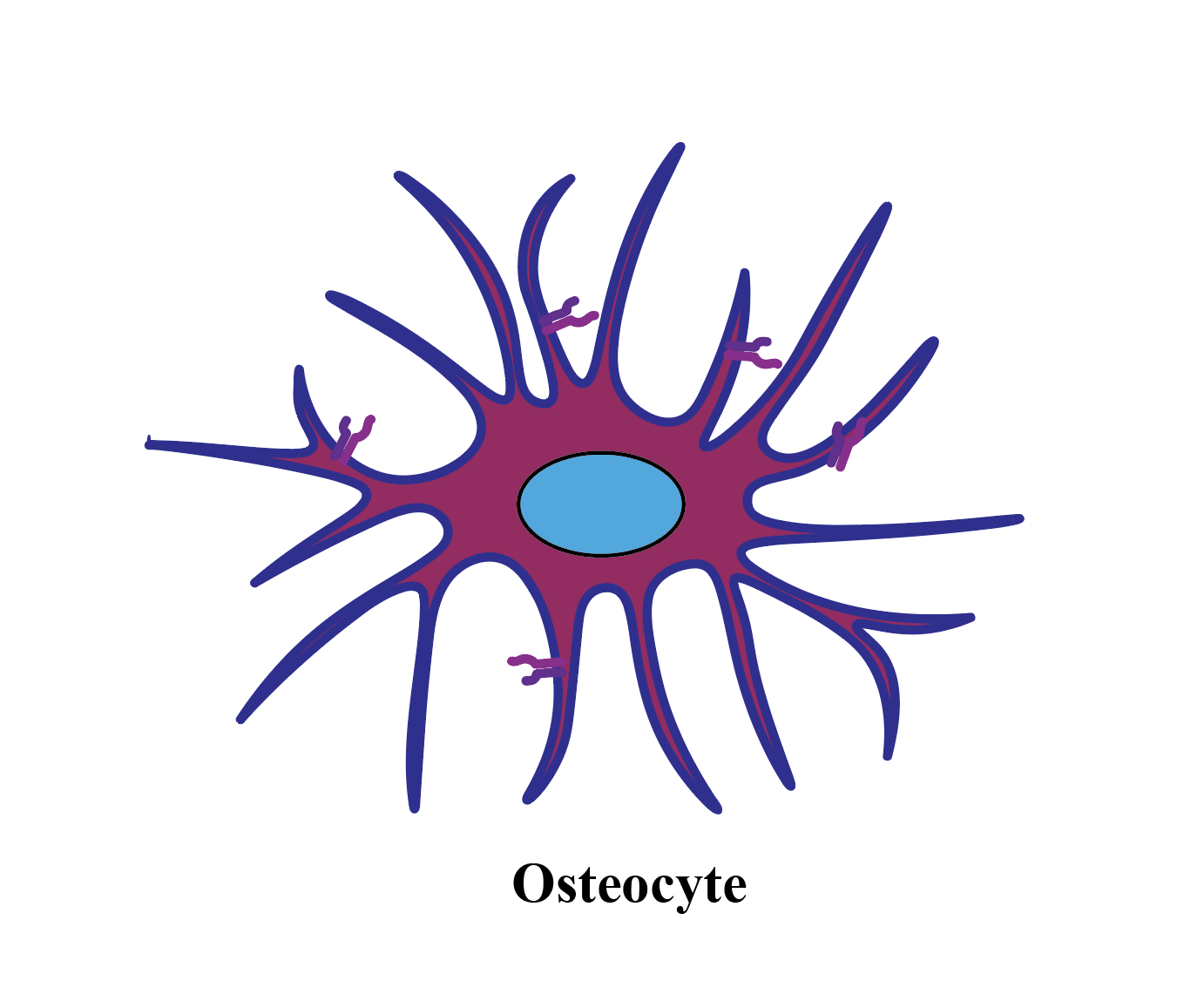
Osteocytes
Primary bone cells that maintain bone tissue; they are found inside lacunae.
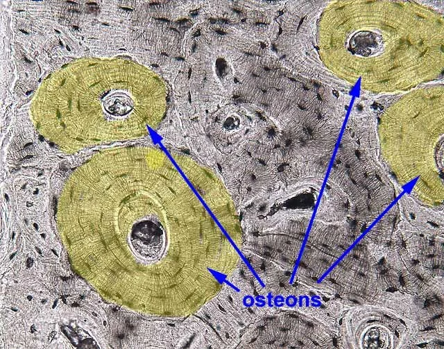
Osteon (Haversian system)
Functional unit of compact bone; a cylindrical structure made of concentric lamellae surrounding a central canal.

Outer circumferential lamellae
Rings of bone tissue that wrap around the outer perimeter of a bone.
Perforating (Volkmann’s) canals
Canals that run across the lamellae and carry blood vessels between the outer bone and osteons.

Periosteum
Connective tissue membrane that covers and protects the outer surface of a bone. Richly supplied with blood vessels.
Compact (cortical) bone
Dense, solid bone tissue found mainly on the outside of long bones; protects deeper structures.
Diaphysis
The middle portion, or shaft, of a long bone.

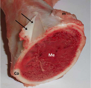
Epiphyseal line
Remnant of the growth plate in an adult bone; located between the epiphysis and diaphysis.

Epiphyses (Ends)
The ends of a long bone; include the proximal and distal epiphyses. Composed mostly of spongy bone. Contain red marrow.

Distal epiphysis
The end of a long bone farther from the body; has compact bone on the outside and spongy bone on the inside.
Proximal epiphysis
The end of a long bone closer to the body; has compact bone on the outside and spongy bone on the inside.
Medullary cavity
Hollow space inside the diaphysis of a long bone; normally contains yellow bone marrow.

Spongy (cancellous) bone
Looser bone tissue that looks sponge-like; spaces between its trabeculae can store substances.

Articular surface
Smooth surface of a bone that forms a joint with another bone; covered with articular cartilage.
Long bones
Longer than wide. Mostly compact bone with spongy at the ends. Ex: Femur, humerus, phalanges.
Short bone
Generally cube-shaped. Contain mostly spongy bone with a thin outer layer of compact bone. Ex: Carpals and tarsals.

Sesamoid
Type of short none that forms in a tendon. Ex: Patella - knee cap.
Flat bones
Thin and flattened. Usually slightly curved. Thin layers of compact bone around spongy bone. Ex: Skull bones, ribs, sternum, and mandible.

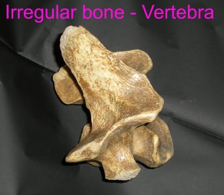
Irregular bones
Complicated shape. Ex: Vertebrae, facial bone, hip
Epiphyseal plate (Growth plate)
Dics of hyaline cartilage at the epiphysis- diaphysis junction.
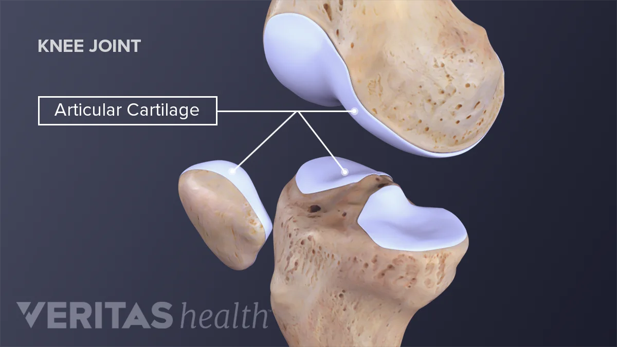
Articular Cartilage
Hyaline cartilage covering the external surface of the epiphyses. Decrease friction at joint surfaces.
Medullary cavity
Cavity of the diaphysis. Contains red bone marrow (for blood cell formation) in infants. Contains yellow bone marrow (mostly fat in adults)

Lacuna
Cavities containing osteocytes

Intramembranous
Bone develops within a fibrous membrane: Skull bones and clavicle.
Endochondral ossification
Bone develops by replacing a hyaline cartilage model; occurs in most bones of the skeleton.
Interstitial bone growth
Growth that increases the length of a bone.
How does interstitial bone growth increase bone length?
Chondrocytes on the epiphyseal side divide; older chondrocytes closer to the diaphysis calcify and die; osteoblasts deposit new bone on the diaphysis side.
Epiphyseal plate closure
At maturity, chondrocyte division slows and osteoblasts catch up. The epiphyseal plate completely ossifies and becomes the epiphyseal line.
Growth hormone
Stimulates activity of the epiphyseal plates and promotes bone growth during childhood.
Estrogen and testosterone
Cause growth spurts during puberty and eventually contribute to closure of the epiphyseal plates.
Appositional bone growth
Growth that increases the thickness (diameter) of a bone.
How does appositional bone growth increase bone thickness?
Osteoblasts deposit bone on the outer surface while osteoclasts remove bone from the inner surface, enlarging the medullary cavity.

Bone remodeling
The continuous process of new bone formation and bone reabsorption; involves osteoblasts and osteoclasts.
How does mechanical stress affect bone remodeling?
Bone is deposited in response to stress from muscle pull, weight, and gravity.
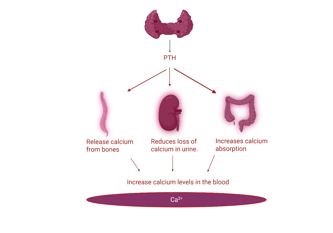
How does PTH affect bone remodeling?
When blood calcium is low, PTH activates osteoclasts to break down bone and release calcium into the blood.
How does calcitonin affect bone remodeling?
When blood calcium is high, calcitonin promotes calcium storage in bone by supporting osteoblast activity.

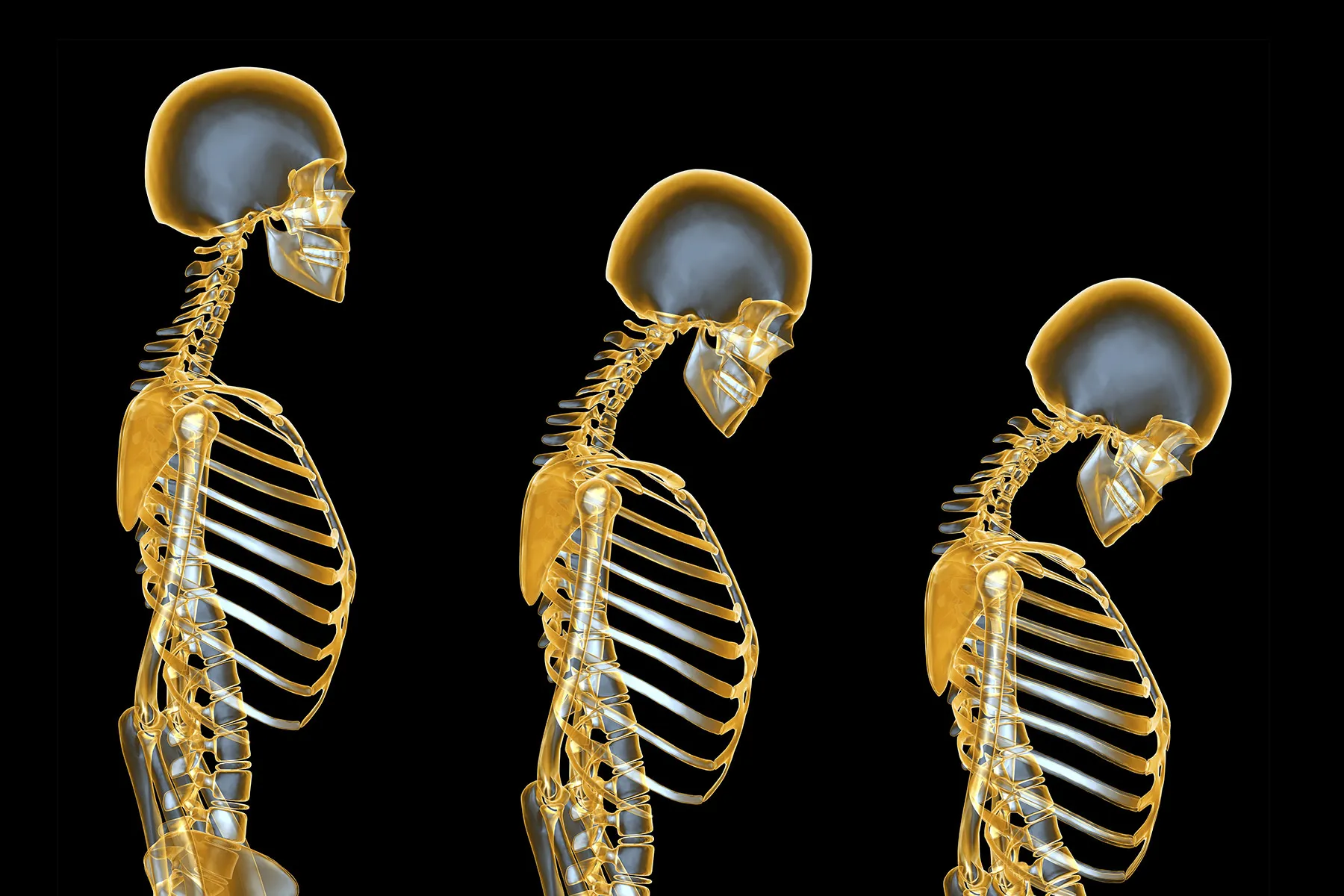
Osteoporosis
A condition in which bone reabsorption occurs faster than bone formation, resulting in porous, light, and fragile bones.
Who is at risk for osteoporosis?
Both men and women can develop osteoporosis, but it is more common in postmenopausal women.
Risk factors for osteoporosis
Decreased estrogen or testosterone, poor diet with insufficient calcium and vitamin D, lack of exercise, genetics, and smoking. Bone density typically reaches its peak around age 35.
Simple (closed) fracture
A fracture is internal in which the broken bone does not penetrate the skin.

Compound (open) fracture
A fracture in which the broken bone penetrates the skin, increasing the risk of infection.
