CT Part Of the CAMRT
 Call Kai
 Learn
Learn Practice Test
 Spaced Repetition
 Match
Match Flashcards
 Knowt Play
1/153

There's no tags or description
Looks like no tags are added yet.
Name | Mastery | Learn | Test | Matching | Spaced | Call with Kai | Chat |
|---|
No analytics yet
Send a link to your students to track their progress
154 Terms
The focus-to-detector distance describes _____________________________________If this distance is increased, the patient dose will __________
the distance between the x-ray source (CT tube) and the detector
array, decrease
the MDCT has a higher dose for the patient because of:
-Decrease in the focus-to-detector distance.
-Use of a cone beam
-Increases in the number of phases of acquisition
-Thinner section widths
The CT dose index (CTDI)
approximate measure of the dose received in a single CT section or slice
CTDIw is
a weighted index that Approximate's radiation dose on the basis of variations that occur across the field of view
CTDIw does not account for the
effects of helical scanning on patient radiation dose
CTDIvol is
used to approximate the radiation dose for each
section obtained during a helical scan
MSAD _______________ when slice thickness is greater than image
spacing—overlapping scans
increases
dose length product (DLP)
the measurement of dose for the entire series of CT images. equal to the calculated dose per section mulitplied by the length of a CT acquisition along the z-axis.
Dose notification describes
an automated software feature
that informs the technologist when the prescribed technical
settings for an individual CT acquisition may result in a
CTDIvol or DLP that is higher than a preset recommended
value.
Recommendations for the reduction of pediatric dose include
-Eliminate CT scans for inappropriate indication.
-Reduce multiphase scanning (precontrast, delays, and so on).
-Reduce mA. and Reduce kVp
-Increase pitch.
-One single-acquisition phase is often enough
Physiologic and pathologic processes that may increase the
risk of adverse reaction or other untoward outcome from
iodinated contrast include:
Asthma.
Environmental and/or food allergies.
Renal disease.
Multiple myeloma.
Diabetes mellitus.
Pheochromocytoma.
Sickle cell disease.
Hyperthyroidism.
Significant cardiac disease.
Anxiety
Contraindications to IV iodinated contrast agents include:
a. Allergy to iodine.
b. Prior severe allergic reaction to an iodinated contrast agent.
c. Renal insufficiency/failure.
Prospective ECG Triggering (Also known as sequential or cine-mode scanning):
Acquires images only in those portions of the cardiac cycle expected to have the lowest cardiac motion
CT Coronary Calcium Screening uses a ______ detector-row CT scanner with a _____second gantry rotation time is the minimal requirement for a coronary calcium measurement
four, 0.5
In CT abdomen imaging The DFOV should be
just large enough to include the skin surface over the key areas being imaged
Normal CT attenuation of the unenhanced liver is between ______________ HU
38 and 70
In healthy subjects the liver is at least _____HU greater than the spleen
10
Measurements from a CT image of the abdomen show the liver density is 35 HU whereas the density of the spleen is 65 HU. Which is a likely diagnosis?
a. Metastatic disease of the liver
b. Cavernous hemangioma
c. Fatty infiltrate
D. Ruptured spleen
Fatty infiltrate-
The pancreas is typically located between T12 and
L2
When initial scans fail to differentiate the margins of the pancreas from the duodenum, the patient is often given additional ______CM and slices are obtained with the patient in the _________________
oral, right decubitus position
•Enhancement phases of the urinary tract :
Corticomedullary phase, _____________ seconds after CM bolus
30-70 seconds
•Enhancement phases of the urinary tract
Nephrogram phase, ______ seconds after CM bolus
100-120 seconds
•Enhancement phases of the urinary tract
Excretory phase, ______________ (time) after CM bolus
3 minutes
Two unique features of adenomas on CT are used to differentiate benign from malignant lesions
-Intracellular lipid (fat) content
-CM washout
Routine Abdomen Pelvis clinical indications:
Suspected abdominal mass, tumor staging, abscess
Routine Abdomen Pelvis start and end locations
Start location: Just above diaphragm End location: Just below symphysis pubisArterial Venous Liver
clinical indications: Evaluation of suspected hypervascular hepatic tumors, including hepatocellular carcinoma and metastases from carcinoid, islet cell carcinoma, thyroid carcinoma, renal cell carcinoma, breast carcinoma, melanoma, and sarcomas
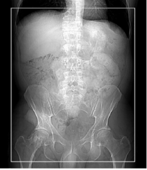
Routine Abdomen Pelvis IV contrast:
125 mL at 3.0 mL/s; 50 mL saline flush. Scan delay = 65 seconds
Routine Abdomen Pelvis Oral Contrast:
675-mL barium sulfate suspension (1.5 bottles Readi-Cat 2). An additional 225 mL (the remainder of the second bottle) given just before scanning
Arterial Venous Liver
clinical indications:
Evaluation of suspected hypervascular hepatic tumors, including hepatocellular carcinoma and metastases from carcinoid, islet cell carcinoma, thyroid carcinoma, renal cell carcinoma, breast carcinoma, melanoma, and sarcomas
Arterial Venous Liver start and end locations
Start location: Just above diaphragm End location: At iliac crest (include entire liver)
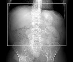
Arterial Venous Liver IV contrast & scan delay:
: 125 mL at 4 mL/s; 50 mL saline at 4.0 mL/s Scan delay: 35 seconds
Arterial Venous Liver Oral contrast
VoLumen or water; 450 mL 30 minutes prior; 225 mL 10 minutes prior, 225 mL just before scan (in scan room)
Arterial Venous Pancreas clinical indications:
clinical indications: Evaluation of suspected hypervascular hepatic tumors, including hepatocellular carcinoma and metastases from carcinoid, islet cell carcinoma, thyroid carcinoma, renal cell carcinoma, breast carcinoma, melanoma, and sarcoma
Arterial Venous Pancreas start and end locations
Start location: Just above diaphragm End location: At iliac crest (include entire liver)
Arterial Venous Pancreas IV contrast and scan delay
contrast: 125 mL at 4 mL/s; 50 mL saline at 4.0 mL/s Scan delay: 35 seconds
Arterial Venous Pancreas Oral Contrast
VoLumen or water; 450 mL 30 minutes prior; 225 mL 10 minutes prior, 225 mL just before scan (in scan room
Mesenteric CT
clinical indications:
Ischemic bowel, bleeding, tumor resection
Mesenteric CT IV contrast:
125 mL (370 concentration) at 4 mL/s; 50 mL saline at 4.0 mL/s Scan delay: Smart Prep; set monitor location at the level of the celiac artery
Mesenteric CT Oral contrast:
VoLumen or water; 450 mL 60 minutes prior; 450 mL 30 minutes prior, 225 mL 20 minutes prior, 225 mL just before scan (in scan room)
Enterography
clinical indications:
Crohn disease, inflamed bowel
IV contrast: Enterography
125 mL (370 concentration) at 4 mL/s; 50 mL saline at 4.0 mL/s Scan delay: 65 seconds
Oral contrast Enterography
: VoLumen or water; 450 mL 60 minutes prior; 450 mL 30 minutes prior, 225 mL 20 minutes prior, 225 mL just before scan (in scan room)
Appendicitis/Diverticulitis CT IV CM & Scan delay
IV contrast: 125 mL at 3 mL/s; 50 mL saline at 3.0 mL/s Scan delay: 65 seconds
Appendicitis/Diverticulitis CT Oral contrast:
Oral contrast: 675-mL barium sulfate suspension (1.5 bottles Readi-Cat 2). An additional 225 mL (the remainder of the second bottle) given just before scanning
Colonography start and end locations
Start location: Just above diaphragm End location: At lesser trochanter
Colonography contrast media
Rectal contrast: Inflate colon with CO2. (Check scout for air; if not sufficient, administer additional CO2 and repeat scout.
Adrenal Mass (With Delay) start and end locations
Start location: Just above diaphragm End location: Just below kidney
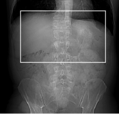
Adrenal Mass (With Delay) contrast?
1st a non contrast scan then administer IV contrast: 150 mL at 3 mL/s; 50 mL saline at 3.0 mL/s Scan delay: 60 seconds
Then wait 15 min for a delayed phase
Renal Mass start and end locations
Start location: 2 cm above kidneys End location: 2 cm below kidneys
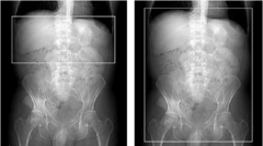
Renal Mass contrast?
1st scan without cm then for the second scan IV contrast: 100 mL at 3 mL/s; 50 mL saline at 3.0 mL/s Scan delay: 150 second
Renal Stone start and end locations
Start location: 2 cm above kidneys End location: Symphysis pubis
CT Urogram
clinical indications
: Hematuria, known or suspected urothelial disease such as transitional cell carcinoma
CT Urogram start and end locations
Start location: 2 cm above kidneys End location: Just below symphysis pubis
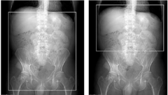
Urogram contrast ?
1st scan no cm 2nd scan IV contrast: 125 mL at 3 mL/s; 200 mL saline at 1 mL/s Scan delay: 600 seconds
Routine chest CT-Clinical indications
Infection, mass, empyema, evaluation of abnormalities discovered on chest radiographs, evaluation of known or suspected congenital thoracic anomalies, evaluation of trauma
Routine Chest CT-Start and end locations
Start location: Just above lung apices End location: Just below costophrenic angles (note: for known or suspected lung cancer, end just below adrenal glands)
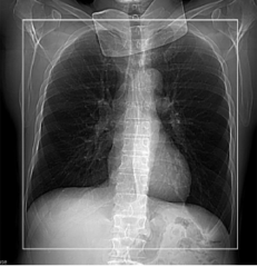
Routine Chest CT-IV contrast:
80 mL at 3.0 mL/s. 50 mL saline flush. Scan delay = 35 seconds
High-Resolution Chest CT—ILD (Interstitial Lung Disease)
Clinical indications:
Asbestos exposure, inhalation injury, interstitial disease, diffuse pulmonary disease, suspected bronchiectasis, suspected small airway disease, sarcoid, scleroderma
High-Resolution Chest CT—ILD (Interstitial Lung Disease) For first series start and end locations are:
Start location: Just above lung apices End location: Just below costophrenic angles
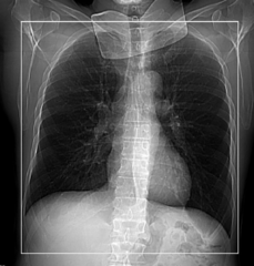
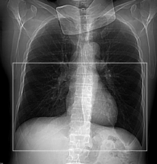
High-Resolution Chest CT—ILD (Interstitial Lung Disease) For second series start and end locations are: Start location: Carina End location: Just below costophrenic angles
Start location: Carina End location: Just below costophrenic angles
Tracheobronchial CT-clinical indications:
Suspected congenital tracheobronchial anomalies, assessment of tracheal narrowing, detection or confirmation of tracheomalacia, suspected foreign body aspiration
Tracheobronchial CT- Start and end locations
Start location: 7 cm below the carina End location: 1 cm above the epiglottis
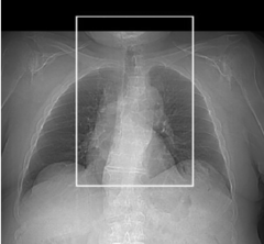
CTA—Chest for Pulmonary Embolism- Start and end locations
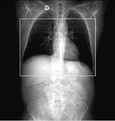
Start location: Just below lowest hemidiaphragm End location: Lung apices (scans are inferior to superior)
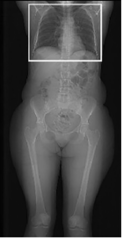
CTA—Chest for Pulmonary Embolism IV contrast and delay
IV contrast: 120 mL (370 concentration) total, split bolus; 70 mL at 4.0 mL/s. Scan delay = Smart Prep; set monitor location at the level of the main pulmonary artery, initiate the scan at first sight of contrast in the main pulmonary artery (~70 HU); 25-second pause after first 70-mL injection is complete, then 50 mL at 3 mL/s
CTA—Chest Aorta (Retrospective Gated)
clinical indications:
Blunt trauma, aortic dissection, aneurysm rupture, atherosclerotic occlusive disease, congenital vascular anomalies
CTA—Chest Aorta (Retrospective Gated) start and end locations
Start location: 2 cm above aortic arch End location: 2 cm below celiac artery
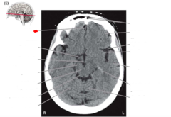
Axial Plane (transverse plane):
Divides body into superior and inferior portions. To view, imagine you are standing at patient's feet and looking toward head
Patient's right is on your left and vice versa
Anterior surface at top of image
Coronal Plane:
Divides the body into anterior and posterior portions
Sagittal Plane:
Divides body into right and left portions. Planes pass through body from superior to inferior and anterior to posterior
falx cerebri
separates the two cerebral hemispheres
Epidural Space
Located above dura mater and below cranium (between skull and dura matter)
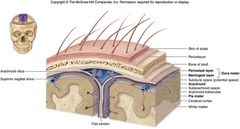
Subdural space
located between the dura mater and arachnoid mater; contains lubricating serous fluid
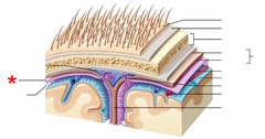
Arachnoid Space
Here there is fluid that helps transmitting nerve impulses
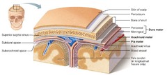
Subarachnoid space
located between the arachnoid and pia mater; contains a significant amount of cerebrospinal fluid
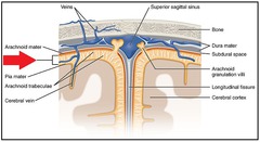
gray matter
a portion of the CNS consisting of cytons (cell bodies), their dendrites and synaptic connections
choroid plexus
A highly vascular portion of the lining of the ventricles that secretes cerebrospinal fluid.
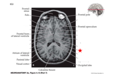
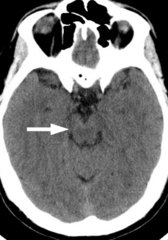
Midbrain
A small part of the brain above the pons that integrates sensory information and relays it upward.
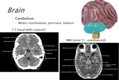
Cerebellum
the "little brain" at the rear of the brainstem; functions include processing sensory input and coordinating movement output and balance
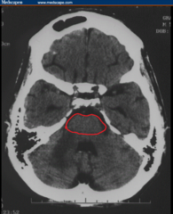
Pons
A brain structure that relays information from the cerebellum to the rest of the brain
ischemic stroke
more common
Caused by an obstruction of an artery leading to or in the brain, preventing oxygenated blood from reaching parts of the brain that the artery feeds
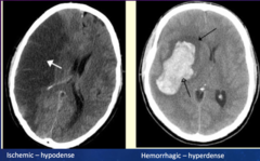
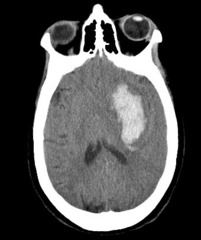
Hemorrhagic stroke
More severe
Occurs when a vessel in the brain suddenly ruptures. Blood begins to leak directly into brain tissue and/or CSF
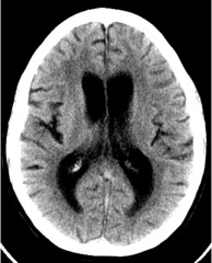
hydrocephalus
abnormal accumulation of fluid (CSF) in the brain
Aqueductal stenosis (AS) is a the most common cause of congenital obstructive hydrocephalus
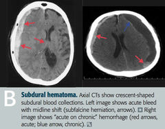
subdural hematoma
collection of blood under the dura mater
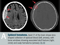
epidural hematoma
collection of blood above the dura mater
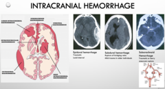
Subdural Intracranial Hemorrhage
bleeding between the dura mater and arachnoid mater
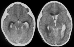
Intraventricular hemorrhage
bleeding that occurs from vessels along the ventricles; the bleeding in this case would be directed into the ventricles
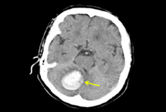
Cerebellar hemorrhage
hemorrhage within the cerebellum
Meningitis
inflammation of the meninges of the brain and spinal cord
Arachnoid cyst
benign cysts within the brain that do not communicate with the ventricular system
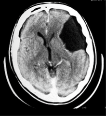
Head CT's uses Thin slices to help reduce ______ artifacts caused by the dense bone of the skull. Often seen at the _________ ___________
hardening, posterior fossa
Routine head uses axial or helical mode?
When would you consider using the other mode for a head?
Routine head studies are most often done using the step-and-shoot method (i.e., axial mode)
Helical mode is primarily used for studies that require 3D reformations or for CT angiography (e.g., circle of Willis studies)
Intracranial Hemorrhage radiographical appearance on CT:
-Hyperdense to normal brain tissue for the first 3 days
-Hyperdense center surrounded by concentric areas of hyperdense and hypodense tissue from 4 to 10 days
-Isodense center surrounded by areas of hypodense tissue from 11 days to 6 months
-Hypodense to normal tissue after 6 months
When imaging a neck in CT, it will Most often be done in _____ mode. IV contrast is used, unless contraindicated.
The goal is to allow sufficient time after contrast administration for mucosa, lymph nodes, and pathologic tissue to enhance, yet acquire images while the vasculature remains opacified
helical
Why would a physician order a CTA of the head and the neck?
-To Accurately measure stenosis of the carotid and vertebral arteries and their branches
-To Evaluate the circle of Willis for completeness using 3D reformation
and Detect other vascular lesions
When using CT to image the spine, we inject CM through____________ then use A delay of _______________ between the injection and scanning is recommended to allow the CM to dilute. (CM that is too dense may mask intradural structures)
Intrathecal Administration, 1 to 3 hours
When suspecting stroke would you use contrast media or not?
A noncontrast CT of the brain is routinely performed to differentiate ischemic stroke from hemorrhagic stroke
It is best to use contrast media for a head when the patient is suspected of having
an AVM, a neoplasm, inflammatory processes or when imaging the pituitary gland
CT Orbits uses axial plane, thin slices and images are acquired parallel to the
IOML
CT imaging of the sinus should include axial images parallel to the
hard palate
True or false? CT imaging of the sinus acquisition should include maxillary sinus to frontal sinus
True
Scan delay for a head can be up to
5 min