Brain, Behavior, & Cognition- Exam 1
1/171
There's no tags or description
Looks like no tags are added yet.
Name | Mastery | Learn | Test | Matching | Spaced |
|---|
No study sessions yet.
172 Terms
Invasive Measures
Recording the electrical activity of neurons via microelectrodes implanted directly in the brain during brain surgery
Implanted microelectrodes and voltammetry probes detect electrical activity
Microdialysis probes measure neurotransmitter concentrations
Electroencephalography – recording electrical activity on the scalp
Noninvasive Measures
Positron Emission Tomography (PET) – Radioactive tracer of metabolic activity (i.e. water, glucose)
Functional MRI (fMRI) provides whole-brain maps of brain oxygenation
Magnetic Resonance Imaging (MRI) – Similar to an X-ray image, but with a magnetic field/radio waves
what are the divisions of the Nervous System?
Central Nervous System
brain
spinal cord
Periphal Nervous System
afferent
somatic
visceral
special
efferent
somatic
autonomic
sympathtic
parasympathetic
enteric
Somatic
Relates the brain/body to the external world
Sensory and Motor inputs
Voluntary and involuntary action
What would be an afferent component of this system? How about an efferent component
Sympathetic Nervous System
Fight, flight, freeze – arouses the body
Reactions to threats and/or opportunities
Think about physiological changes that occur during stress
Parasympathetic Nervous System
“Rest and Regenerate”- returns the body to homeostasis
Body wants to dispel tension – cannot remain “activated” all the time
Think about physiological changes that occur after a large meal
Enteric
More neurons reside in the human gut than in the spinal cord (why?!)
Innervation from Vagus nerve (Cranial Nerve X) but can operate even when it is severed
Regulation of digestion, contraction of stomach muscles, secretion of enzymes
Also has a curious connection to mood/emotion
Spinal Cord
Structure which encases many nerves and coordinates the inputs and outputs of the peripheral nervous system
The spine is segmented – each segment handles inputs to a specific region (dermatomes)
Each segment contains a central canal surrounded by gray matter and white matter
Gray matter – Neuronal cell bodies and localized connections
White matter – Long-distance connections between neurons (we will learn why it is white when we discuss neuronal signaling)
Sensory input and motor output are separated:
Sensory input enters via the dorsal nerve root
Motor output exits via the ventral nerve root
Central Nervous System (CNS) –
Brain & Spinal Cord
Peripheral Nervous System (PNS) –
Connects CNS to rest of the body (skin, muscles, internal organs)
Afferent pathways go from
the Peripheral Nervous System (PNS) to the Central Nervous System (CNS)
Efferent Pathways go from
the Central Nervous System (CNS) to the Peripheral Nervous System (PNS)
Enteric Nervous System
More neurons reside in the human gut than in the spinal cord (why?!)
Innervation from Vagus nerve (Cranial Nerve X) but can operate even when it is severed
Regulation of digestion, contraction of stomach muscles, secretion of enzymes
Also has a curious connection to mood/emotion
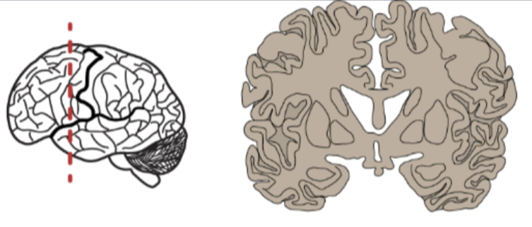
Frontal Plane Image
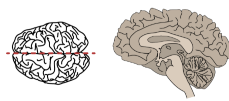
Sagittal Plane Image
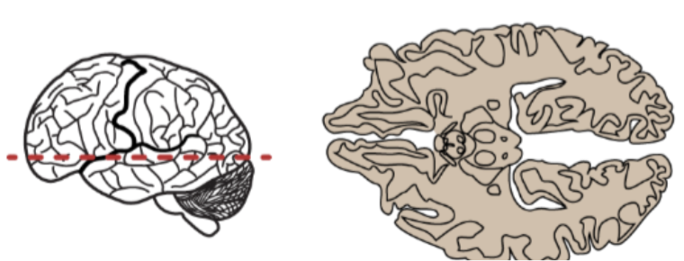
Horizontal Plane Image
Jerison’s Encephalization Quotient
Ratio of actual brain volume compared to the expected brain size for a species of equivalent size
Human EQ ~ 7.4-7.8
Crude measure of expected intelligence and complexity among animals
The developing brain has three distinct bulges which eventually develop into
forebrain, midbrain, and hindbrain
Forebrain later divides into
telencephalon and diencephalon
Hindbrain later divides into
metencephalon and myelencephalon
The midbrain is also called the
mesencephalon
Sonic Hedgehog pathway
influential for cell differentiation
Primarily expressed in the prenatal human cortex
Disruptions in the Sonic hedgehog gene pathway can lead to
seizure disorders, language or cognitive impairment, down syndrome, hyperactivity, and/or schizophrenia
Three main structures to brainstem:
medulla, pons, midbrain
Medulla:
linked to breathing, heart rate, blood pressure; relays signals between cerebellum and cerebrum (cerebral cortex)
Pons:
linked to arousal, sleep, breathing, swallowing, bladder control, eye movement, facial expressions, hearing, equilibrium, and posture
Cranial Nerves
in brainstem
10 of the 12 cranial nerves come from the brainstem (except for I and II which connect to the cerebrum)
2 sets of paired nerves which transmit sensory and motor information from CNS to PNS
6 of the 12 control the eye – why?
Facial nerve is also referred to as the
intermediate nerve
The Midbrain coordinates
survival and reproduction:
Appetitive Behavior
Agonistic Behavior
Reproductive Behavior
In the midbrain, the Ventral Tegmental Area is the
Reward system and motivation (dopamine & substantia nigra)
In the midbrain, the Reticular Formation is the
Network of midbrain cells which regulate consciousness
Cerebellum as “the little brain”
Complex motor control center
Links memory & emotion
Super densely packed tissue – surface area equal to ~80% of cortex surface area
Many neurons packed densely into folds (lobules)
“Fractured” Somatotopy
Much more advanced region than initially thought
“Forward model” may also apply to social processing, not just movement
Intentional movement is predictive
Hypothalamus:
Homeostasis – Body’s tendency towards “set points” and equilibrium
Controls the “basic drives” – hunger, thirst, sexual arousal, temperature regulation, sleep
The hypothalamus is the master control gland of the neuroendocrine axes via
signaling with the pituitary gland
Thalamus:
Relay station of sensory data (EXCEPT smell)
Relays sensory data to respective areas of the cerebral cortex
Relays motor signals from cerebellum and basal ganglia (in the cortex)
Also serves association areas in the cortex (integrative areas not solely dedicated to sensory or motor functions) – crucial role in motor planning
Importance in integrated function
Frontal Lobe General Function
higher level cognitive functions (behavior& emotional regulation& planning)
Frontal Lobe Key Structures
primary motor cortex
prefontal cortex
orbitofrontal cortex
brocas area
Parietal Lobe General Function
primary sensory area; spatial awareness & perception
Parietal Lobe Key Structures
primary somatosensory cortex
precuneus
temporal lobe general functions
responses to the environment; communication/language'; memory access; emotions e
temporal lobe key structures
limbic system
wernicke’s area
temporal gyri
auditory cortex
occipital lobe general function
visual processing area; depth perception; color vision
occipital lobe key structures
visual cortexes
Four Lobes of the Cortex are
frontal
parietal
temporal
occipital
Corpus Callosum:
The bridge between hemispheres
plays a crucial role in interhemispheric communication, allowing information to travel between the left and right sides of the brain
Sasai et al. driving study – “functional split”
Aimed to produce a “functional” rather than “anatomical” split within the brain
Normal, healthy brains underwent driving simulation task(s):
Driving while listening to GPS directions
Driving while listening to radio broadcast
Identification of distinctive “driving network” and “listening network” in the split task condition
Integration of networks was much higher in the GPS task
Ability to switch between high and low integration networks corresponded to overall better driving performance
The limbic system
Interconnected regions that integrates environmental stimuli (sensory input) with internal environment
Key structures include hypothalamus, amygdala, hippocampus, some nuclei of brainstem and midbrain, certain circuits of the cortex (via basal ganglia)
The hippocampus acts as the
memory center of the brain (more on this in our memory lecture)
The amygdala triggers
emotional responses such as fear
The Neuron & its parts
collect information (dendrites)
integrate information (soma)
conduct electrical signals (axon)
transmit signals (axon terminals)
Hodgkin & Huxley: Squid Giant Axon & Voltage Clamp
Attempted to measure viscosity of axoplasm with mercury
Initial experiment was initially a failure – researchers pivoted focus
Inserted fine capillary electrode to measure membrane potential – Led to first recording of intracellular action potential
Discovery of voltage gated channels that controlled cell membrane potential
Voltage Clamp Method
Allows researchers to keep membrane potential constant to measure activity of ion channels
0 mV is able to overcome threshold
Sodium is coming in, potassium is coming out
When we isolate the channels the TTX blocks sodium
TEA blocks potassium so sodium channel works fine but there is no outward of K+
Membrane potentials
Overall charge of the neuron is determined by differences in concentration between Na+ and K+ ions
As resting state, this difference is about -70mv
Sodium ions exist largely outside the cell at rest, while potassium is inside the cell
These concentrations are maintained via the sodium potassium pump
Cell as has a passive potassium leak channels selectively permeable to K+
Sodium Potassium Pump
Binds 3 sodium ions and a molecule of ATP
Splitting of ATP provides energy to change the shape of the channel. The sodium ions are driven through the channel
The sodium ions are released to the outside of the membrane, and the new shape od the channel allows two potassium ions to bind
Released of phosphate allows the channel to revert to its original form, releasing the potassium ions on the inside of the membrane
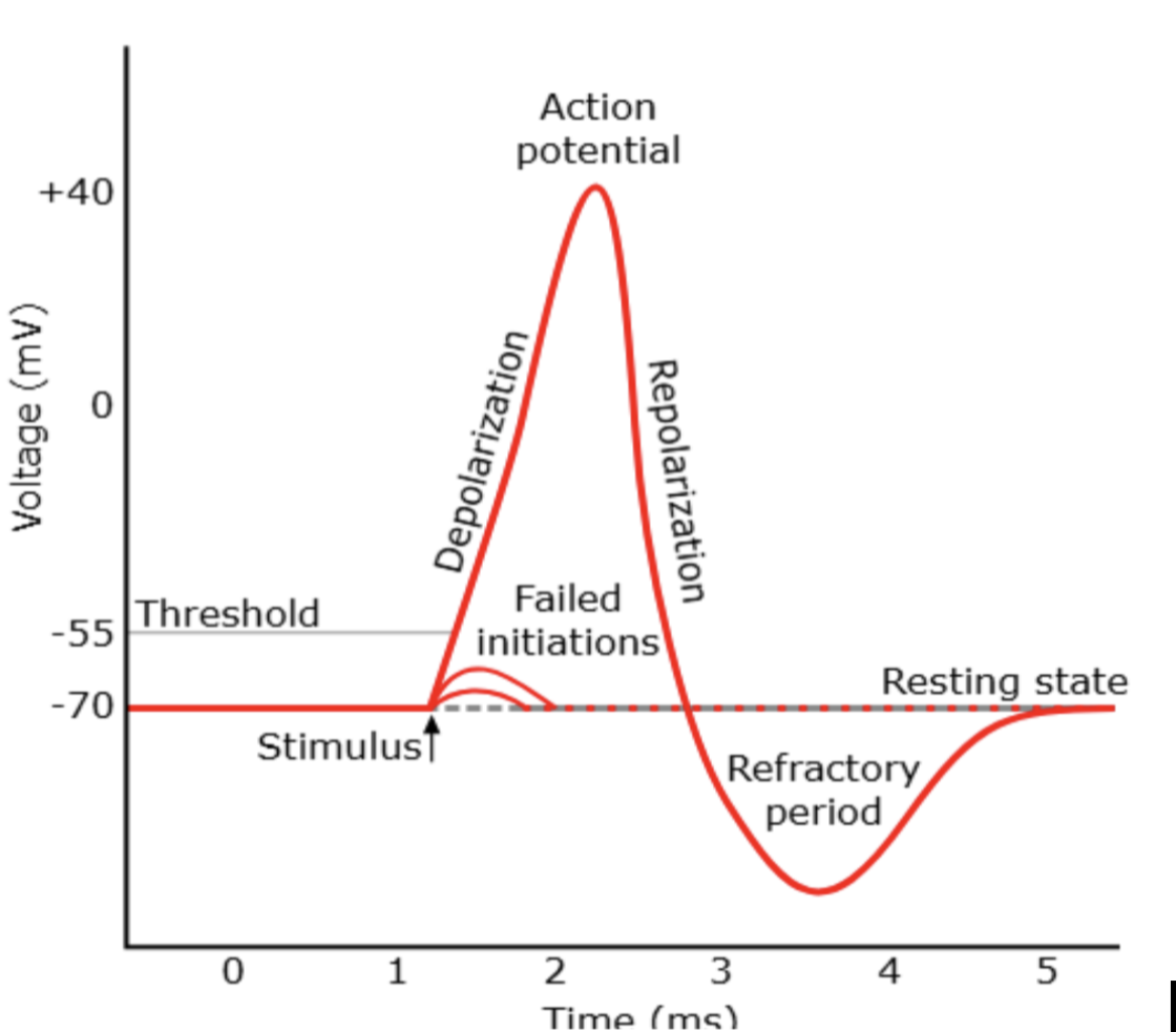
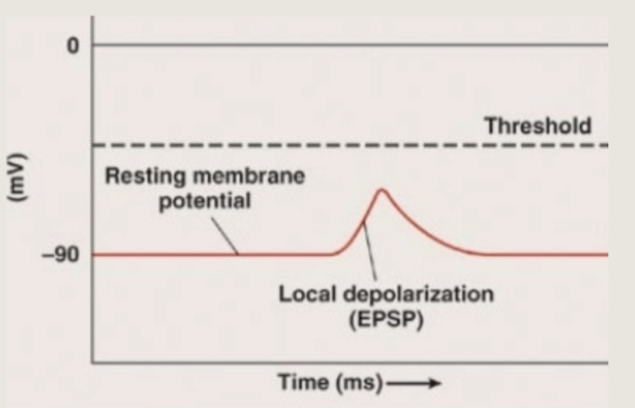
Events of Action Potential
Upon sufficient stimulation voltage gated ion channels will open, causing sodium ions to leak into the cell
The rush of positively charged ions causes the voltage to rise (become more positive) from -70mV
Depolarization occurs as the cells charge gradually moves from -70 towards the positive values (up to about 40mv)
As voltage becomes positive, potassium gated ion channels will open, causing potassium ions to leak out of the cell
This causes the cells positive charge to decrease (become more negative)
Repolarization occurs as the cells charge gradually moves from about +40 mv down to below- 70 mv. This overshoot is known as hyperpolarization
A refractory period occurs due to hyperpolarization neurons require a short cooldown before firing another action potential
Depolarization occurs as
the cells charge gradually moves from -70 towards the positive values (up to about 40mv)
Repolarization occurs as
the cells charge gradually moves from about +40 mv down to below- 70 mv. This overshoot is known as hyperpolarization
A refractory period occurs due to hyperpolarization,
neurons require a short cooldown before firing another action potential
Graph of normal action potential
Influence of ESPS
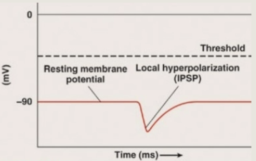
Influence of IPSP
After an action potential occurs the electrical signal travels down the length of the
myelin sheath
a lipid layer that acts like insulation of a wire
The mylein sheath contains gaps called
nodes of ranvier
contain their own sodium & potassium channels
To ensure the potential reaches the full length of the axons
the action potential resets at each node of ranvier- causing the signal to arrive to the axon hillock at full capacity
Mylenate only axons that need it- the ones that travel the farthest
Saltatory propagation
In response to a signal the soma end of the axon becomes depolarized
The depolarization spreads down the axon meanwhile the first part of the membrane repolarizes. Because Na+ channels are inactivated and additional K+ channels have opened, the membrane cannot depolarize again
The action potential continues to travel down the axon
The Events at the synapse
Upon reaching axon hillock- action potential stimulates voltage gated calcium channels
Calcium ions cause vesicles with neurotransmitters to move towards the membrane and fuse with the membrane
The vesicle fuses with the membrane via SNARE proteins and neurotransmitters are released via exocytosis
The cell sending signals is the presynaptic neuro
The cell receiving signals is the postsynaptic neuron
The neurotransmitter Acetycoline (ACh) has the function of
Motor function (muscles); learning and memory
the neurotransmitter Dopamine has the function of
Reward system; movement regulation
the neurotransmitter endorphins has the function of
Pain relief; released during exercise; elevates mood
the neurotransmitter norepinephrine has the function of
Arousal; fight/fight response
the neurotransmitter serotonin has the function of
Natural mood booster, sleep/wake cycles; lower serotonin levels linked to depression
the neurotransmitter GABA (gamma aminobutyric acid) has the function of
Lowers arousal and reduces anxiety; primary inhibitory neurotransmitter
the neurotransmitter Glucamate has the function of
Primary excitatory neurotransmitter of the Central Nervous System (CNS)
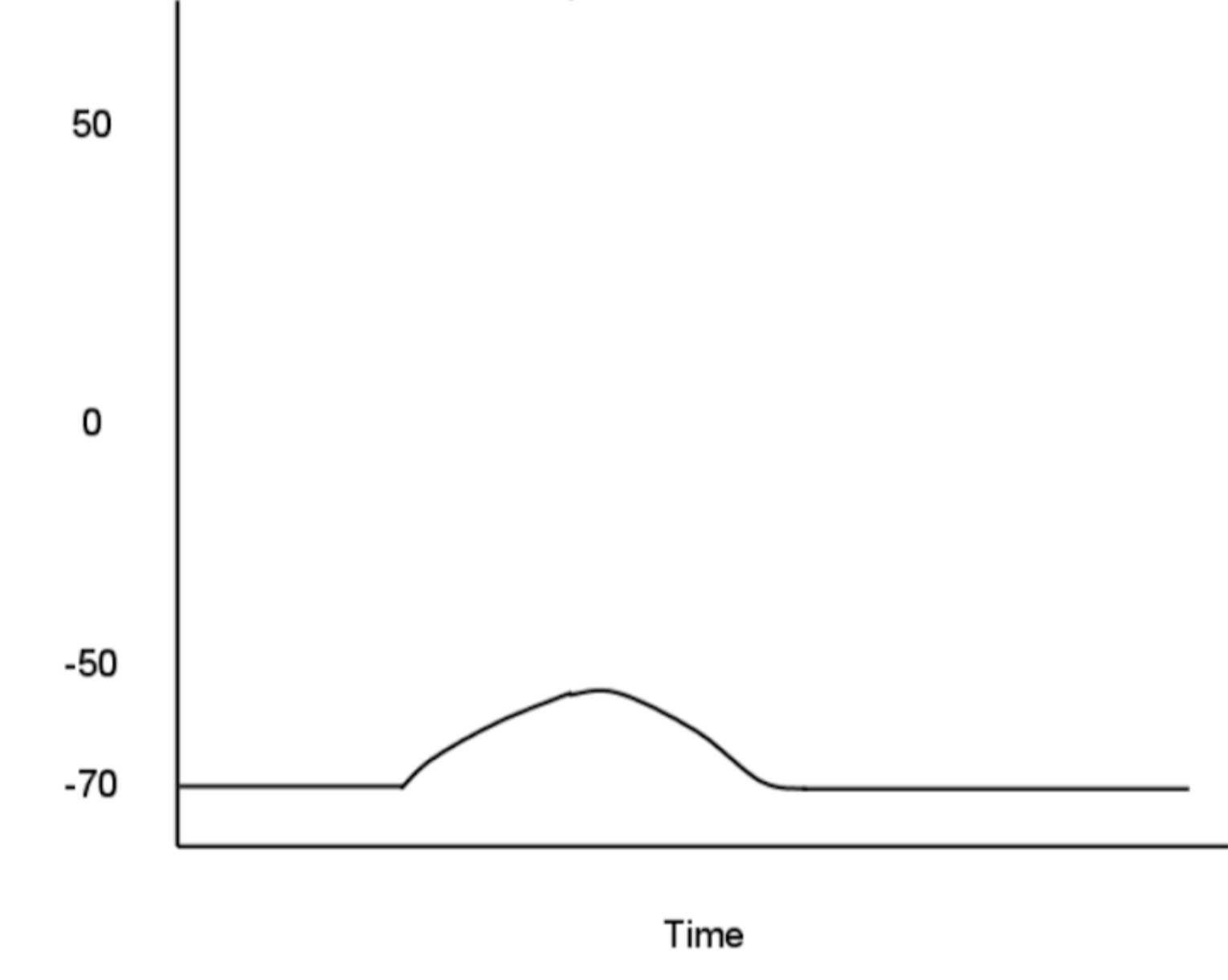
What would happen if the nervous system poison Tetrodotoxin (TTX) was present
______ is a neurotoxin that selectively blocks voltage-gated Na+ (sodium) channels… so…
Sodium is what makes it go up (depolarization)
Since action potential is contingent on sodium
the action potential doesn't really happen
falls flat
What would the graph look like if the nervous system poison Tetrodoxin (TTX) is present
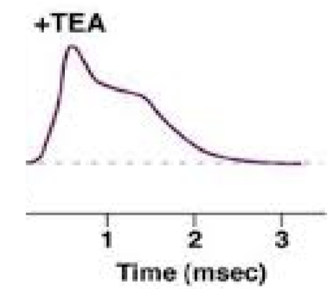
What would happen if the nervous system poison Tetraethylammonium (TEA) was present
Able to depolarize but since only the K+ (potassium) channels are blocked the repolarization process is delayed
Slowly starts to level out
Not a dramatic level of spiking down
Takes a lot longer for repolarization to occur
What would the graph look like if the nervous system poison Tetraethylammonium (TEA) is present
Archetypes of neural networks
Feedforward excitation
Feedforward inhibition
Convergence
Divergence
Lateral inhibition
Feedback excitation
Feedback inhibition
Feedforward excitation
Neuron relays information to its neighbor
Feedforward inhibition
A presynaptic cell excites an inhibitory interneuron which then inhibits the next cell
Lateral inhibition
A presynaptic cell excites inhibitory interneurons, inhibiting neighboring cells in the network
important for vision
Feedback excitation is involved
in learning/memory
Feedback inhibition is important
for circadian rhythm
Convergence
One postsynaptic cell receives convergent input from many presynaptic cells
Divergence
any individual neuron can make divergent connections to many different postsynaptic cells; crucial for stretch reflex
Structural neuroplasticity
the brain changes as a result of learning/experience
Functional neuroplasticity
neuronal remodeling following dysfunction/injury
Homouncolous Model
Way of visually representing how visual and sensory cortex map out on body
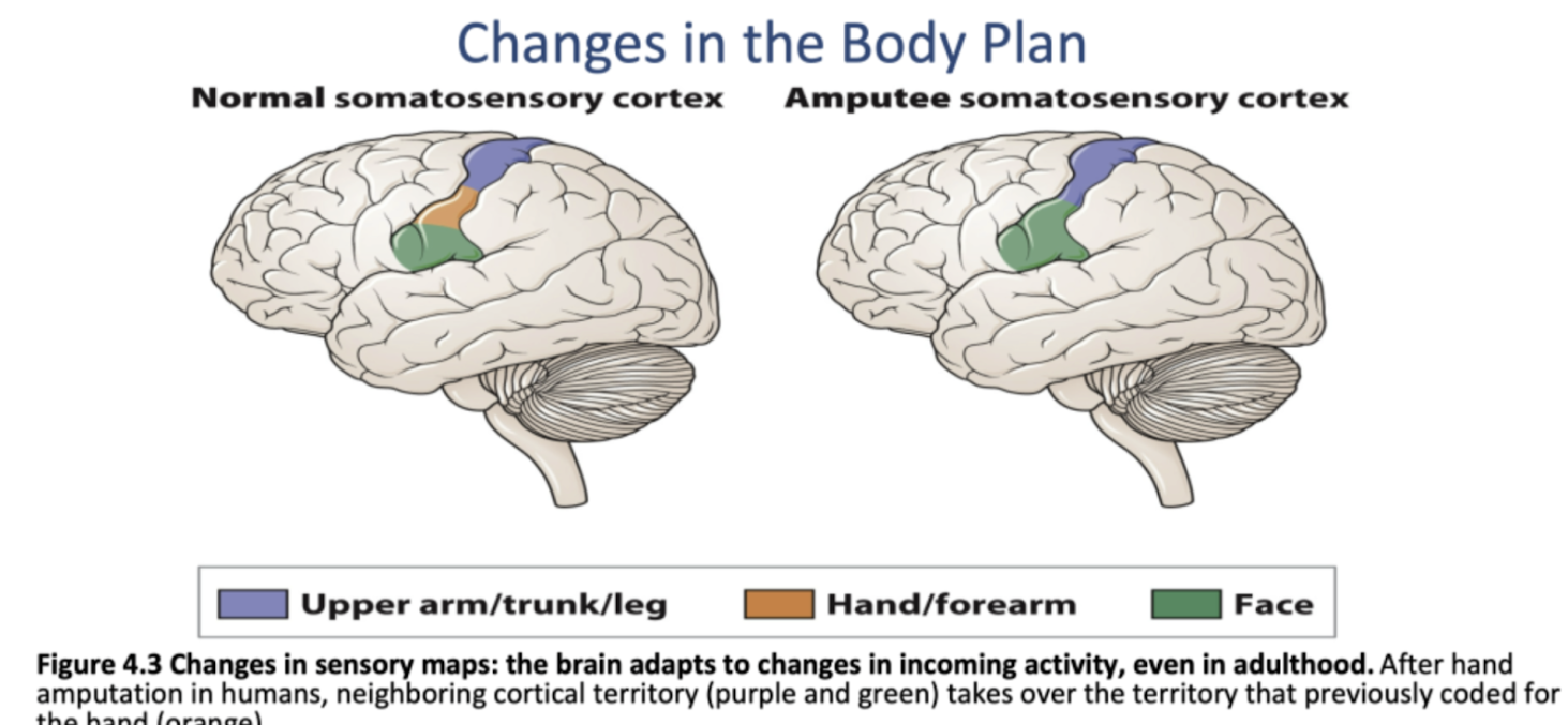
Body plan changes are reflected in the brain
This includes both somatosensory and motor cortex
Not a part of the brain, but a way of representing cortex’s
Phantom sensation
the phenomenon where amputees report sensations in their missing limbs
Phantom limb pain
magnitude of pain correlates with extent of remodeling in the cortex
treatment- mirror
D.M Case Study
Right arm amputee (above elbow)
Reported to be able to feel and move her right forearm, hands, and fingers
Touch on the right side of face was also felt in missing limb
Chin and jawline are next to hand in the homunculus
Somatosensory Cortex rewiring
Making use of available tissue in brain
Plasticity during youth vs. adulthood
Brains are most “plastic” during youth
Our brain is like a sponge- we don't have any innate functions
we have to be primed to learn more
more than other animals
not born with innate wiring
Adults are pushed to focus and hone skill
cholinergic neurons provide focus and inhibit unwanted plasticity
Critical/Sensitive Periods
babied/children need to learn- cholinergic neurons are more primed for activation
Adults are pushed to focus and hone skill- cholinergic neurons provide focus and inhibit unwanted plasticity
“Flexibility vs skill” tradeoff
Experience expectant neuroplasticity
the brain contains wiring predisposed for “expected” experiences
Experience dependant neuroplasticity
additional growth that varies greatly within individual experiences
Sperry’s Frogs (Chemoaffinity Hypothesis)
Brains innate wiring is due to predefined chemical signaling
Experience independent hypothesis- this mechanism is not influenced by an animal's experience of the world
Held & Hein’s Kitten Study – Procedure and Takeaway
One of the cats was guiding and could walk around while the other could not walk; only see where it was being pulled bc of the first cat
vision develops in response to understanding of causal relationship between motor actions and visual field
The way you interact with the world changes how your vision develops
Vision tested in response to
Visually guided paw placement
Depth perception (visual cliff)
Blink (“flinch”) in response to approaching object
The way you engage in the world leads to development of normal vision