Organization of the Body Wall
 Call Kai
 Learn
Learn Practice Test
 Spaced Repetition
 Match
Match Flashcards
 Knowt Play
1/16

There's no tags or description
Looks like no tags are added yet.
Name | Mastery | Learn | Test | Matching | Spaced | Call with Kai | Chat |
|---|
No analytics yet
Send a link to your students to track their progress
17 Terms
Overview (what it is & structures)
The “body wall” is the portion of a person’s body that encloses the large body cavity housing the major viscera.
The limbs are also considered to be parts of the body wall.
Body wall structures include the skin, the skeletal framework of the body, the skeletal muscles that attach to that framework, and a variety of connective tissue structures (e.g., fascia, septa) that organize the deeper structures.
Internally the body wall is separated from the “body cavity” by an additional fascial layer termed the “internal fascia”.
layers of the body wall
skin
epidermis
dermis
subcutaneous tissue
fatty (superficial) layer
membranous (deep) layer
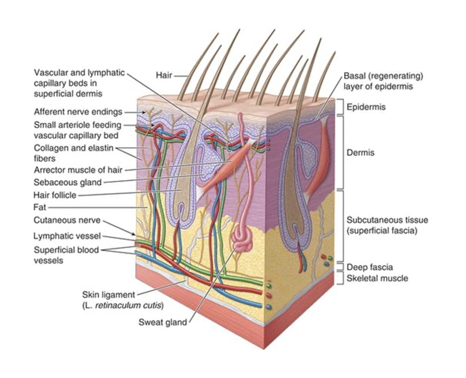
epidermis
epithelium, supplied with sensory nerve endings
most superficial layer
dermis
collagen and elastic fibers, give strength to skin
Subcutaneous tissue
also called superficial fascia (most common in course), hypodermis
Fatty (superficial) layer: loose connective tissue/fat (yellow appearance)
Contains sweat glands, superficial blood vessels, cutaneous nerves, lymphatic vessels.
Body fat storage and insulation to retain heat.
Membranous (deep) layer
thin
more connective tissue than fat
wispy-like appearance
thermoregulation
arrector pili muscles
sweat glands
arterioles within dermis
arrector pili muscles
erect the hair (goosebumps)
smooth muscles in the dermis
Contraction of muscle when cold, air becomes trapped between the erect hairs to retain heat.
Compresses sebaceous glands to secrete oily product onto skin.
sweat glands
evaporation of sweat from skin allows for cooling
arterioles within the dermis
stem from blood vessels in subcutaneous layer
dilate to fill superficial capillary beds to radiate heat (turn skin red color)
constrict to decrease blood in superficial capillary beds to minimize surface heat loss (turn skin blue color)
layers of the body wall: limbs
Deep fascia:
dense connective tissue deep to subcutaneous tissue.
keeps everything contained
Investing fascia:
Covers (invests) muscles to allow to smooth movements.
Intermuscular septum (septa = plural):
Thick sheets that attach to bone to separate muscles into “compartments” (muscles will have similar action and neurovascular supply).
Retinaculum (retinacula = plural):
Thickened sheet that holds tendons in place.
typically in the wrists and ankles where there are a lot of tendons
Muscle – covered in (deep) investing fascia.
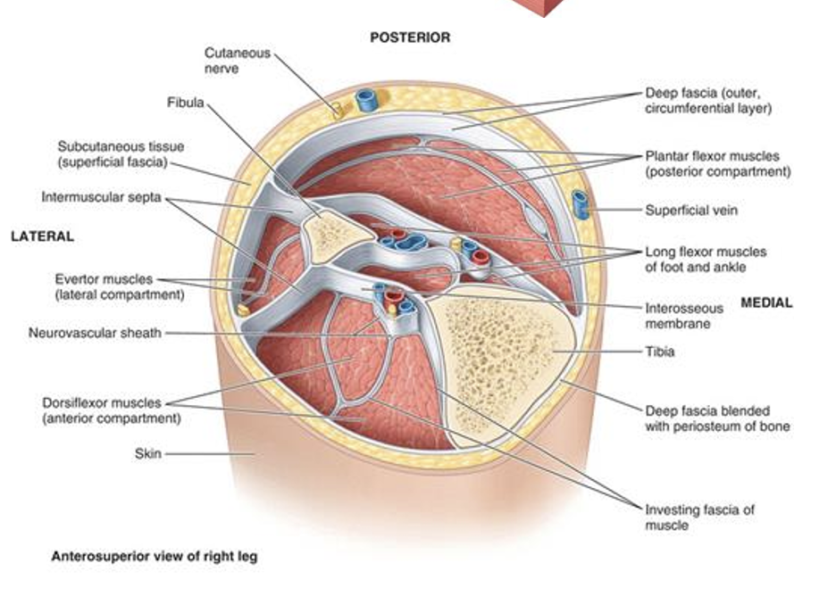
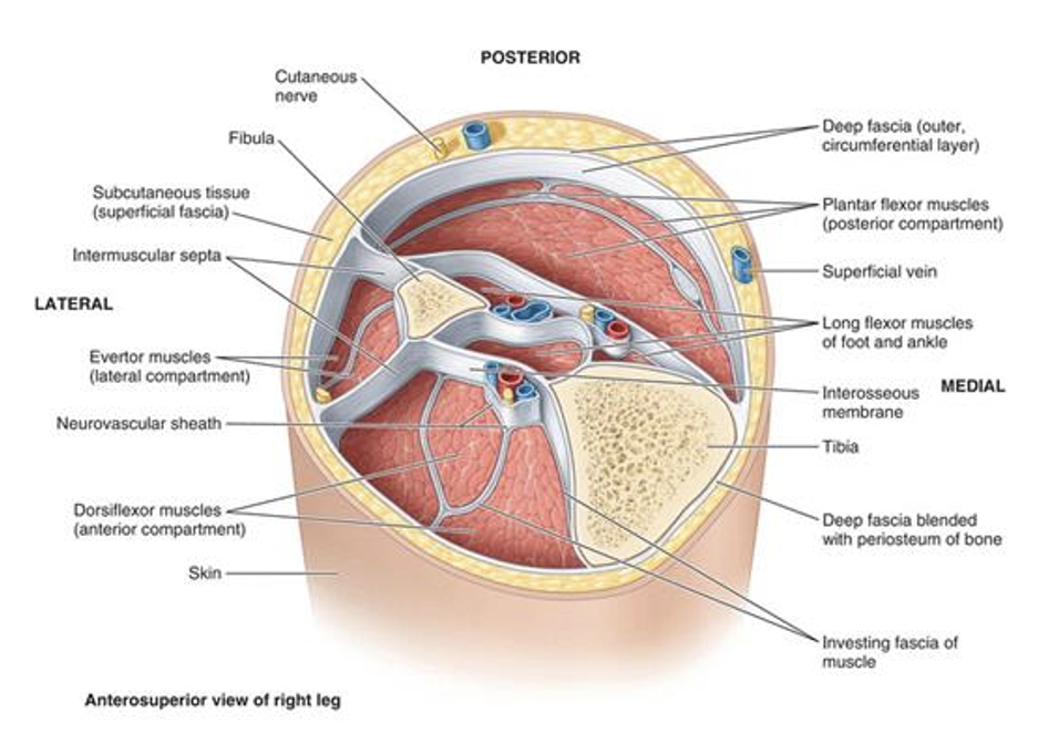
Fascia Comparison
Superficial fascia:
Composed of loose connective tissue and fat.
Between dermis and deep fascia.
Provides most of body’s fat storage, thickness varies.
Deep fascia:
Composed of dense connective tissue.
Deep to superficial fascia.
Extensions:
Invest muscles of neurovascular bundles (investing fascia).
Divide muscles into compartments (intermuscular septa).
Hold tendons in place during movement (retinacula).
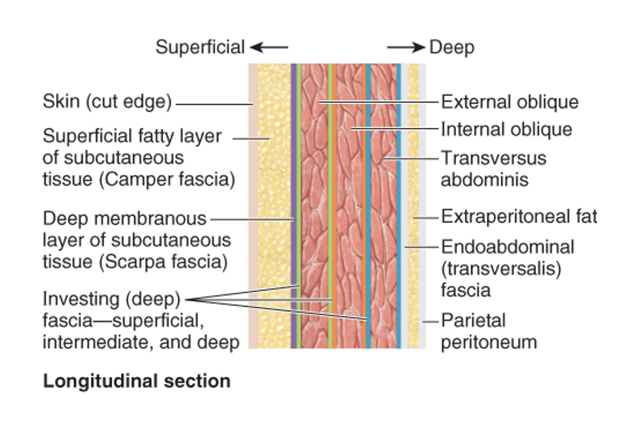
ventral body cavity
Ventral Body Cavity:
Skin.
Superficial fascia.
Fatty layer.
Membranous layer.
Layers of muscle with deep investing fascia.
Internal fascia.
Serous membrane.
serous membranes
Reduce friction between moveable organs and the walls of their compartments.
Form a serous sac that allows for smooth movements of heart, lungs, GI organs.
Continuous sheets that covers organs and compartment walls.
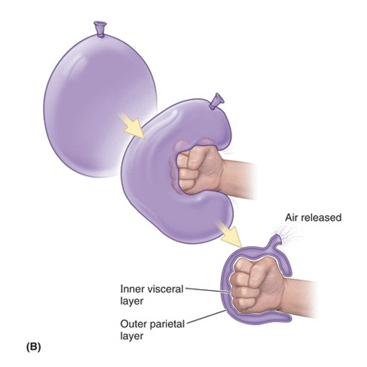
3 layers of serous membrane
Visceral layer (organ).
Parietal layer (wall).
Cavity (space).
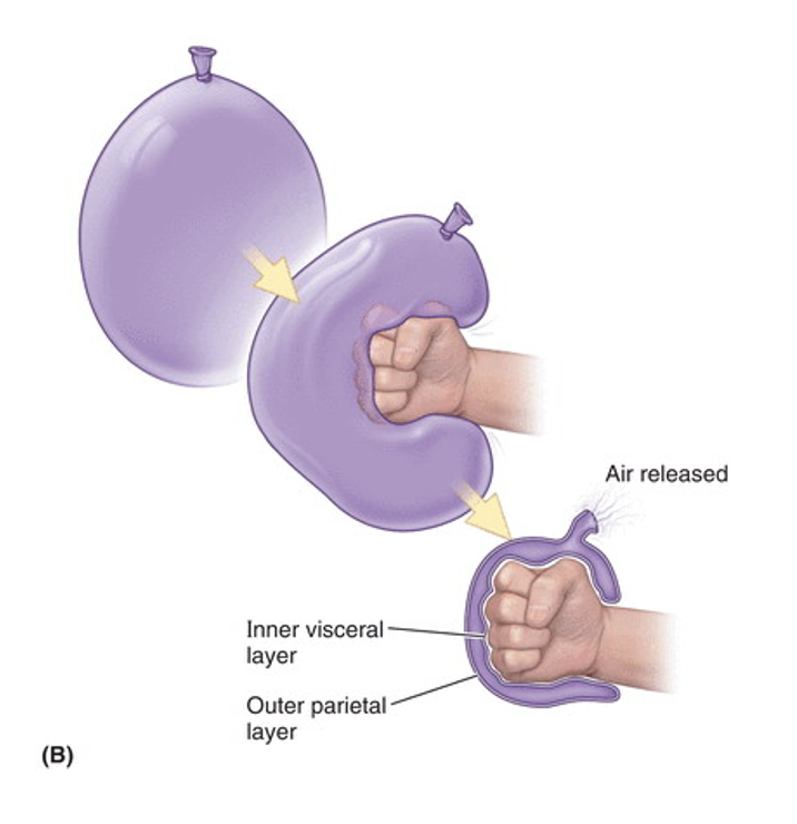
3 types of serous membranes
Pleura (lungs).
Pericardium (heart).
Peritoneum (GI tract).
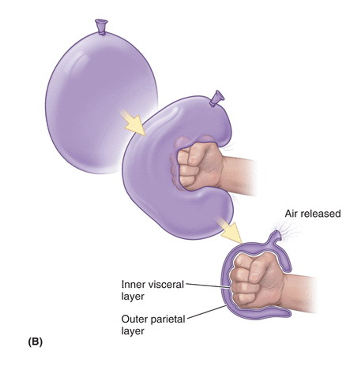
bursae
Closed sacs of serous membrane.
Secretes fluid to lubricate an internal surface.
Found in locations subject to friction.
Normally collapsed, potential spaces.
When excess fluid accumulates, or the wall is interrupted, they become real spaces (pathological, abnormal).
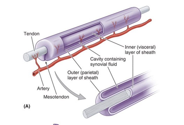
types of bursae
Subcutaneous – between skin and bony prominences.
Subfascial – beneath deep fascia.
Subtendinous – between tendons and bone.
Synovial tendon – elongated around tendons.