Special Senses
1/19
There's no tags or description
Looks like no tags are added yet.
Name | Mastery | Learn | Test | Matching | Spaced | Call with Kai |
|---|
No analytics yet
Send a link to your students to track their progress
20 Terms
General Senses
have receptors scattered throughout the body
Nociceptors- free nerve endings that convey the sense of pain
thermoreceptors- temperature sensitive
mechanoreceptors- establish an action potential in response to physical deformation.
Tactile receptors- May measure “touch”
baroreceptors/pressoreceptors- mechanoreceptors might measure change in pressure
Proprioreceptors- sense of body position and motion
senses can also be classified based on the location of stimulus
exteroreceptors- receptors that respond to a stimulus outside the body such as touch or temperature
teloreceptors- detect change that occur away from the body
include eye and the ear
Interoceptors/enteroceptors/visceral receptors- detect changes within the body
Special senses
receptors are localized in special, complex sensory structures
chemical senses- receptors for these senses respond to specific molecular stimuli
olfaction, gustation, and “common chemical” sense
Common chemical- sense of chemical irritants
receptors found in mucous membranes of humans
respond to onion fumes, tear gas, and acid fumes
Olfaction- sense of smell
neurons from the nasal epithelium convey impulses through the cribriform plate of the ethmoid bone to the olfactory bulb
olfactory hairs (dendrites) are exposed to the surface of the nasal epithelium
mucus is secreted by glands in the epithelium to dissolve the substances we smell (odorants)
Gustation- sense of taste
receptors found on tongue, soft palate, and pharynx
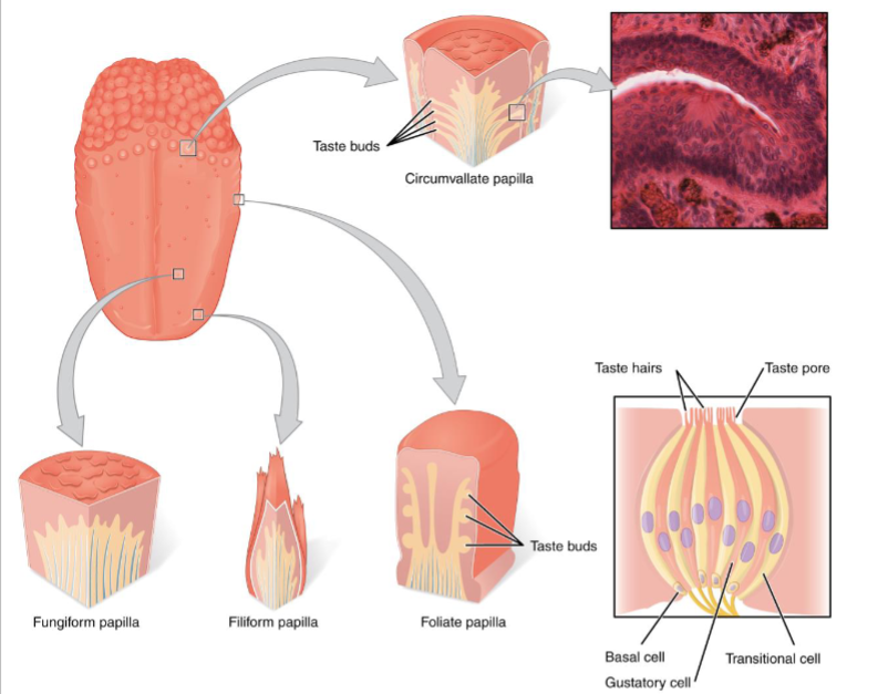
Taste buds found on the tongue associated with papillae (small elevations) of the tongue
Papillae
circumvallate/ vallate papillae- largest of the papillae
located at the very back of the tongue and form a “V” shape
fungiform papillae- large bumps on the tongue
filiform papilla- smallest papillae
when they get excessively long, a person is said to have a “hairy tongue”
people are instructed to scrub tongue with a toothbrush to wear papillae down
Circumvallate, fungiform, and foliate papillae are associated with taste buds. Filiform are not
foliate papillae- posterior, lateral border of tongue is irregularly shaped in children
looks like edge of a leaf
as person gets older, tongue flattens and broadens, and this surface disappears
Primary tastes
flavor comes from odor produced by food
has specific receptors on the tongue
more than sweet, salt, sour, and bitter
chemical senses are especially prone to sensory adaptation
sensory adaptation- decrease in the intensity of a sensation as time passes
strong smell but we then get nose blindness
The eye
ophthalmology- study of structure, function, and diseases of the eye
superciliary (supraorbital) ridge- ridge above eye/eyebrow
helps keep perspiration out of the eye
deflect overhead sunlight
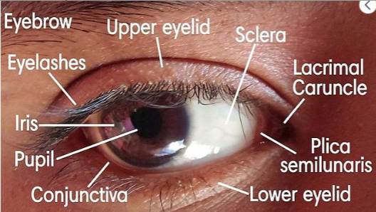
palpebrae- eyelids
protect eye and keep it moist
palpebral fissure- space between the upper and lower palpebrae
commissures- corners of the palpebral fissure
medial commissure/ medial canthus- corner next to the nose
lacrimal caruncle- small pink mass of tissue in medial canthus
lateral commissure/lateral canthus- corner of the palpebral fissure that is away from the nose
conjunctiva- mucous membrane that covers the visible surface of the eye and lines the underside of the eyes
lacrimal glands- superior and lateral to the eye
produce tears that wash across the surface of the eye toward the medial commissure
helps to clean surface of eye and keep it moist
contain lysozyme (bactericide enzyme) that helps prevent bacterial infections
tears drain through nasolacrimal ducts into nasal cavity
why excess production of tears cause “sniffling”
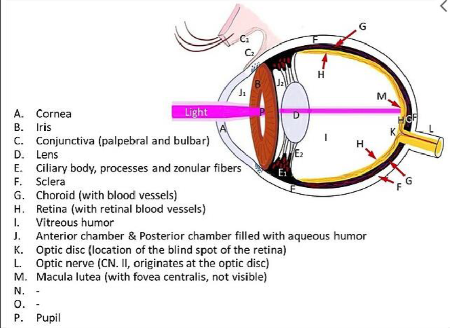
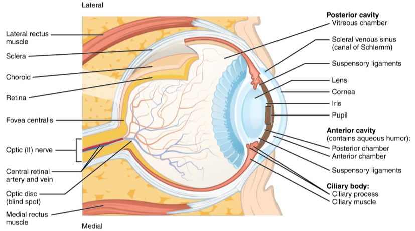
Wall of eye
fluid filled structure composed of 3 tunics/coat (layers) in the wall
fibrous tunic- outermost layer of the eye is tough
composed of an anterior, transparent cornea and a posterior, white sclera
sclera “white of the eye”
vascular tunic/uvea- middle layer of eye
iris- most anterior part of the eye that gives people different eye colors
pupil- opening in the iris that allows light to enter the internal part of the eye
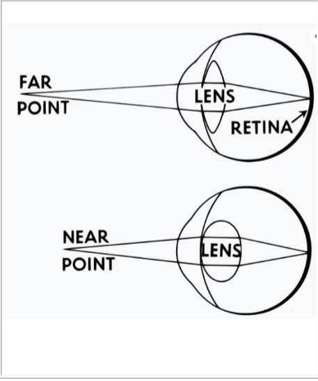
Ciliary body- behind iris is muscular structure that is shaped like a donut
alters shape of the lens and allow the eyes to focus on objects that are different distances from the eye
lens- in hole of donut; focuses light on the back of the eye
Choroid- posterior part of the uvea
many blood vessels and supplies nutrients to the nervous, inner layer of the eye
Nervous tunic/retina- inner layer of the posterior portion of the eye
photoreceptors- nervous receptors sensitive to light
Ciliary body
muscle shaped like a donut that can alter the shape of the lens
lens has natural tendency to become round, but is attached to ciliary body by suspensory ligaments
ligaments pull on lens and make it have a lesser curvature
When look at distance object, lens is pulled into a relatively flat shape
as you move an object closer, tension of the ligaments is lessened and the lens becomes more round
new shape bends light to a greater degree and the image stays focused on the retina of the eye
accommodation- increasing curvature to maintain focus
as we age, eye loses some of its elasticity and its ability to get as round
presbyopia- cant focus on near objects
why a person holds phone book or newspaper farther
Eye cavities
lens and ciliary apparatus divide the inerior part of the eye into 2 cavities (segments)
anterior cavity- front of lens
aqueous humor-filled with a thin, runny fluid
aqueous humor can flow from the posterior chamber through pupil to the anterior chamber
anterior chamber- anterior cavity is divided by iris into this in front of iris
posterior chamber- behind iris
Chamber and cavity are not the same!!!
Posterior cavity/vitreous chamber- behind the lens.
vitreous humor/body- posterior cavity is filled with a thick, jelly-like substance
glaucoma- pressure can build up in the eye if aqueous humor is produced faster than it is reabsorbed. increase in pressure within eye.
pigment
pigment- some molecules absorb certain wavelengths (colors) of light
photopigments- pigment molecules that change structure when they absorb light.
said to be BLEACHED when their structure is altered.
altered protein initiates a nerve impulse
pigment has to be rejuvenated/returned to original form
photopigments of the eye are found in specialized photoreceptors in the retina of the eye
Photoreceptors- have rods and cones
rods- more numerous
do not allow us to distinguish different colors, but do not require much light
cones- more important
allow us to distinguish different colors and form a sharper image
require more intense light, so rods are an important part of our vision in dim light
Rods
photopsin- general term for visual pigment
rhodopsin/visual purple- visual pigment in rods
absorbs light, is bleached,
molecules split into trans retinal (molecule) and opsin (protein)
this event initiates a nerve impulse
trans retinal- carotenoid which means that is relative to vitamin A.
Retinal isomerase- enzyme changes the shape of trans retinal
cis retinal- new molecule
spontaneously recombines with the opsin to reform rhodopsin
Bright room= pigment in rods is bleached faster than it can be rejuvenated
dark room= slowly see better as your rods become functional
Cones
events in cone vision are similar to those in rod vision
one of three different pigments.
pigments absorb different wavelengths of light
so different wavelengths, colors, affect different cones
allows us to distinguish between different colors
red, green, and blue
different pigments have different proteins associated with trans retinal.
if a person is missing one or more of the three pigments, he would be “color blind”
reason cones are more important is the location of the cones in our retina.
macula lutea- lens focuses light on a part of our retina that has only cones
fovea centralis, central fovea, or central depression- depression in the macula lutea where the cones have the greatest concentration.
this area of the retina forms the sharpest image
Vision terms
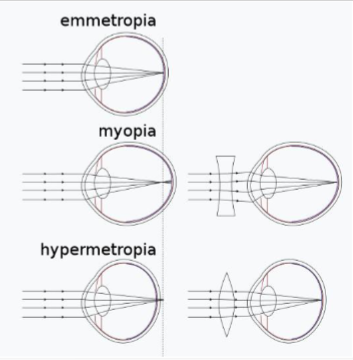
emmetropia- normal vision
myopia- image is formed in front of the retina; “nearsighted”
hyperopia (hypermetropia)- image is focused behind the retina; “farsighted”
astigmatism- irregular curvature of the cornea or lens
Other terms with eye and vision
optic disc- site on the retina where the optic nerve is attached
“blind spot”- no photoreceptors here
medial to the macula lutea
two reasons you do not have a “blank” are in your visual field
light that is striking the optic disc on one eye is striking the lateral part of the retina on the other eye. so light from that part of the visual field is striking photoreceptors
even when you close one eye, no blank area. because the brain fills in the area with an image of what it believes should be in that area.
Snellen chart- tests visual acuity, or ability to form sharp image
20/40- if lowest line you can read lists 40 feet for a person with normal vision
20/25- see the line that lists 15 feet
Ishihara chart- image with lots of color dots
person with normal color vision sees one word or image.
person who is color blind sees a different word or image
miosis- pupil constriction
mydriasis- pupil dilation
The outer ear
pinna/auricle
part of ear on the sides of the head
external auditory meatus-
pinna channels sound waves, like a funnel into these
cerumen(earwax)- canal has tiny hairs and this
tympanum, tympanic membrane, ear drum- EAM ends at a thin membrane
sound waves striking the tympanum cause it to vibrate
tympanum separates the outer ear and the middle ear

The middle ear
middle ear/tympanic cavity- medial (deep) to the tympanic membrane
auditory ossicles- there is a space with three tiny bones
malleus/hammer- most lateral of these bones
touches the tympanic membrane
incus/anvil- next ossicle
stapes/stirrup- most medial ossicle, smallest bone in the body
When tympanic membrane vibrates, causes malleus to vibrate
malleus causes the incus to vibrate, and the incus makes the stapes vibrate
vibrations of the stapes are transferred into the inner ear through OVAL WINDOW (opening)
Canal that connects the middle ear with the nasopharynx
Eustachian tube, auditory tube, or pharyngotympanic tube
passage allows air pressure to equalize on both sides of the tympanic membrane
why your ears “pop” when you swallow as you drive in the mountains or fly in an airplane
as external pressure changes when you change altitude, you need to alter the pressure in the middle ear to compensate
Auditory tube- passage is also a route taken by bacteria to cause otitis media (middle ear infections). Many children get this
Two muscles associated with middle ear
tensor tympani & stapedius
when muscles contract, inhibit movement of ossicles
helps protect the delicate structures of the inner ear from loud noises
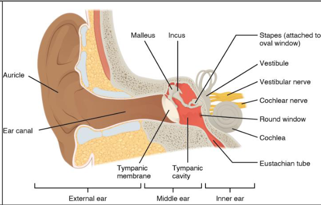
The inner ear/labrynth
has osseous labrynth (outer bony) and an inner membranous labrynth
perilymph- fluid between the bone and the membrane
chemically similar to the CSF
Endolymph- inside membranous labrynth
chemically similar to intracellular fluid
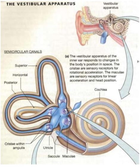
vestibule- middle, enlarged part of the inner ear
semicircular canals- 3 tubes above vestibule
cochlea- below vestibule; snail shaped
Inner ear responsible for much of our sense of balance and equilibrium. also responsible for hearing
Equilibrium in the ear
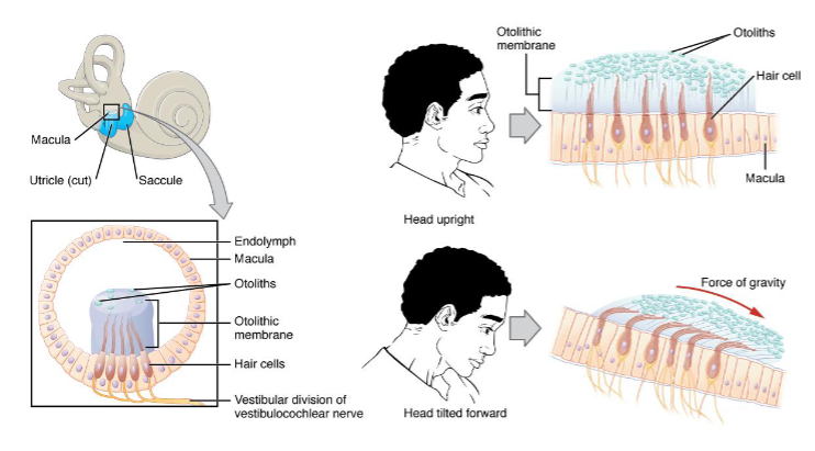
vestibule and semicircular canals associated with the sense of equilibrium
utricle and saccule- vestibule of osseous labyrinth contains portions of the membrane labyrinth
macula- each of these parts of the membranous labyrinth has thickened wall
otolithic membrane- cells in the maculae have tiny hairs that extend into a thick, jelly-like mass
gravity causes otolithic membrane to bend the nerve hairs
if you tild head, these slide across the hairs
conveys a sense of the orientation of the head
Static equilibrium- sense of body position
dynamic equilibrium
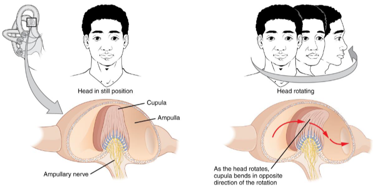
semicircular canals- bony loops of the inner ear
semicircular ducts- membranous labyrinth within the canals
3 semicircular canals are located on three different planes
ampullae- there are swelling or enlargements at the base of each of the 3 duscts
cristae- thickenings in the ampullae
cupula- cells with nerve hairs in the cristae and hairs are covered with this gelatinous mass
when head moves, inertia causes a current of fluid (endolymph) in a semicircular dict
current bends the nerve hairs
since ducts are located on 3 different planes, direction of head movement determines which ampullae are affected
allows us to maintain equilibrium as the body is moving
Dynamic equilibrium- this equilibrium of motion
Sound waves
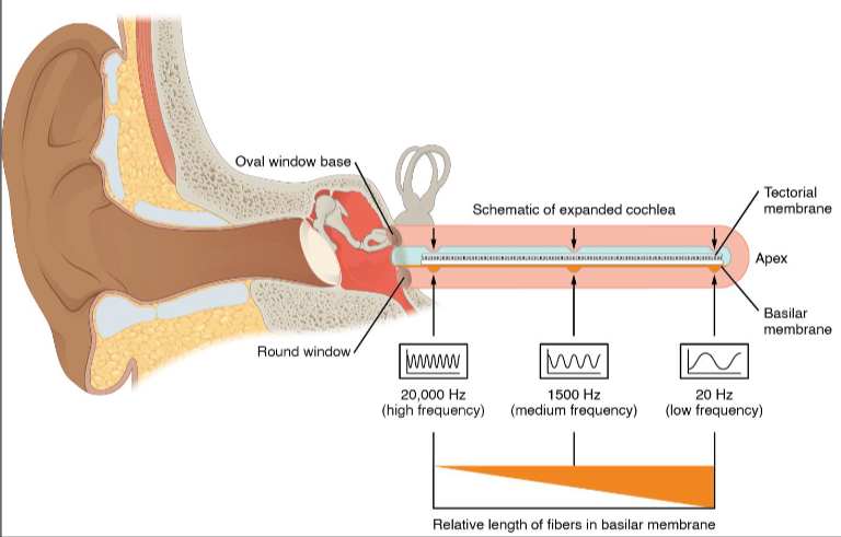
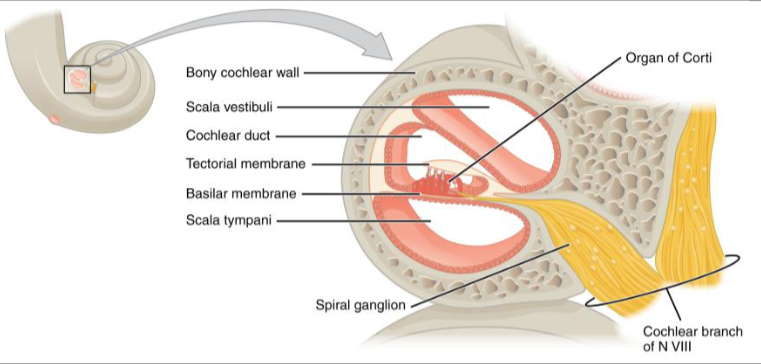
sound waves converted into nervous impulses in the snail-shaped cochlea
Illustration has cochlea unwound to see perilymph (gray shading) is in a passage that begins at the oval window
oval window- where vibrations of the stapes creates waves in perilymph of inner ear
FENESTRA VESTIBULI/ FENESTRA OVALIS
helicotrema- channel with perilymph goes around “end of snail”
Fenestra cochlea/fenestra rotundum- comes back to the lower part of the vestibule where there is another opening covered by a membrane
scala vestibuli- channel containing perilymph from the oval window to the helicotrema
scala tympani- channel from helicotrema to the round window
Waves are created in the perilymph at the oval window and the waves are dissipated at the round window
organ of Corti- spiral organ in the inner ear which has hair cells (stereo cilia) in a basilar membrane
tectorial membrane rests on the hair cells
waves in the perilymph create waves in the endolymph
waves cause basilar membrane to vibrate which pushes the nerve hairs into the tectorial membrane
creates nerve impulses
Pitch- wavelength of the sound waves determines which hairs on the organ of Corti get bent
louder sounds cause bigger waves, so more nerve hairs are stimulated