Conjunctiva
1/36
There's no tags or description
Looks like no tags are added yet.
Name | Mastery | Learn | Test | Matching | Spaced |
|---|
No study sessions yet.
37 Terms
Causes for acute red eye
@CIKSA
conjuntivitis
iridocyclitis
keratitis
sub conjunctival hemorrhage
Acute congestive glaucoma
DD for red eye
Conjunctivitis, iriditis,keratitis,scleritis,episcleritis,sub conj hemm,Blepharitis, Corneal abrasion fb, chemical burn, glaucoma
Causes of conjunctival chemosis
Acute inflammation of conjunctiva,eyeball
Obstruction to circulation:orbital tumor, dysthyroid eye disease, pulsating exopthalmos,
Blood abnormalitis: Anaemia, urticaria, angioneurotic edema, nephritis
Sub conjunctival hemorrhage causes
-Trauma
-Straining
-Fragility of bvs:DM,HTN, Atherosclerosis,Nephritis,
-Severe conjunctivitis
-Serious head trauma
-Severe pressure on thorax
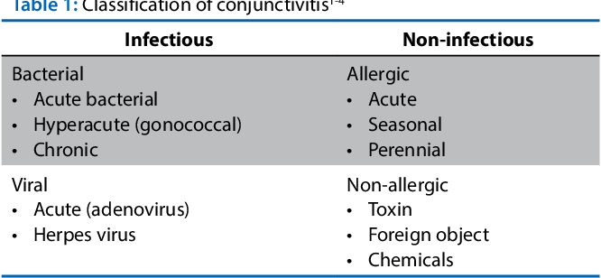
Conjunctivitis classsification
-ONSET
-DISCHARGE
-CONJUNCTIVAL RESPONSE
-AETIOLOGY;
Acute conjunctivitis(24 hrs-3weeks)
Staphylococcus aureus
Streptococcus pyogens
Hemophilus influenza
Hyper acute conjunctivits(12-24 hrs)
Neisseria gonorrhoea
Neisseria meningitidies
Chronic
Staphylococcus aureus
Moraxella lacunata
Enteric bacteria( Gram -ve bacilli)
Most common causative organism of viral conjunctivitis(iP;?)
Adenovirus
IP_8 days
Causes of viral conjunctivits
HE A Passionate MAN
Herpes simplex
Enterovirus
Adenovirus
Paramyxovirus
Myxovirus
Arborvirus
New Castlevrius
Forms of adenoviral conjunctivitis
-Epidemic keratoconjunctivitis(8,19)
-Non specific acute follicular conjunctivits(most common)(1-11,19)
-Pharngoconjunctivl fever(3,4,7)
-Chronic relapsing adenoviral conjunctivitis
Hyperendemic trachoma
Chlamydia trachomatis(A,B,Ba,C)
Pretrachoma
D to K
Acute inflmmatroy stage of trachoma signs
Conjunctival signs:
Congestion of upper tarsal +forniceal conjunctiva
Follicles
Pappillary hyperplasia
COrneal signs:
Herbert follicles: Follicles at limbal areas
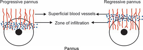
Progressive pannus
Superficial keratitis
Phase of cicatrization signs
Conjunctival sign:
a. Conjunctival scarring: irregular scar, star shaped scar, linear scar-at sulcus subtarsalis-Arlt’s line.
b. Concretions; hard masses pinpoint to 2mm due to dead epithelial cells and mucus from glands of henle
c.xerosis,psuedocyst,symblepharon
Corneal signs:
Herbert pits:pval circular scars from healing of herbert follice\les
Regressive pannus
Corneal opacity
aru: lid;madarosis, entropion, ankyloblepharon, ptosis, trichiasis
lacrimal apparatus;chronic dacrocystitis, chronic dacroadenitis
Progressives vs regressive pannus
Progressive -area of infiltration ahead of vascularization
Regressive; vessels extend a short distance beyond the area of infiltration
WHO grading of trachoma
@FISTO
follicles;>5 follicles each follicle>0.5mm in upper tarsal conjunctiva
inflammatory intense; inflammatory thickening of upper tarsal conjunctiva>half of normal depth
Scarring
Trichiasis; one eyelash rubs eyeball/recent removal of inturned eyelashes
Corneal opacity; Coreal scarring that ibscures at least part of pupillary margin
Inclusion bodies of trachoma is called as
Halberstaedter Prowazeke
Treatment of choice of trachoma
Azithromycin ODX 4 days(20mg/kg up to 1gm)
Opthalmia neonatorum
Bilateral inflammation of conjunctiva occuring in an infant of less than 30 days old.
Etiology of ON
CHemical: AgNO3,ab used for prophylaxis(Creeds methos)
Infectious; Gonococcus, Chlamydia(d to k), Staphylococcus, streptococcus hemo,pyo, herpes simplex , NLD obstruction
Management of bacterial conjunctivitis
Topical;Chloramphenicol(1%), Tobramycin, Framycetin, Gentamycin(0.3%) OD
No response; quniolone ab;Ciprofloxacin, ofloxacin, moxifloxacin,gatifloxacin
Antinflammatory and analgesics
Treatment of viral conjunctivitis
Supportive treatment: Cold compression and sunglasses
decongestans and lubricants (methly celluose, hydroxymethylcelluloe,polyvinyl alcohol)
topical antiviral: Adenine arabinosides, cidofovir
topical ab
topical antiinflam
cycloplegics
Topical vasoconstrictors(Nafazolin)
Classification if allergic conjunctivitis
@VP le GAS chodxa
Vernal keratoconjunctivitis/Spring catarrah/ Warm weather conjunctivitis
Phlyctenular conjunctivitis
Giant Papillary conjunctivitis
Atopic Conjunctivits
Simple allergics; Season, Perennial
Type of reaction vernal ?
Type 1 hypersensitivity reaction
Symptoms of VK
Intense burning/itching
stringy ropy discharge
lacrimation
heaviness of eyelid
mild photophobia
Conjunctival signs in VK
-Palpebral form;
Hard flat topped papillae arranged in cobblestone or pavement stone in upper tarsal conjunctiva. papillae enlarge form cauliflower like mass called as giant papillary
Limbal form:
Dusky red triangular congestion of bulbar conjunctiva at inter palpebral site
gelatinous thickened accumulation of tissue around limbus
Horner tranta’s spots:white raised dots around the limbal area contains eosinophils.
Mixed form;both palpebral and limbal
Corneal signs in VK
Punctate keratitis
Ulcerative VK- shield ulcer
Corneal Plaques
Pseudogerontoxon
subepithelial scarring
Shield ulcer
formed due to deposition of calcium phosphate and mucin in micro erosions.
Maxwell Lyon Sign
Pseudomembrane formation due to execess mucus deposition in papillae. Can lead to keratoconus
Treatmen of VK
Topical anti inflammatory: MAN CTS
Mast cell stabilizers only; Na cromoglycolate, Nedocromil Na
Mast cell stabilizers+antihistaminics: Azelastine, ketotifen, Olopatadine
NSAIDS:Diclofenac,Ketorolac
Cyclosporine
Tacrolimus
Steroids: Fluorometholone, Medrysone
Topical lubraicant:Methyk cellulose
Topical mucolytics;Acetylcystine
Signs in atopic keratoconjunctivitis
Dennie MOrgan fold’ skin folds under the eye due to excessive rubbing
Hertoghe’s sign; Loss of lat 3rd of eyebrows
Madarosis: Loss of eyebrows and eyelashes
Inferior tarsal and forniceal conjunctivitis
Phlyctenular conjunctivitis definition
characteristic nodular affection in the form of allergic response of the conjunctiva and the corneal epithelium to some endogenous allergens to which they have becomes sensitized.
ENdogenous allergens for PK
Proteins of Staph aureus
Moraxella axenfeld
M TB
Parasite
Phlyctenular conjunctivitis
Simple; small whitish nodule surrounded by hyperemia near limbus.
Necrotizing
Milliary
Phlyctenular keratitis
Ulcerative; sacrofulous; formed by breakdown of small limbal phlycten
fascicular; forms ring ulcer. paralllel leash of bv
miliary
Diffuse infiltrative: central infiltration of cornea
‘