epithelial and connective tissues (with pictures!!)
 Call Kai
 Learn
Learn Practice Test
 Spaced Repetition
 Match
Match Flashcards
 Knowt Play
1/38

There's no tags or description
Looks like no tags are added yet.
Name | Mastery | Learn | Test | Matching | Spaced | Call with Kai | Chat |
|---|
No analytics yet
Send a link to your students to track their progress
39 Terms
simple cuboidal epithelium
Function: secretion and absorption
Location: kidney tubules and ovary surface

simple columnar epithelium
Functions: absorption; secretion of mucus, enzymes, and
other substances; ciliated type propels mucus or reproductive cells by ciliary action
• Location = Non-ciliated
form lines digestive tract, gallbladder, ducts of some glands, ciliated form lines small bronchi, fallopian tubes, and uterus
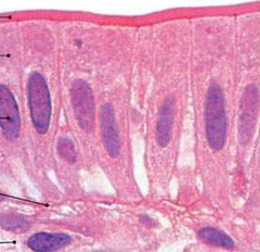
pseudostratified (ciliated) columnar epithelium
Function - secretion of mucus; propulsion of mucus by cilia • Locations = Non-
ciliated type is in ducts of male reproductive tubes and ducts of large glands; ciliated variety lines trachea and most of upper respiratory tract

stratified squamous epithelium
Specific types = Keratinized - contain the protective
protein keratin; surface cells are dead and full of keratin; Non-keratinized - forms moist lining of body openings
• Function - Protects underlying tissues in areas subject to
abrasion
• Location = Keratinized -
forms epidermis, Non-keratinized - forms lining of esophagus, mouth, and vagina

simple squamous epithelium
Specific types = Keratinized - contain the protective
protein keratin; surface cells are dead and full of keratin; Non-keratinized - forms moist lining of body openings
• Function - Protects underlying tissues in areas subject to
abrasion
• Location = Keratinized -
forms epidermis, Non-keratinized - forms lining of esophagus, mouth, and vagina

stratified columnar epithelium
Function - protection and secretion • Location = found in male urethra and large ducts of some
glands

stratified cuboidal epithelium
Function - protection
• Locations = forms largest ducts of sweat glands and
forms ducts of mammary glands and salivary gland

transitional epithelium
Function - stretches and permits distension of urinary
bladder causing thinning (from 6 to 3 layers when filled with urine)
• Location = Lines ureters, urinary bladder and part of
urethra

areolar connective tissue
stain clearly
paint brush strokes
------------------------------
Description = Gel-like matrix with all three fiber types, cells
include - fibroblasts, macrophages, mast cells, white blood cells
• Functions = Wraps and cushions organs,
holds and conveys tissue fluid, important role in inflammation
• Location = widely distributed under
epithelia, packages organs, surrounds capillaries

adipose tissue
doesn't uptake color as well as others
----------------------------------
•Functions = provides reserve food fuel, insulates
against heat loss, supports and protects organs
• Locations = under skin,
around kidneys, behind eyeballs, within abdomen and in breasts

reticular connective tissue
geometric RECT-angle style
-----------------------------------
• Function - form a soft, internal skeleton (stroma) - supports
other cell types
• Location - lymphoid organs (lymph nodes, bone marrow,
and spleen)

regular connective tissue
packed with collagen fibers and follows a parallel pattern
----------------------------------
• Functions = Attaches muscle to bone (tendons), attaches
bone to bone (ligaments); withstands great stress in one direction
• Locations = Tendons and ligaments, aponeuroses (sheet-
like tendons), fascia (membranes) around muscles, nerves, and large vessels

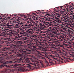
elastic connective tissue
takes up so much dye looks like purple squiggly lines
--------------------------------
Hyaline Cartilage
glassy or hazy , fibers do not stain clearly but cells do!
-------------------------------
• Functions = Supports and reinforces, resilient cushion,
resists repetitive stress
• Locations = Fetal skeleton, ends of long bones, costal
cartilage of ribs, cartilages of nose, trachea, and larynx

elastic cartilage
elastic stains very dark purple but reveals very big cells
------------------------------------
• Functions = Maintains shape of structure, allows great
flexibility
• Locations = Supports external ear, epiglottis

fibrocartilage
hideous fuchsia pink
--------------------------------
• Function = Tensile strength and ability to absorb
compressive shock
• Locations = Intervertebral discs, pubic symphysis, discs of
knee joint

osseous (bone) tissue
looks like a tree or an OY-ster
--------------------------------
• Functions = Supports and protects organs, provides levers
and attachment site for muscles, stores calcium and other minerals, stores fat, marrow is site for blood cell formation
• Location = Bones

blood cells
LARGE purple dots are white blood cells
Pink are blood cells
LITTLE purple ones are platelets

epithelial tissue def.
sheets of cells covering body surface or lining a body cavity
connective tissue
provide support
muscle tissue
includes skeletal, cardiac, smooth
---------------------------------------
Functions = Propels substances along internal passageways,
involuntary control
• Location = mostly walls of hollow organs (digestive,
urinary, uterus, and blood vessels
nerve
main component of the nervous system: provides control
-----------------------------------
• Functions = Transmit electrical signals from sensory
receptors to effectors
• Location = Brain, spinal cord, and nerves
Epithelial tissue FACTS
-cellularity: composed almost entirely out of cells
-polarity: apical(upper free) and basal (lower attached) surfaces
-avascular (no blood vessels) but innervated (supplied by nerves)
*supported by connective tissue
-highly regenerative
endothelium (simple squamous) does?
slick lining of hollow organs
mesothelium (simple squamous) does?
lines peritoneal, pleural and pericardial caviities
Unicellular Exocrine Glands (The
Goblet Cell)
Scattered throughout epithelial
lining of intestines and respiratory tubes, between columnar cells
protects and lubricates many internal body surfaces

Cell Junctions
factors holding epithelial cells together
Tight junctions (zona
occludens)
belt-like proteins in plasma membrane of adjacent cells are fused
close
off extracellular space

Desmosomes
anchoring junction; two disc-like plaques connected across intercellular space

Gap junctions = nexus
tunnel-like passageway between two adjacent cells

Basal Lamina
microvilli
extensions of plasma membrane to increase surface area

Basal lamina and reticular layer
form the basement membrane

skeletal muscle
skeletal muscles, bicep, tricep

cardiac muscle
walls of heart, involuntary, uninucleated, branches off

smooth muscle
walls of hollow organs, involuntary, long and skinny

apical
top
avascular
no blood exchange
inervated
has nervous system exchange