Clinical Manifestations of and Diagnostic Tests for Joint Diseases
 Call Kai
 Learn
Learn Practice Test
 Spaced Repetition
 Match
Match Flashcards
 Knowt Play
1/43

Earn XP
Description and Tags
Lecture 2
Name | Mastery | Learn | Test | Matching | Spaced | Call with Kai | Chat |
|---|
No analytics yet
Send a link to your students to track their progress
44 Terms
Animal with joint disease commonly present with a history of ____ or ____.
lameness
gait abnormality
Lameness involving one joint/limb is typically?
traumatic or developmental
Lameness involving multiple joints/limbs is typically?
degenerative or inflammatory
Non-inflammatory joint disease has what predominant cell type on cytology?
monocytes
Inflammatory joint disease has what predominant cell type on cytology?
neutrophils
List categories of non-inflammatory joint diseases.
developmental (UAP, FCP, OC/OCD)
degenerative (hip dysplasia)
neoplastic
traumatic processes
List categories of inflammatory joint diseases.
infectious (Borrelia burgdorferi, Staphylococcus, Ehrlichia spp)
non-infectious (immune mediated) —> erosive (rheumatoid arthritis), non-erosive (SLE)
Is this inflammatory or non-inflammatory joint disease:
very painful joints; multiple joints affected
shifting leg lameness
systemic disease —> fever, lethargy, inappetence, exercise intolerance
infammatory
Is this inflammatory or non-inflammatory joint disease:
low grade chronic discomfort
fluctuating/intermittent lameness
reluctance to exercise
no systemic signs of illness
multiple joints affected
“warms out” of lameness
non-inflammatory
What should you be assessing during physical exam for suspected joint disease?
goal is to localize region of pain or inflammation
assess animal’s posture and gait
manipulate and palpate the spine and the muscles, bones, and joints of each limb
give equal consideration to a systemic exam (auscultation and abdominal palpation)
List differentials for pain upon palpation of bones.
trauma
panosteitis
hypertrophic osteodystrophy
osteomyelitis
bone neoplasia
List differentials for pain upon palpation of muscles.
myositis
strain/sprain injuries
List differentials for pain upon palpation of the neck.
spinal cord or vertebral abnormalities such as IVDD, intracranial disease, meningitis, polyarthritis
List differentials for pain upon palpation of the spine.
spinal cord or vertebral abnormalities such as IVDD, discospondylitis, intervertebral facet joint inflammation
A decreased joint ROM, crepitation, and joint instability on physical exam suggests what?
articular damage and wear
osteophytes
peri-articular changes
tendon/ligament damage
What is the largest and easiest of joint to tap on a dog?
radiocarpal joint
Where is effusion most obvious on the carpus?
cranial and medial aspects of joint
Where is effusion most obvious on the elbow?
lateral aspect between lateral epicondyle and olecranon
How can you detect effusion in the stifle?
patellar ligament becomes indistinct and medial joint pouch protrudes
Where is effusion most obvious on the tarsus?
craniomedial, craniolateral, caudolateral, and caudomedial aspects, adjacent to the collateral ligaments
What is the minimum views we want when taking joint radiographs?
2 orthogonal views 90 degrees to each other
What imaging modalities can be used for joints?
radiography
CT
MRI
Ultrasound
Which imaging modality is used for initial diagnositc evaluation when only one joint is clinically affected, or joint palpation reveals crepitation, instability, or a restricted ROM?
radiography
What is the most common polyarthropathy in dogs?
immune mediated polyarthropathy (IMPA)
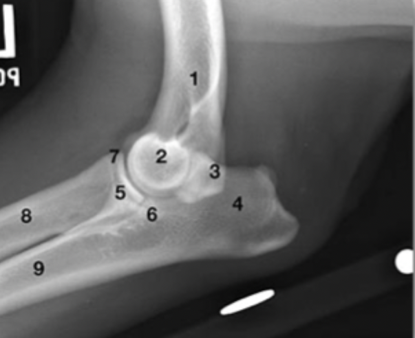
What is labeled by 1?
humeral diaphysis
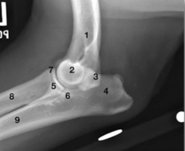
What is labeled by 2?
humeral condyle
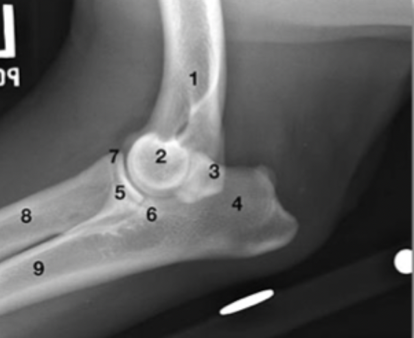
What is labeled by 3?
anconeal process with superimposed epicondyle of the humerus
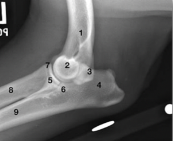
What is labeled by 4?
tuber olecranon
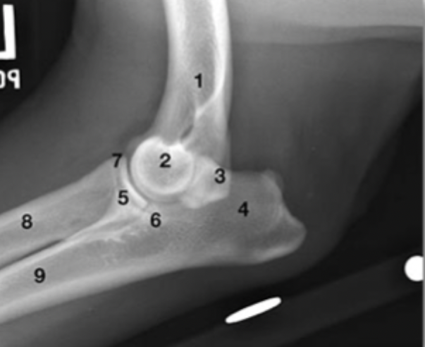
What is labeled by 5?
medial choronoid process of ulna with superimposed radial head
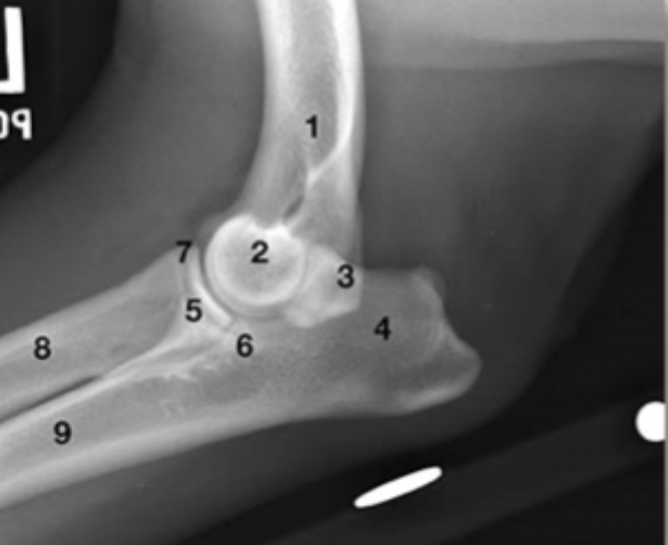
What is labeled by 6?
lateral choronoid process of the ulna
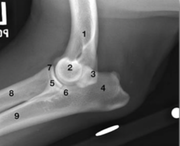
What is labeled by 7?
cranial aspect of the radial head
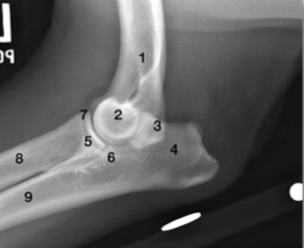
What is labeled by 8?
proximal diaphysis of radius
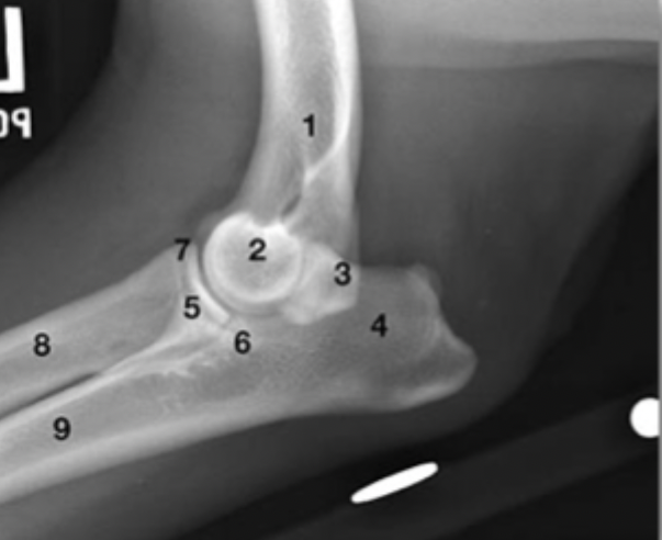
What is labeled by 9?
proximal diaphysis of ulna
By what age should the growth plate be closed for the anconeal process?
6 months
If polyarthritis is suspected, synovial fluid should be analyzed from how many joints?
three to four, including at least one carpus, one hock, and one stifle
Describe normal synovial fluid.
thick, viscous fluid secreted by synovium —> should form a strand of at least 1 inch when pulled apart
clear to slightly yellow in color
high content of HA and additional molecules such as lubricin
What is the primary function of synovial fluid?
lubricate articular surfaces and facilitate movement within the joint
What cell profile should be seen with normal synovial fluid on a 40x field for cytology?
no more than 3 WBCs per high dry power field
mixture of large and small mononuclear cells
<10% neutrophils
You are presented with a synovial fluid cytology with the following characteristics:
1000-6000 WBC/uL
0-12% PMN
What type of joint disorder do you suspect?
Degenerative
You are presented with a synovial fluid cytology with the following characteristics:
40,000-280,000 WBC/uL
90-99% PMN
What type of joint disorder do you suspect?
Septic
You are presented with a synovial fluid cytology with the following characteristics:
4000-370000 WBC/uL
15-95% PMN
What type of joint disorder do you suspect?
Nonerosive immune mediated
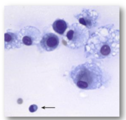
This is synovial fluid from a canine stifle.
What are these cells?
Is this cytology consistent with normal, inflammatory or noninflammatory joint disease?
Name a common joint disease that is consistent with this cytology?
Mononuclear/macophage-type cells
Non-inflammatory
Ruptured CCL
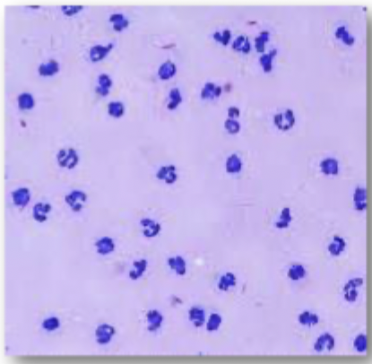
This is synovial fluid was taken from the carpus of a dog with a fever.
What are these cells?
Is this cytology consistent with normal, inflammatory or noninflammatory joint disease?
What are two differential diagnoses for this patient based on this cytology?
Neutrophils
Inflammatory
Tick borne / infectious disease OR immune mediated such as rheumatoid arthritis
What other tests should be done for joint cases in order to rule out systemic disease?
radiographs of abdomen and thorax
bloodwork —> tick borne disease panels, immune panels (ANA, LE, RF)
cultures of blood, urine, bone