3-MOM
 Call Kai
 Learn
Learn Practice Test
 Spaced Repetition
 Match
Match Flashcards
 Knowt Play
1/227

There's no tags or description
Looks like no tags are added yet.
Name | Mastery | Learn | Test | Matching | Spaced | Call with Kai | Chat |
|---|
No analytics yet
Send a link to your students to track their progress
228 Terms
- Attach to mandible and effect its movement
- Mastication
- Speech
- Resting posture of the mandible
What are the functions of the muscles of mastication?
- Depression
- Elevation
- Protrusion
- Retrusion
- Lateral deviation
Motions of the mandible:
Depression
What is the following movement:
Open the mouth
Elevation
What is the following movement:
Close the mouth
Protrusion
What is the following movement:
Move mandible anteriorly
Retrusion
What is the following movement:
Move mandible posteriorly
Lateral deviation
What is the following movement:
Move the mandible to the side
temporalis m.
masseter m.
which two muscles of mastication can be palpated externally?
lateral pterygoid m.
medial pterygoid m.
which two muscles of mastication can be palpated internally?
temporalis m.
masseter m.
lateral pterygoid m.
medial pterygoid m.
What are the four primary muscle of mastication?
CN V3 (mandibular division of the trigeminal nerve)
innervation for the muscles of mastication:
medial pterygoid n.
which nerve for muscles of mastication does not come of the anterior division of V3, but the trunk?
The inferior head of lateral pterygoid is larger than the superior head
The lateral pterygoid has 2 heads, which one is larger?

internal pterygoid
The medial pterygoid is also referred to as the...
external pterygoid
The lateral pterygoid is also referred to as the...
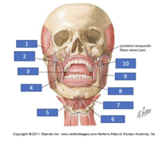
Temporalis m.
Identify the structure at #1
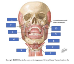
External pterygoid m.
Identify the structure at #2

Tongue
Identify the structure at #3

Buccinator m.
Identify the structure at #4

Mylohyoid m.
Identify the structure at #5
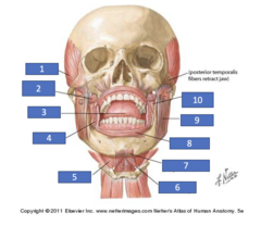
Digastric m. (anterior belly)
Identify the structure at #6
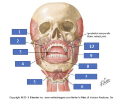
Geniohyoid m. (deep to other mm.)
Identify the structure at #7

Orbicularis oris m.
Identify the structure at #8
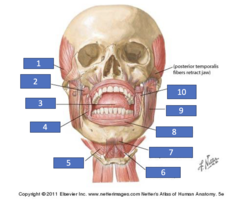
Masseter m.
Identify the structure at #9

Internal pterygoid m.
Identify the structure at #10
pterygoid part of maxillary artery (2nd part)
blood supply for the muscles of mastication come from the:
anterior belly of digastric m.
myohyoid m.
geniohyoid m.
buccinator
Tensor vili palatini
Accessory muscles of mastication?
CN VII
what nerve pierces through the parotid gland?
masseter m.
what muscles lies deep to the parotid gland?
buccinator m.
what muscle does the parotid duct pierce?
masseter m.
the transverse facial artery will supply the anterior surface of what muscle?
transverse facial a.
what artery runs superior to the parotid duct?
Masseter m.
The facial artery is anterior to what muscle at the inferior border of the mandible?
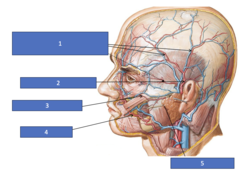
Frontal, middle, & parietal branches of superficial temporal a.
Identify the structure at #1
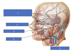
Superficial temporal a.
Identify the structure at #2

Transverse facial a.
Identify the structure at #3

Facial a.
Identify the structure at #4

Parotid gland and duct
Identify the structure at #5
facial a.
what artery runs through the antegonial notch to supply structures in the face?
superficial temporal a.
the transverse facial artery is a branch of the:
external carotid a.
the facial artery is a branch of the:
facial a.
what artery runs inferior to the masseter muscle?
Masseter is lateral to ramus of mandible
Where is the masseter located in relation to the ramus?
anterior to zygomaticotemporal suture
the superficial fibers of the masseter m. arise:
along entire zygomatic arch
the deep fibers of the masseter m. arise:
zygomatic process of maxilla
3rd fibers of the masseter m. would arise from:
lateral ramus of mandible
the masseter m. inserts onto the:
Superficial fibers insert near the inferior border mandible
Deep fibers insert more superiorly
Where do superficial fibers of the masseter m. insert? Where do deep fibers insert?
masseter m.
medial pterygoid m.
what two muscles form a mandibular sling for the mandible?
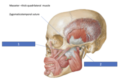
Superficial masseter
Identify the structure at #1

Deep masseter
Identify the structure at #2
masseteric a.
transverse facial aa.
facial a.
arterial supply to the masseter m. is done by:
mandibular notch
the masseteric nerve and artery pass through what structure to reach its destination?
•Elevates mandible
•Deep fibers retract mandible
•Slight ability to laterally deviate mandible
muscle actions of masseter muscle:
Lateral pterygoid m.
What is the main muscle for lateral deviation of the mandible?
- Coronoid process
- Ascending ramus below the plane of occlusion into the retromolar pad (can be traumatized with inferior alveolar nerve block)
what is the point of insertion for the temporalis m.?
Temporal fossa and deep surface of temporal fascia
what is the origin of the temporalis m.?
1. Temporalis superficial investing fascia
2. Superficial investing fascia extending from the masseteric fascia (deep)
Potential space between the 2 layers
What are the two layers of fascia superficial to the temporalis muscle? What do they create?
Important when considering the spread of infection along fascial planes
- Tendon of temporalis muscle and its 2 layers of fascia extends into the oral cavity
What is important to know about the potential space between the 2 layers of fascia of the temporalis muscle?
- Anterior fibers elevate
- Middle fibers elevate and retract
- Posterior fibers retract
muscle actions of temporalis muscle:
Posterior fibers of temporalis m. along with gravity
What is responsible for maintaining the resting posture of the mandible?
trismus
restricted motion of jaw or opening of the mouth:
temporalis m.
trismus affects what muscle of masticaiton?
anterior and posterior deep temporal nn.
innervation for temporalis m.:
deep to muscle: anterior and posterior deep temporal aa.
superficial to muscle: superficial temporal a. & middle temporal a.
blood supply for temporalis m.:
Coronoid process
Ascending ramus
Retromolar pad
Temporalis insertion points:
Superior temporal line
Identify the structure

Inferior temporal line
Identify the structure

Mandibular notch (clinically referred to as sigmoid notch)
Identify the structure

Coronoid process
Identify the structure
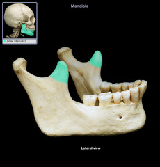
Condylar process
Identify the structure

Temporal fossa
Identify the structure
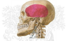
Infratemporal fossa
Identify the structure

Angle (gonial angle)
Identify the structure

Zygomatico-temporal suture
Identify the structure at the blue box

pterygoid part of maxillary artery
the anterior and posterior deep temporal aa. are branches of the:
Zygomatic process of maxilla
Identify the structure at #1

Zygomatic bone
Identify the structure at #2
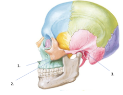
Zygomatic process of temporal bone
Identify the structure at #3

Pterygomaxillary fissure
Identify the structure at #1

Infratemporal surface of maxilla with Posterior Superior Alveolar Foramina
Identify the structure at #2

Pyramidal Process of Palatine bone
Identify the structure at #3
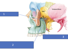
Greater wing of sphenoid
Identify the structure at GWS
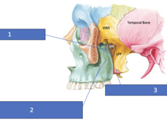
Infratemporal crest sphenoid
Identify the structure at ITC

Sphenopalatine foramen
Identify the structure at SPF

Lateral pterygoid plate
Identify the structure at LPP

Temporal fossa
Temporalis muscle occupies what fossa?
maxilla
zygomatic
temporal
what bones contribute to the zygomatic arch:
True - the superior and inferior heads of the lateral pterygoid
T/F: The lateral pterygoid has 2 heads immediately adjacent to one another
lateral pterygoid n. (V3)
innervation for lateral pterygoid m.
Lateral pterygoid a from the 2nd part of the maxillary artery (muscular or pterygoid portion)
Blood supply of lateral ptyergoid m:
pterygoid part of maxillary artery
the lateral pterygoid a. is a branch of the:
posterior superior alveolar foramina
identify the structure (at the arrows):

Smaller Superior head arises roof Infratemporal Fossa, inferior surface of Greater Wing Sphenoid & Infratemporal Crest
origin for superior head of lateral pterygoid m.:
pterygoid fovea of mandible only OR
Pterygoid fovea and the TMJ disc
insertion for superior head of lateral pterygoid m.:
lateral surface of lateral pterygoid plate
origin for inferior head of lateral pterygoid m.:
pterygoid fovea of mandible
insertion for inferior head of lateral pterygoid m.:
- contracts when large elevators of the mandible are contracting
- helps coordinate closing movement of the mandible
muscle action for superior head of lateral pterygoid m.
results in protrusion and depression of the mandible
muscle action: bilateral contraction of inferior head of lateral pterygoid m.
draws the condyle on the side which is contracting medially which results in a lateral deviation of the mandible to the opposite side
muscle action: unilateral contraction of inferior head of lateral pterygoid m.