TCP Week 7 (Eye Emergencies and Vision Loss)
 Call Kai
 Learn
Learn Practice Test
 Spaced Repetition
 Match
Match Flashcards
 Knowt Play
1/169

There's no tags or description
Looks like no tags are added yet.
Name | Mastery | Learn | Test | Matching | Spaced | Call with Kai | Chat |
|---|
No analytics yet
Send a link to your students to track their progress
170 Terms
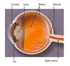
eye anatomy review
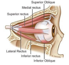
CN III, IV, and VI - innervates EOMs
Cornea - CN V1, very painful
Sclera - white
Lens
Retina - posterior eye. axons of ganglion cells become the optic nerve.
Choroid - VERY VASCULAR. highest blood flow in the body to supply the retina
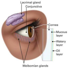
meibomian glands produce
lacrimal glands produce
lipid part of tears
watery part of tears
what disease affects the optic nerve
glaucoma
what disease involves extra ocular muscles
thyroid eye disease (TED)
CN II
optic nerve, formed from the retina
CN V1 innervates
innervates the cornea
CN III innervates
superior rectus
inferior rectus
medial rectus
inferior oblique
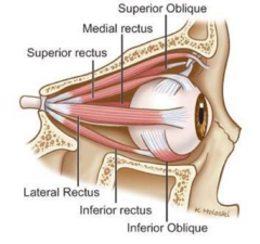
CN IV innervates
superior oblique
the ONLY CN that exits posteriorly
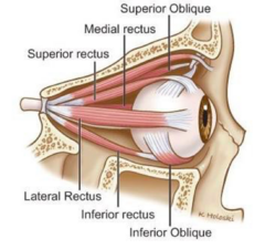
CN VI innervates
lateral rectus
clouding of the lens (cataracts) occurs due to?
- aging
- sun exposure
- diabetes
- steroids
The three tunics of the eye are?
1) A fibrous tunic, consisting of the sclera behind and the cornea in front;
2) A vascular pigmented tunic, comprising, from behind forward, the choroid, ciliary body, and iris
3) A nervous tunic, the retina.
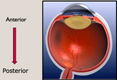
the visual pathway
cornea --> lens --> retina --> optic n. --> optic chiasm --> optic tract --> lateral geniculate nucleus --> thalamus --> occipital lobe
vision is processed in the occipital lobe
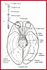
what sits below the optic chiasm
pituitary gland
pathology of this = bitemporal hemianopsia
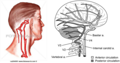
eye is supplied by
eye = carotid a. supplies
opthalmic lobe = vertebrobasilar a. supplies
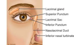
lacrimal drainage
Lacrimal gland --> punctum --> lacrimal sac
CN supply
CN 1 - Frontal lobe
CN 2 - Thalamus
CN 3-4 Midbrain
CN 5-7 Pons
CN 8-12 Medulla
• 8 gives eye info about head position
history taking for eye
DRFLOPS
Duration
Relief
Frequency
Location
Onset
Pain
Severity
eye complaints include
Ocular trauma - 27%
• 73% of trauma group had corneal abrasion
Conjunctivitis - 15% (allergy, viral, bacterial)
Retinal problems - 3.6% (usually trauma related)
Glaucoma - 2% (m/c AA, hispanic, latino)
episcleritis associated with
rheumatoid arthritis
THE RED EYE - differentiating feature
•Pain
•Itching
•Photophobia
•Systemic symptoms
•Discharge
•Visual loss
Corneal, pupil, and intraocular pressure changes
history questions and what ddx might they lead you towards?
Is vision affected? Acute change in vision is usually indicative of a visual pathway or globe problem
Foreign body sensation? Denotes active corneal process
Photophobia?
• Objective signs - difficulty opening eyes/trying to block out the light
• Patients with iritis have photophobia w/o foreign body sensation
Contact lens use? Red eye with discharge in a contact lens wearer would increase suspicion of keratitis
Trauma? Finger poke, object, blunt trauma
Discharge?
•Watery or purulent?
•Conjunctivitis - viral, bacterial, allergic
•Keratitis - opaque discharge with signs of photophobia and foreign body sensation
EXAMINATION OF THE EYE
acuity, visual fields, EOMs
Visual acuity: Smallest line read with one half of the letters correct
Confrontation visual fields: Visual field defects can represent pathology from occipital cortex to the optic nerve.
Extraocular movements: Can be impaired by restriction, interrupted or decreased innervation, or trauma.
EXAMINATION OF THE EYE
Pupillary reactions
Anisocoria can be acutely emergent or result of previous trauma
EXAMINATION OF THE EYE
lids and adenexa
•Periorbital skin, eyebrows, lashes, palpebral and bulbar conjunctiva, and lacrimal apparatus
•Trauma, infection, dysfunction, deformity, crepitus, or proptosis
•Palpate for subcutaneous emphysema (blow-out fractures) and step off deformities (trauma)
EXAMINATION OF THE EYE
Conjunctiva and Sclera
Subconjunctival edema, injection, inflammation, discharge, trauma, hypopyon, hyphema, foreign bodies.
EXAMINATION OF THE EYE
Cornea
Traumatic injuries, foreign bodies, hand tools.
EXAMINATION OF THE EYE
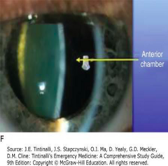
anterior chamber
•Hypopyon or hyphema
•Examine with a slit lamp
EXAMINATION OF THE EYE
lenses
Assess for opacities, lacerations, and subluxations
EXAMINATION OF THE EYE
IOP assessment
•Eye-care tonometer
•Tono-pen
EXAMINATION OF THE EYE
fundoscopic exam
May be difficult in acutely emergent conditions (iritis, keratitis, angle-closure glaucoma)
SLIT LAMP
- Provides a magnified view of the eye
- A thin slit can demonstrate the cornea, iris, and lens
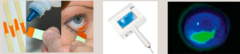
WOOD'S LAMP
•Handheld ultraviolet light used to detect corneal abrasions, corneal ulcers and foreign bodies.
•Light appears as light purple or violet and in the presence of fluorescein will fluoresce damaged tissue.
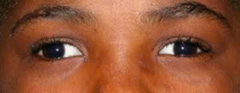
STRABISMUS - ocular misalignment, 2 types
Ocular misalignment
• Manifest strabismus - deviation present under binocular viewing
• Latent strabismus - deviation present after occlusion of one eye
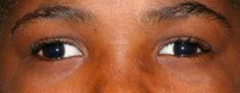
STRABISMUS - H&P and treatment
History & PE - visual acuity, cover-uncover test
Treatment: Patching or surgical correction
The Eye Emergency (CN III)
R CN III-downgaze
R CN III up-gaze
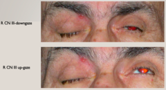
CN III palsy is a ____ until proven otherwise
aneurysm!! (likely at circle of willis - PCOM a.)
Cranial Nerve 3 palsy - Pupil involved ophthalmoplegia
lid will be down (ptosis)
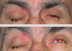
Cranial Nerve 3 palsy - Pupil involved ophthalmoplegia
Medical emergency if pupil is dilated !!!!
•Complete pupil involvement - Send to ER .... 20% die within 48 hrs
•Relative pupil involvement (poor light reflex, slightly larger size)
•Pupil spared
Presentation of CN 3 palsy
- Ptosis - Levator muscle is affected
- "Down and Out"
- Limited ocular motility
- Complete or relative sparing of the pupil, in general a good sign
- May be painful
Cranial Nerve 3 palsy - Pupil involved ophthalmoplegia
complications
•90% of aneurysmal third-nerve palsies have pupil involvement.
•1/2 dx w/ aneurysmal third-nerve palsies have a subarachnoid hemorrhage within 2 wks
•1/2 of those patients who hemorrhage will die soon after
**A lack of anisocoria in an isolated third-nerve palsy does not rule out aneurysm or a compressive lesion**
Cranial Nerve 3 palsy - Pupil involved ophthalmoplegia
imaging
Catheter Cerebral Angiography is the best imaging tool, however, it may cause stroke or myocardial infarction in 1% - 2% of patients.
EXTERNAL EYE: lid laceration
•Needs thorough evaluation
•Evaluate for globe rupture, FB, penetration of the orbit, damage to eye structures
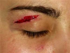
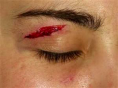
EXTERNAL EYE: lid laceration
treatment
- Superficial lacerations: suture with 6-0 nylon
- Significant laceration warrants ophthalmology consult
OPEN-GLOBE INJURY: ZONE OF INJURY
zone 1
opening of globe is limited to cornea or corneo-scleral limbus
OPEN-GLOBE INJURY: ZONE OF INJURY
zone 2
those that involve the anterior 5mm of the sclera (not more posterior than the pars plana)
OPEN-GLOBE INJURY: ZONE OF INJURY
zone 3
those that extend the full thickness into the sclera more than 5mm posterior to the limbus
THE RED EYE ddx
benign
Benign - stye, chalazion, hordeolum, blepharitis, subconjunctival hemorrhage, conjunctivitis (allergic, viral, bacterial),
dry eye syndrome, episcleritis
THE RED EYE ddx
urgent
Urgent - corneal abrasion, corneal foreign body, contact lens over wear (if not better in 24 - 48 hours).
Should be seen in 24 - 48 hours - iritis, viral keratitis, and scleritis.
THE RED EYE ddx
emergent
Emergent - Angle-closure glaucoma, hyphema, hypopyon, bacterial keratitis
EXTERNAL EYE COMPLAINTS
•Blepharitis
•Hordeolum
•Chalazion
•Entropion (lid in)
•Ectropion (lid out)
•Lid lacerations
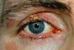
Blepharitis
define, presentation, tx
Inflammation of the eyelids
Erythema, crusty with scaling debris that clings to lashes.
Eyelid hygiene, warm compresses, baby shampoo
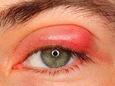
Hordeolum
define, presentation, tx
stye!
Staph infection of the glands
Erythema, tender nodule on lid
Warm compresses, topical antibiotics (bactericidal preference!)
Chalazion
define, presentation, tx
Chronic granulomatous inflammation
Painless nodule within the eyelid
Glucocorticoid injection,
compresses, surgery
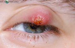
Periorbital/Preseptal cellulitis
treat with?
when the lid swells, treat to prevent orbital cellulitis
oral antibiotic
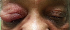
blepharitis treatment
•Lid scrubs
•Heat
•Tobradex - topical
Ectropion
management
Lid eversion
•Aggressive lubrication, artificial tears, gels, and ointments
•Surgical management if cornea becomes compromised from dryness and scarring.
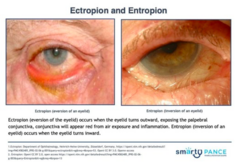
Entropion
management
Lid inversion
•Eyelashes can cause corneal and conjunctival damage
•Treated with artificial tears/lubrication or contact lenses to protect the surface of the eye
•Botulinum toxin is helpful with spastic entropion.
•Surgical management
Dacrocystitis
organisms
Streptococcus pneumoniae
Staphylococcus species
Haemophilus influenzae
Dacrocystitis
presentation
•Risk of orbital cellulitis if untreated
•Pain, redness, swelling just below medial canthus
Dacrocystitis
treatment
Empiric antibiotic until culture available, ophthalmology consult.
Periorbital/Preseptal cellulitis
organisms
staphylococcus aureus
group A streptococcus
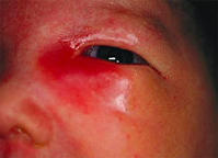
Periorbital/Preseptal cellulitis
what is it? tx?
structures anterior to orbital septum
treatment: Augmentin or 2nd or 3rd generation cephalosporin
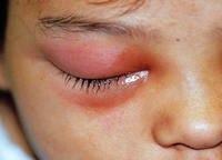
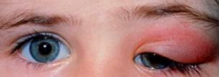
Periorbital/Preseptal cellulitis
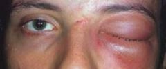
orbital cellulitis
orbital cellulitis
organisms
Streptococcus anginosus
Streptococcus pneumoniae
Haemophilus influenzae
Moraxella catarrhalis
S aureus
orbital cellulitis
presentation
Serious bacterial infection within the orbit
Fever, painful red eyelid, eye weakness, pain with eye movements, and proptosis
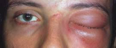
orbital cellulitis
diagnosis and treatment
Diagnosis - clinical diagnosis confirmed with CT of orbits and sinuses with contrast.
Admission - ophthalmology consult
- Vancomycin plus 3rd generation cephalosporin IV
CHANDLER'S CLASSIFICATION OF OCULAR AND ORBITAL INFLAMMATION
Stage 1: Inflammatory edema
Stage 2: Orbital cellulitis
Stage 3: Subperiosteal abscess
Stage 4: Orbital abscess
Stage 5: Cavernous sinus thrombosis
Life-threatening**
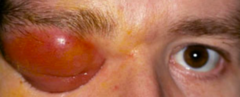
CONJUNCTIVITIS
•Allergic --> itchy
•Viral --> clear, runny
•Bacterial --> purulent
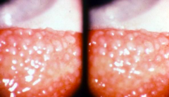
conjunctiva follicles
Sexually Transmitted Disease's on the "Ocular-Genital" Axis:
- Syphilis
- Chlamydia
- Pediculosis
- Gonorrhea (image)
- HIV
- Herpes (HSV)
- Hepatitis B
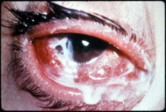
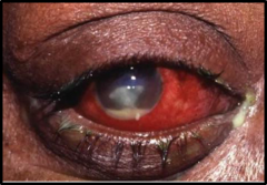
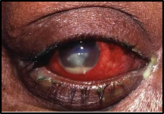
infectious keratitis - white circle/infiltrate is WBC (neutrophils, macrophages). can cause ulcer.
mcc = over wearing contact lens
CONTACT LENS COMPLICATIONS
Infectious keratitis
Painful red eye, may have reduced visual acuity, eyelid swelling and photophobia.
Corneal defects can be seen with fluorescein staining
Hypopyon
CONTACT LENS COMPLICATIONS
treatment
stop using contact!
•Urgent ophthalmology referral
•Fluoroquinolone (moxifloxacin)
•Do not patch eye!
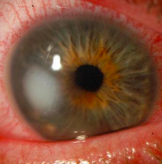
CORNEAL ULCER
Serious condition involving multiple layers of the cornea
Ulcer develops due to breaks in the epithelium
CORNEAL ULCER
etiology
Can be bacterial, viral, fungal, or parasitic.
•S. pneumoniae and S. aureus
•Contact lens users can develop Pseudomonas infection
•Herpes simplex 1 and Herpes zoster viruses common in US
CORNEAL ULCERS
s/sx
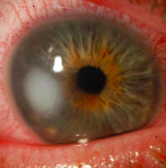
symptoms
•photophobia, pain, and foreign body sensation
signs
•eyelid swelling and redness, tearing, conjunctival injection, corneal haziness
•slit lamp examination reveals flare and cells from iritis and occasionally a hypopyon
hypopyon
pus in the anterior chamber of the eye
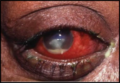
CORNEAL ULCERS
treatment
Treat aggressively with topical antibiotics and oral analgesia
• Fluoroquinolone - Ciprofloxacin or ofloxacin - one drop every hour
• Topical steroids are relatively C.I. but may decrease the incidence of scarring and perforation
Emergent ophthalmology consult!
CORNEAL ULCERS
complications
complications include corneal scarring, corneal perforation, glaucoma, and cataracts.
herpes zoster ophthalmicus
cause and sx
Herpes zoster eruption in any branch of trigeminal nerve (CN V)
Pain, red eye, blurred vision, photophobia, watery discharge.
Dendritic lesions
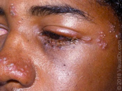
herpes zoster ophthalmicus
complications
keratitis, anterior uveitis, ocular nerve palsies, and acute retinal necrosis
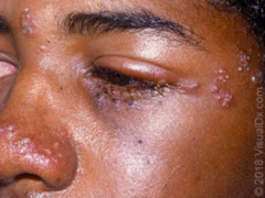
herpes zoster ophthalmicus
treatment
•Ophthalmology consult
•Antiviral agents
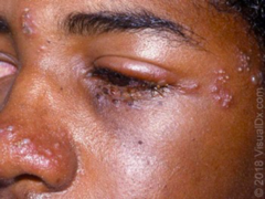
herpes zoster vs herpes simplex
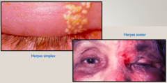
herpes simplex keratitis
•Vision threatening emergency
•Recurrent corneal infections
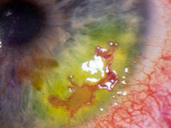
herpes simplex keratitis
s/sx, PE
Pain, visual blurring, watery discharge
Exam: chemosis, decreased corneal sensation, dendritic lesions of the cornea, corneal ulcers
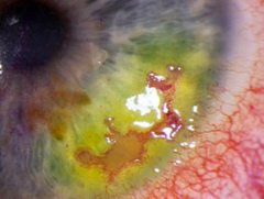
herpes simplex keratitis
treatment
Treatment: Ophthalmology referral
Topical or oral antivirals
SCLERITIS
associated with
Associated with systemic diseases: RA, vasculitis, infection, local trauma, sarcoidosis, IBD
SCLERITIS
s/sx
•The sclera is comprised of the anterior portion and a posterior portion.
•Pain, constant and boring, painful ocular movements, photophobia, globe tenderness to palpation
SCLERITIS
Diagnostics
CBC, CMP, UA, ESR, CRP may also need RF, ANA, ANCA
CXR for nodules and infiltrates (sarcoidosis and tuberculosis)
SCLERITIS
treatment
ophthalmology consult
SCLERITIS COMPLICATIONS
•Corneal changes
•Glaucoma
•Cataracts
•Posterior segment complications
SEVERE PAIN
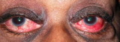
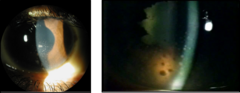
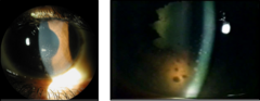
ANTERIOR UVEITIS (aka iritis)
Inflammation of the uvea (middle portion of the eye)
Caused by inflammatory and infectious processes
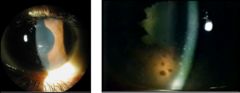
ANTERIOR UVEITIS
s/sx, PE findings
•Pain and redness
•Slit lamp exam shows cells and flare in the anterior chamber
ANTERIOR UVEITIS
treatment
•Glucocorticoids to reduce inflammation
•Ophthalmology consult
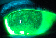
DRY EYE - keratoconjunctivitis sicca
presentation and causes
Burning foreign body sensation, conjunctival injection, and photophobia.
Decreased tear production.
• Medications
• Sarcoidosis
• Sjogren's syndrome
DRY EYE - keratoconjunctivitis sicca
treatment
frequent application of artificial tears or lubricant eye drops
SUBCONJUNCTIVAL HEMORRHAGE
Focal, red region on the ocular surface
May be caused by trauma, contact lens use, elevated venous pressure, hypertension, coagulopathy
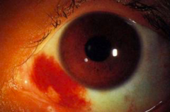
SUBCONJUNCTIVAL HEMORRHAGE
treatment
•Treat underlying issue.
•Self-limiting.
SUBCONJUNCTIVAL HEMORRHAGE
complications/reason
can be due to INR being off with warfarin use (coagulopathy)
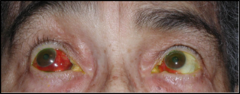
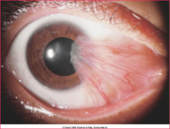
PTERYGIUM
m/c where? what is it?
symptoms?
More common in tropical regions, due to prolonged sun exposure
Fibrovascular triangular encroachment on the cornea
Can cause redness and irritation