Eye and Ear Anatomy Flashcards
1/65
Earn XP
Description and Tags
Flashcards about the anatomy of the eye and ear.
Name | Mastery | Learn | Test | Matching | Spaced |
|---|
No study sessions yet.
66 Terms
What are the general senses?
pressure, temperature, pain, and touch
Aka somatic
What are S types of sensory receptors?
• Chemoreceptors
• Pain receptors = nociceptors
• Thermoreceptors
• Mechanoreceptors
• Photoreceptors – rods and cones in eye
What are nociceptors?
Pain receptors
Describe how referred pain feels
pain that may seem to be coming from a different area of the body than the one actually being stimulated
Explain difference between acute and chronic pain fibers.
Acute pain:
Acute pain fibers are thin, myelinated nerve fibers that conduct nerve impulses rapidly (sharp pain, on skin)
Chronic pain:
Chronic pain fibers are thin, unmyelinated nerve fibers that conduct impulses more slowly (dull aching pain, sensed coming from deeper)
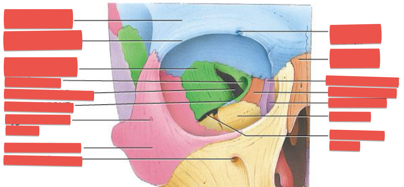
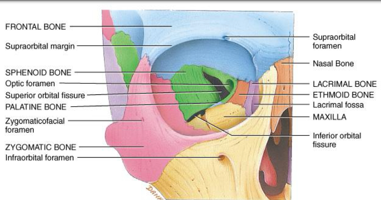
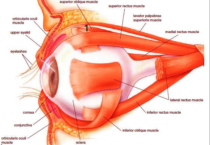
What are the bones of the bony orbit?
Medial wall: maxilla, lacrimal, ethmoid, palatine, sphenoid, Frontal bones.
Lateral wall: zygomatic, frontal.
Superior wall: frontal. Inferior wall: maxilla, zygomatic, palatine bones
eyelids (palpebrae):
Upper/Lower eyelid, Upper eyelid continual blinking keep the eye lubricated & moist.
Meibomian gland keeps the eyelid from sticking together. Remember it sounds like my balm
Eyelids are connected at the medial & lateral canthus. Eyebrow and Eyelashes

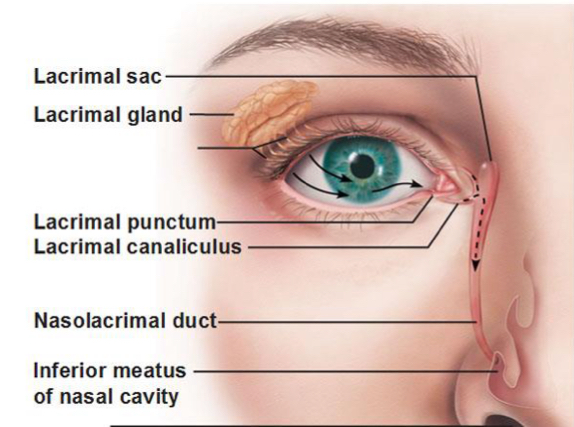
Lacrimal system:
Lacrimal gland: Produces tears to lubricate and cleanse the eye.
Lacrimal punctum: Small openings that drain tears from the eye surface.
Lacrimal canaliculus: Small channels that connect the lacrimal puncta to the lacrimal sac.
Lacrimal sac: Collects tears from the canaliculi.
Nasolacrimal duct: Carries tears from the lacrimal sac to the nasal cavity, draining inferior to the inferior concha/turbinate.
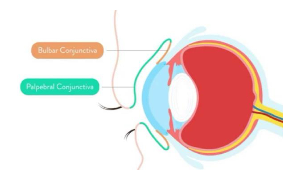
Conjunctiva:
The epithelium covering the inner surface of the eyelid, cover the anterior surface of the eye, and extend to the edge of the cornea. The conjunctiva keeps bacteria and foreign material from getting behind the eye

Describe 2 types of conjuctiva
The palpebral conjunctiva cover the inner surface of the eyelid.
The bulbar conjunctiva (ocular conjunctiva) cover the anterior surface of the eye, and extend to the edge of the cornea
Eyelashes:
Helps filter out foreign matter(including dust and debris). Prevents foreign matter from getting into the eye
What are 4 components of lacrimal apparatus?
1. The lacrimal gland (superolateral) with its ducts = secretes tears
2. The lacrimal canaliculi
3. The lacrimal sac
4. The nasolacrimal duct
Muscles of Eyelid:
Orbicularis oculi m. – closes eye tight innervated with CN VII (Facial nerve)
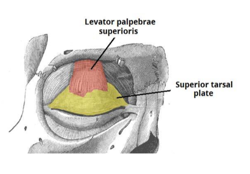
Levator palpebrae superioris m:
innervated by CN III (oculomotor nerve), elevates superior eyelid
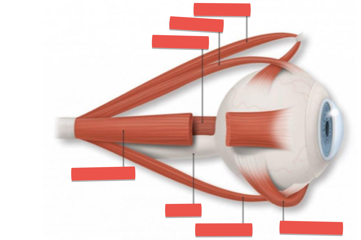
What muscles are involved in movement of the eye (rectus)
Superior rectus m. – innervated by Oculomotor n. (CN III), Inferior rectus m. – innervated by Oculomotor n. (CN III), Medial rectus m. – innervated by Oculomotor n. (CN III), Lateral rectus m. – innervated by Abducens (abducent) n. (CN VI)
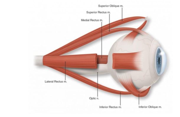
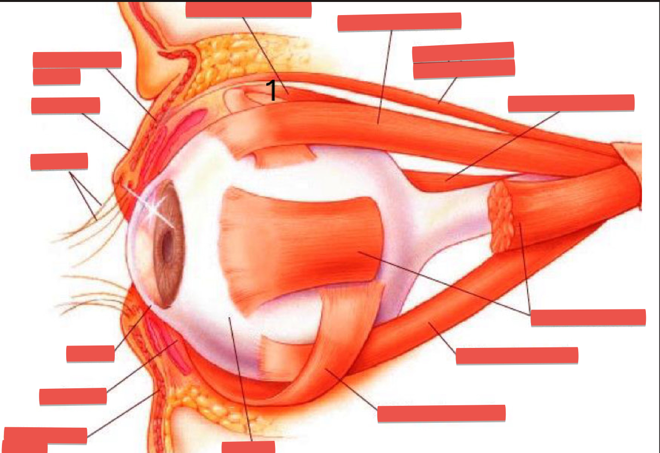
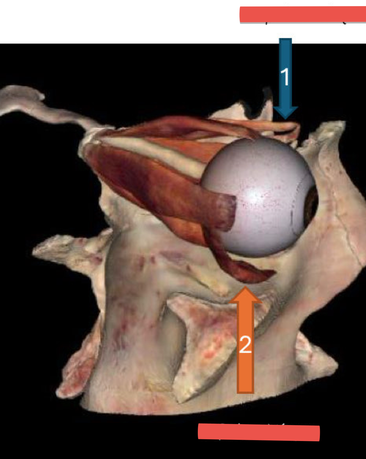
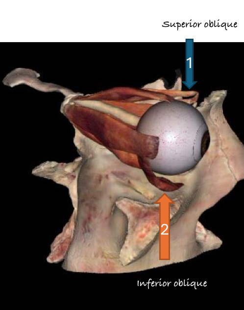
Oblique:
Superior oblique m. – innervated by Trochlear n. (CN IV), Inferior oblique m. – innervated by Oculomotor n. (CN III)
LR6:
Lateral Rectus muscle is innervated by the 6th cranial nerve (Abducens nerve)

SO4:
The Superior Oblique muscle is innervated by the 4th cranial nerve (Trochlear nerve)
O3:
All other extraocular muscles (Superior, Inferior and Medial Recti, and Inferior Oblique) are innervated by the 3rd cranial nerve (Oculomotor)
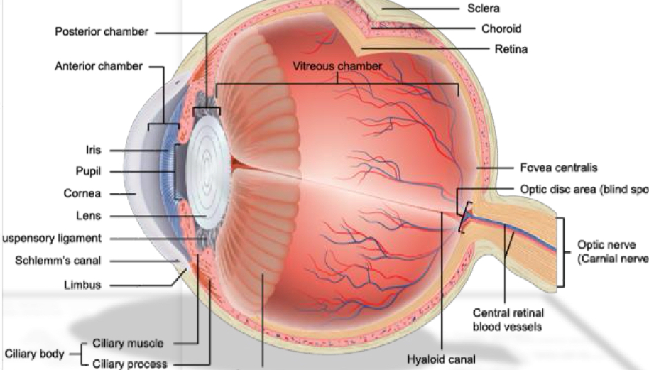
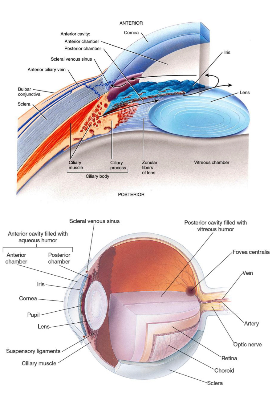
What are the structures in the Fibrous tunic (outer layer) of the eye?
Sclera (white, opaque): Provides protection and shape to the eyeball.
Cornea (clear, avascular): Refracts light entering the eye; transparent to allow light to pass through.
What are the structures in the Vascular tunic (middle layer) of the eye?
• Iris: Controls the amount of light entering the eye(pupillary size).
• Ciliary body: Produces aqueous humor and controls the shape of the lens for accommodation.
• Choroid: Provides blood supply to the retina and absorbs scattered light.
What are the structures in the Nervous tunic (inner layer) of the eye?
Retina: neural layer responsible for photoreception (rods and cones). Optic Nerve (CN II): Carries visual information from the retina to the brain.
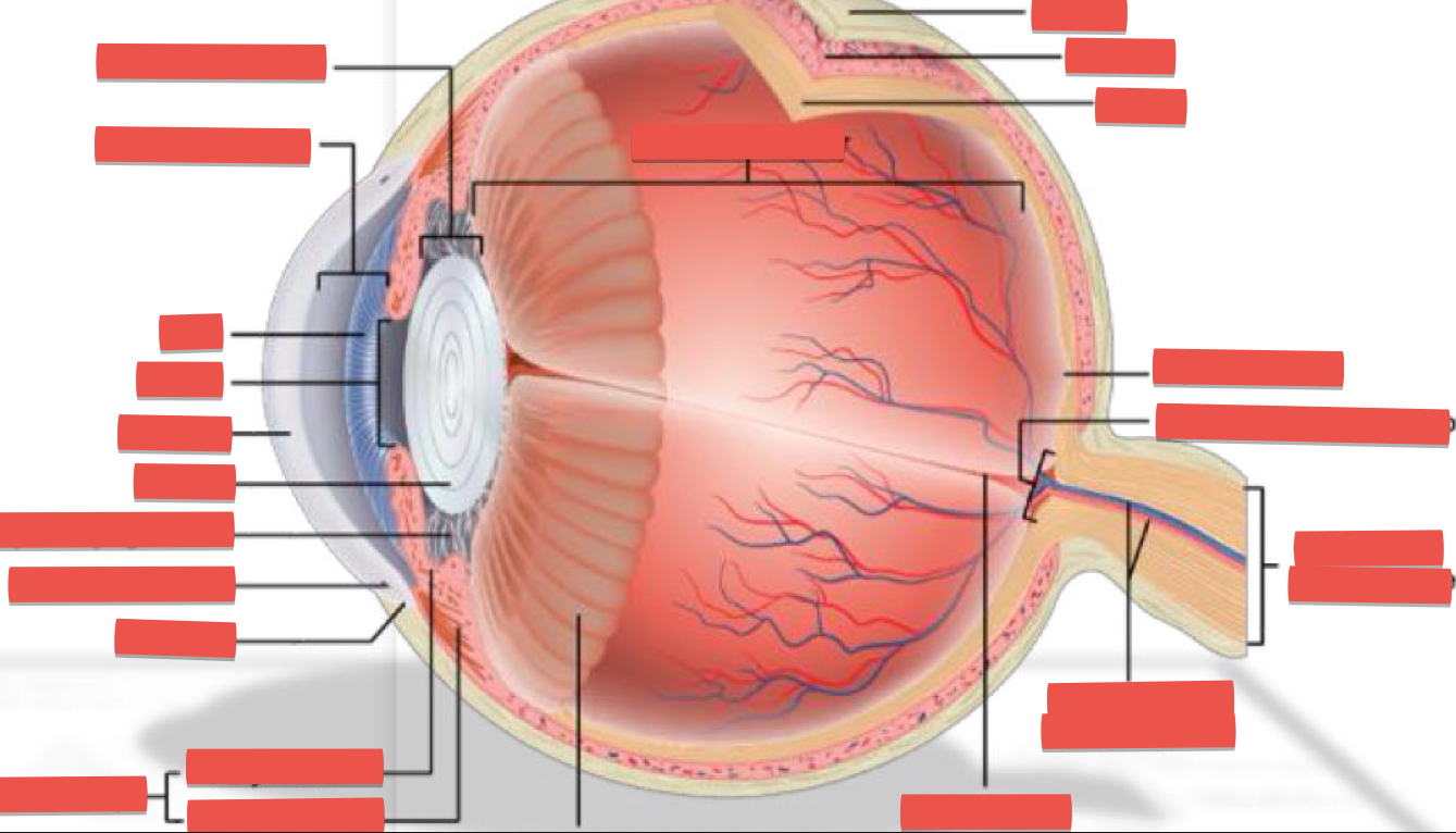
Canal of Schlemm:
at angle of iris and sclera/corneal junction, is route that aqueous fluid of anterior chamber escapes (Blockage of Canal of Schlemm results in glaucoma)
True or false: sclera does cover cornea
False - Sclera doesn’t cover cornea
What is the function of cornea?
Aids the lens with focusing
Uveal tract(uvea):
the choroid, ciliary body, and iris
What is the function of the Iris?
Lies between the cornea and lens
The iris divides the anterior cavity into an anterior and posterior chamber
Where is the aqueous humor secreted?
Circulates from the posterior chamber by ciliary body through the pupil into the anterior chamber
What divides the eye into posterior and anterior cavity?
Ciliary body and the lens
What does anterior and posterior cavity contain?
• The Anterior Cavity : subdivided into anterior chamber and posterior chambers (by the iris) that's filled with fluid Aqueous humor
• The Posterior Cavity is filled with Vitreous humor
What are functions of sphincter pupillae muscle?
Contraction causes constriction of the pupil (miosis).
Controlled by the parasympathetic nervous system and innervated by CN III (Oculomotor nerve).
Causes pupil to constrict in response to bright light or focusing on near objects (aka myosis).
What are functions of dilator pupillae muscle?
• Contraction causes pupil dilation.
• Controlled by the sympathetic nervous system.
• Causes pupil to dilate(mydriasis) in response to dim light or emotional arousal.
• It is innervated by sympathetic fibers, not CN III like the sphincter pupillae.
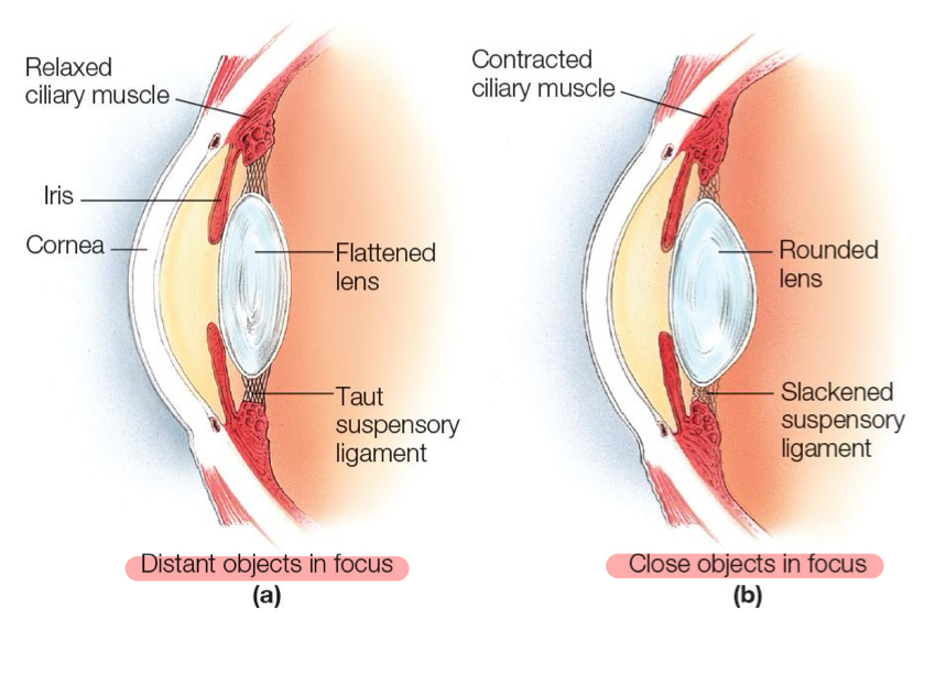
Which part of eye assists with focusing on near objects?
The lens becomes more rounded (increases in curvature) to focus on near objects. This is due to the contraction of the ciliary muscles, which allows the lens to adjust its shape for optimal focus at close range.
What is the fundus and its structures?
The interior surface of the eye visible during an exam. Key structures: retina (captures visual information), optic disc (optic nerve exit point), macula (detailed vision), fovea (highest visual acuity), and retinal blood vessels.
Describe what retina contains and purpose.
• Contains the visual receptor cells = photoreceptors
• A central depression (the fovea centralis) in the portion of the retina that produces the sharpest vision
What is the optic disc?
the optic disc is the point where the optic nerve exits the eye
What ave 2 types of cells retina is made of?
Rods and cones
Rods:
•Function: Specialized for scotopic vision (vision in low light).
•Color Sensitivity: Do not detect color; they provide black and white (monochromatic) vision.
•Light Sensitivity: Extremely sensitive to light can respond to a single photon.
•Location in Retina: Most densely packed in the peripheral regions of the retina.
•Quantity: Humans have about 120 million rods.
•Role: Crucial for night vision, motion detection, and peripheral vision
Cones:
•Function: Specialized for photopic vision (vision in bright light).
•Color Sensitivity: Responsible for color vision. There are three types:
•S-cones: Sensitive to short wavelengths (blue light)
•M-cones: Sensitive to medium wavelengths (green
light)
•L-cones: Sensitive to long wavelengths (red light)
•Light Sensitivity: Less sensitive to dim light than rods.
•Location in Retina: Concentrated in the fovea centralis, the central part of the retina.
•Quantity: Humans have about 6 million cones.
•Role: Provide sharp, detailed central vision and color
discrimination.
What does macula contain?
The macula located at the posterior, lateral pole of eye is very rich in cone cells
What are ganglion cells?
Their axons form the optic nerve, transmitting signals to the brain
What are 2 primary layers of optic cup?
Inner layer: Develops into the neural retina, which contains photoreceptors, bipolar cells, and ganglion cells responsible for visual processing.
Outer layer: Becomes the retinal pigment epithelium (RPE), which supports photoreceptor function and absorbs excess light.
Define cup to disc ratio (cdr)
ratio of the diameter of the optic cup to the diameter of the optic disc.
•A normal CDR is typically 0.3 or less.
•A higher CDR (e.g., >0.6) may indicate optic nerve damage, often associated with glaucoma.
Explain the light pathway in more detail
- Light Enters the Eye: Light passes through the cornea, which begins to focus the light.
- Anterior Chamber: Light then travels through the anterior chamber, which is filled with aqueous humor.
- Pupil: Light passes through the pupil (the opening in the iris), which controls the amount of light entering the eye.
- Lens: Light passes through the lens, which further focuses light onto the retina. The lens adjusts its shape to focus on objects at varying distances.
- Vitreous Humor: Light travels through the vitreous humor, a clear, gel-like substance that fills the posterior cavity of the eye.
- Retina: Light reaches the retina, where photoreceptor cells (rods and cones) convert light energy into electrical signals.
- Optic Nerve: The electrical signals travel along the optic nerve.
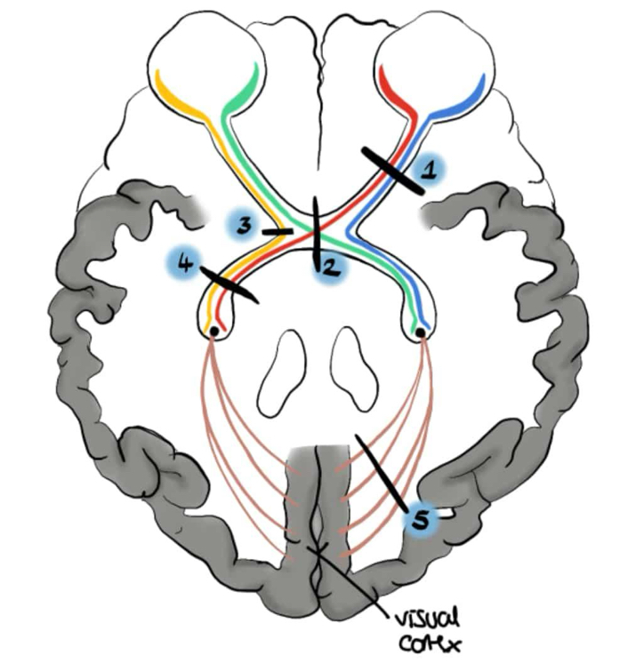
- Optic Chiasm: The optic nerves from each eye meet at the optic chiasm, where some nerve fibers cross over to the opposite side of the brain.
- Optic Tract: The nerve fibers continue along the optic tract to the lateral geniculate nucleus (LGN) in the thalamus.
- Visual Cortex: Finally, the signals reach the visual cortex in the occipital lobe, where the brain processes the visual information to create an image.
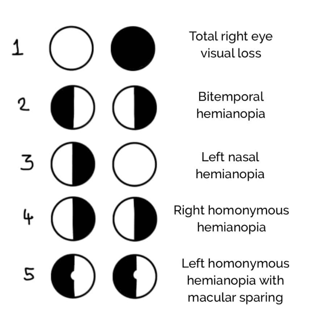
When does bitemporal hemianopsia?
If you cut the optic chasm
What is function of external ear?
Collect sounds
Describe helix and concha in more detail
• Helix: outer rim of the ear, collects and directs sound waves into the ear canal.
• Concha: deep cavity of the auricle that further directs sound into the ear canal and provides protection.
What does the middle ear (tympanic cavity) contain?
Contains auditory ossicles: malleus, incus, stapes- these bones bridge the TM and inner ear to transmit vibrations.
Describe difference between pars tensa and flaccida
• Pars Tensa:
Location: The taut, lower portion of the tympanic membrane.
Function: Responsible for sound transmission due to its tension and larger surface area.
Structure: Contains three layers, including the fibrous layer that provides its tautness; attached to the malleus.
• Pars Flaccida:
Location: The small, slack, upper portion of the tympanic membrane above the malleolar folds.
Function: Has minimal role in sound transmission due to its slackness.
I
Left Homonymous Hemianopia with Macular Sparing:
Loss of the left half of the visual field in both eyes, but central vision(macula) is preserved; often due to damage to the occipital lobe
Tympanic membrane:
Separates the external ear from the middle ear
Transmits sound from air to the ossicles.
Also known as the eardrum
Eustachian tube (auditory tube, pharyngotympanic tube):
Connects the middle ear to the nasopharynx - Equalizes pressure in the middle ear - Allows air to move the TM during valsalva
What are the middle ear muscles and their function?
• Tensor tympani m. (innervated by mandibular branch of CN V): Dampens loud sounds, protecting the inner ear.
• Stapedius m. (innervated by CN VII): Stabilizes stapes, reducing excessive movement and preventing overstimulation of the inner ear. Protects the inner ear from loud noises, especially sudden, high-intensity sounds.
Structures of cochlea
• Oval window: Acoustic energy enters through here, playing a key role in sound transmission.
• Round window: Vibratory acoustic energy exits into the middle ear cavity through here.
What is the inner ear?
The inner ear is a complex part of the ear that contains:
3 semicircular canals: structures for dynamic equilibrium
Cochlea: structure responsible for hearing
Semicircular Canals
Three fluid-filled loops in the inner ear that are oriented in different planes to detect rotational movements of the head. Each canal is responsible for detecting movement in a specific plane:
- Anterior (Superior) Canal: Detects head movements in the sagittal plane, such as nodding up and down.
- Posterior Canal: Detects head movements in the coronal plane, such as tilting the head left and right.
- Lateral (Horizontal) Canal: Detects head movements in the transverse plane, such as shaking the head side to side.
These canals are essential for maintaining dynamic equilibrium, which is the sense of balance during motion.
Describe the path of sound through the cochlear duct in even more detail:
• Scala vestibuli:
– The upper chamber of the cochlea that receives sound vibrations from the oval window and transmits them toward the apex. Perilymph-filled.
• Scala media (Cochlear Duct):
– The middle chamber containing endolymph and the organ of Corti, where sound vibrations are converted into nerve impulses. The organ of Corti houses hair cells which rest on the basilar membrane.
• Scala tympani:
– The lower chamber that carries vibrations from the apex back toward the round window, helping dissipate sound energy; Perilymph-filled.
Explain the flow of sound through the cochlear duct in detail
Sound waves enter the cochlea through the oval window and travel through the scala vestibuli.
They then pass through the helicotrema at the apex of the cochlea and enter the scala tympani, eventually reaching the round window.
The basilar membrane vibrates in response to sound, stimulating the organ of Corti, which converts mechanical signals into nerve impulses
What is organ of corti?
Lined with hair cells called stereocilia. Converts vibration into nerve impulses.
Describe the process of hearing in detail.
- Sound waves enter the ear canal.
- Eardrum vibrates.
- Auditory ossicles amplify vibrations.
- Stapes transmits vibrations to the oval window.
- Different frequencies stimulate different receptor cells in the endolymph.
- Neurotransmitter release stimulates sensory neurons (organ of Corti).
- Sensory impulses travel via cochlear nerve (CNVIII) to the auditory cortex.
- Auditory cortex interprets sensory impulses.
Name 3 semicircular canals
• Superior semicircular canal
• Posterior semicircular canal
• Horizontal semicircular canal
Describe difference between static and dynamic equilibrium.
Static equilibrium is the position of the head is sensed while the head and body are still, maintaining stability.
Dynamic equilibrium is when the head and body move or rotate, the motion is detected, aiding in balance.
Where is the location of the ampulla and its structures?
The organs of dynamic equilibrium are the three semi circular canals in the labyrinth. Inside these canals Is the ampulla, which houses the sensory organs of the semi circular canals.
What are the functions of ampulla?
Responsible for balance.
What is cranial nerve of the car region?
Cranial nerve VIII - vestibulocochlear nerve