Lab Exam #1(Microbiology
1/59
There's no tags or description
Looks like no tags are added yet.
Name | Mastery | Learn | Test | Matching | Spaced | Call with Kai | Chat |
|---|
No analytics yet
Send a link to your students to track their progress
60 Terms
scientific method
1.Observation 2.Hypothesis 3. Experimentation 4. Data collection 5.Analysis and conclusion
Coarse adjustment knob
adjust the height of objective lens
Fine focus adjustment knob
(the distance between the objective lens and the specimen
Substage condenser
brightness and control contrast
Resolution
is wavelength of light and design of condenser
How can resolution be maximized?
Condenser being kept at high can maximize the resolution
parafocal
Specimens to remain in focus when objectives are switched
Cyanobacteria (Anabaena)
o oxidize hydrogen sulfide to sulfur, found in hot springs and stagnant water
Amoeba
shapeless,amorphous form
Paramecium
cilia for locomotion and water-regulating vacuoles
Euglena
unicellular, eukaryotic
Characteristics of fungi
Cell walls, multicellular molds (eukaryotic-true nucleus , heterotrophic, saprophytic(feeding on dead decaying matter, grows fuzzy on plates)
3 general shapes of bacteria
Coccus
Bacillus
Spirillum
steps for preparing a successful smear
1. The bacteria must be evenly and thinly dispersed
2. Let air dry completely
3. The bacteria needs to be attached to the slide
heat fix bacteria
Bacteria becomes heat fixed when organisms are killed and can attach them to the slides being used
why we stain bacteria
easier to view when it is stained
the difference between a positive & negative stain
Positive stain adhere to the cell surface and will be colored with the stain
Negative stain determines cell size and arrangement
Simple stain
stain use only one stain and basic/acidic staining
Differential stain
uses more than one stain and examples of this acid-fast staining, endospore stain
Gram positive cell walls
have a thick peptidoglycan cell wall
Gram negative cell walls
have a thin peptidoglycan cell wall
Steps of gram stain-
1. After performing a smear prep on the slide, saturate the smear with crystal violet for 20 seconds.
2. Rinse with water completely.
3. Saturate the smear with iodine for 1 minute.
4. Rinse with water completely.
5. Decolorize with Gram decolorizer (75% ethanol + 25% acetone) for 10 seconds; if you leave the decolorizer on too long, it will remove the crystal violet out of a gram positive cell as well!!
6. Rinse with water completely.
7. Counterstain with safranin for 1 minute.
8. Rinse with water completely.
9. Carefully blot the slide dry with bibulous paper.
10. Observe the slide using a light microscope, draw under oil immersion.
Gram negative bacteria
be stained the reddish-pink color of safranin
Gram positive bacteria
be stained the purplish color of crystal violet
Staphylococcus epidermidis
spherical cocci,gram positive
Escherichia coli
rod-shaped-bacillus Gram negative
Bacillus subtilis
(rod shaped-bacillus,gram positive
Mycobacterium smegmatis
bacillus,gram positive
What is the purpose of steam in acid -fast stain?
Loosens mycolic acid and secures carbol fuchsin
name & function of the acid found in cell wall of acid-fast bacteria
Mycolic acid and it resists the uptake of simple dyes such as the ones in a gram stain.
Acid-fast organism at end of stain
resist de-colorization by acid alcohol
Not acid-fast organism
decolorized by acid alcohol and are counterstained with methylene blue
Mycobacterium smegmatis
bacillus(shape),is acid fast bacteria
Staphylococcus epidermidis
o spherical cocci(shape), NOT a acid fast bacteria
Two pathogenic acid fast bacteria
1. Mycobacterium tuberculosis
2. Mycobacterium leprae
Steps and reagments in Endospore stain
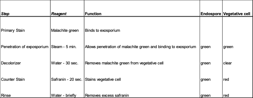
The purpose of the endospore in some bacteria?
Endospore allow bacteria to survive without nutrients for very long periods of time and can resist harsh environmental conditions that are exposure to heat and radiation
· Under what conditions do endospores form?
Endospores form when a bacteria encounter harsh environmental conditions or nutrient depletion.
Endospore
allow bacteria to survive nutrients for long period of time
Vegetative stage
the organism can return to the metabolically active stage
What is the shape of Bacillus subtilis and does it form endospore?
(rod-shaped) bacterium and doesn’t form endospores
· Give an example of at least two pathogenic endospore forming bacteria
Clostridium tetani and Clostridium botulinum
Aspetic technique
minimizes the possibility of introducing contamination.; Prevents contamination of microbial cultures
Disinfection
meaning that the number of living microbes particularly those potentially hazardous to humans has been greatly reduced by application of a chemical or chemical mixture
Sterilization
:- (completely free of living microbes
Pure culture
microbes are genetically identical to one another and free of other species or strains
Mixed culture
comprised of two/more distinct species or strains in a shared environment
· the procedure for performing a broth or slant transfer
Aseptic technique is used for broth or slant transfer
The streak method and why we do this?
Streak method dilutes a population bacteria to an agar surface that separates individual cells. Helps create pure cultures
What do we invert media (plates) for incubation?
· This helps prevent condensation droplets from falling onto the agar surface.
Selective
o : allow growth of only certain types of organism
Example: Phenyl ethyl alcohol agar (PEA): selective for gram +, inhibits gram –
Differential
o which allow growth of closely-related organisms that will appear visibly different on the media due to their growth patterns
Example: Blood agar and TSI
Enriched:
o Contains additional nutrients to enhance growth Example: blood agar
Phenyl ethyl alcohol plate (PEA)
o selective for gram + and inhibits gram;-alcohol dissolves outer membrane of gram negative;selective media
Blood agar
contains 5% sheep blood;cultivation of fastidious bacteria such as Streptococcus, by observation of hemolysis;differential media
Triple sugar Iron Agar (TSI)
o fermentation and production of gas;- fermentation using indicator phenol red; differential media; differentiates gram-enteric
Mac Conkey agar (MAC)
o contain crystal violet; contain lactose and neutral red selective and differential; differentiates for lactose fermenters
Mannitol salt agar (MSA
o contain 8-10% NaCl and contain mannitol and phenol red; selective and differential; differentiates for mannitol fermenters
Eosin Methylene Blue agar (EMB)
selective/differential 2 dyes are toxic(methylene blue and eosin); lactose fermentation produces acids; selects for gram – bacteria, differentiates lactose fermentation.