ADL- Quiz 1: Microscopes and Porifera
1/72
There's no tags or description
Looks like no tags are added yet.
Name | Mastery | Learn | Test | Matching | Spaced | Call with Kai |
|---|
No analytics yet
Send a link to your students to track their progress
73 Terms
Under which objective lens is the field of view the largest?
A) Lowest magnification
B) Medium magnification
C) Highest magnification
A) Lowest magnification
Under which objective lens is the field of view smallest?
A) Lowest magnification
B) Medium magnification
C) Highest magnification
C) Highest magnification
Which objective lens gives you the largest working distance?
A) Low
B) Medium
C) High
A) Low
Which objective lens has the greatest depth of field?
A) Low power
B) Medium power
C) High power
A) Low power
As a rule of thumb, as magnification increases, depth of field ___
A) increases
B) decreases
B) decreases
Which level of magnification requires the most illumination for the best clarity and contrast?
A) Low magnification
B) Medium magnification
C) High magnification
C) High magnification
If you have a specimen that is transparent or thin, would you need to open (allow more light in) or close ( allow less light in) the iris diaphragm?
Close (allow less light in)
When compared to most compound microscopes, dissecting microscopes have a ___ (smaller/larger) working distance, ___ (smaller/larger) depth of field, ___ (smaller/larger) field of view, ___ (lower/higher) magnification, and a ___ (lower/higher) resolution.
1. larger working distance
2. larger depth of field
3. larger field of view
4. lower magnification
5. lower resolution
What is the magnification of the ocular lens?
A) 1x
B) 5x
C) 10x
D) 20x
C) 10x
T/F: To focus on a specimen, start with any objective.
False
T/F: You can touch the slide with the lens, no problem
False
Describe the change in the following as the magnification DECREASES.
A) field of view ___ (increases/decreases)
B) working distance ___ (increases/ decreases)
C) depth of field ___ (increases/ decreases)
D) light intensity requirement ___ (increases/ decreases)
A) increases
B) increases
C) increases
D) decreases
When looking through a compound microscope you should look through the ___.
ocular lens
Which knob can you use/turn to improve the focus when changing objective lenses?
Fine-focus adjustment knob
In a compound microscope, light is captured by ___.
objective lenses
Compound microscopes use ___ (one or two) sets of lenses to obtain higher magnification.
Two
T/F: Compound microscopes are used to view very small samples that cannot be identified with the naked eye.
True
Lenses nearest the eye through which you look
ocular lenses
Lenses of different magnification that work in conjunction with the ocular lenses to magnify the image; located just above the stage
objective lenses
Housing that keeps ocular and objective lenses in proper alignment
body
Revolving housing that supports objective lenses
nosepiece
Supports microscopic body, stage, and adjustment knobs
arm
Move stage up or down to focus image
coarse-focus adjustment knob
Permits precise focusing
fine-focus adjustment knob
Supports slidees
stage
Hold slide in steady; stationary position
stage clips
Move stage to center slide under objective lens
stage adjustment knobs
Lens mounted beneath stage that focuses the light beam on the specimen
condenser
Mounted beneath the stage near the condenser; regulates the amount of light illuminating specimen
iris diaphragm
Moves condenser lens up or down to focus light
condenser adjustment knob
Source of light
illuminator
Supports microscopic unit
base
Rheostat (dimmer switch) that permits further adjustment of light intensity
light intensity adjustment dial
Turns microscope light on or off
power switch
At what level is the basic body plan organized for Porifera?
A) Tissue level
B) Organ level
C) Cellular level
C) Cellular level
List the characteristics of embryonic development for Porifera:
A) Diploblast
B) Triploblast
C) Protostome
D) Deuterostome
E) No embryonic development
E) No embryonic development
What type of symmetry does Porifera have?
A) asymmetry
B) bilateral symmetry
C) radial symmetry
A) asymmetry
List the structures/ways that members of Porifera support themselves.
A) hydrostatic skeleton
B) endoskeleton of mesodermal origin
C) spicules and spongin
C) spicules and spongin
What structures are used to help members of Porifera move around?
A) Motile larvae
B) Water vascular system
C) No structures are used for movement; animals are sessile
D) Hydrostatic skeleton
E) Striated muscles
Motile larvae and no structures are used; animals are sessile.
Describe the nervous system and sensory structures found in Porifera:
A) Nerve network
B) Ocelli
C) Cerebral ganglia
D) Ventral nerve cord
E) Dorsal nerve cord
F) Cephalization
G) Light sensing organs
H) No nervous system
H) No nervous system
Describe the circulatory system for Porifera:
A) Diffusion
B) Open circulatory system
C) Closed circulatory system
D) 3- chambered heart
E) 4-chambered heart
F) No TRUE circulation
Diffusion and no TRUE circulation
Describe the digestive process and any specialized structures for feeding in Porifera:
A) Incomplete gut/ gastrovascular cavity
B) Complete (mouth and anus)
C) Use choanocytes for "uptake"
D) Filter feed
E) Nutrient transport via amoebocytes
F) Intracellular digestion only
G) Extracellular digestion only
Use choanocytes for "uptake," filter feed, nutrient transport via amoebocytes, and intracellular digestion only
List how members of Porifera eliminate waste from their system:
A) Diffusion
B) Protonephridia= flame cell
C) Metanephridia
D) Malpighian tubules
E) Cellular, waste out of osculum
F) Nephridia
G) Green gland
Diffusion and waste out of osculum
Describe how members of Porifera reproduce:
A) Sexual
B) Monoecious only
C) Dioecious only
D) Both monoecious and dioecious
E) Asexual reproduction
Sexual, both monoecious and dioecious, and asexual reproduction
Describe how members of Porifera respire:
A) Diffusion
B) Book lungs
C) Cellular
D) Book gills
E) Simple gills
F) Tube feet that carry oxygen from water
Diffusion and cellular
Flattened cells that collectively make up the outer sponge body (pinacoderm)
pinacocytes
a mobile cell that carries food to other sponge cells and has the ability to transform into any other cell type; becomes egg during sexual reproduction
amoebocytes
Doughnut-shaped cell that carries water into the radial canals
porocytes
Flagellated cell that lines the interior surface of the radial canals and captures food particles for the sponge; creates water current; becomes sperm during sexual reproduction
choanocytes
The three body types in sponges from least complex to most complex are:
A) asconoid, syconoid, leuconoid
B) syconoid, asconoid, leuconoid
C) leuconoid, syconoid, leuconoid
A) asconoid, syconoid, leuconoid
Which type of sponge will be able to attain the greatest size and why?
A) asconoid because it has a high SA:V
B) asconoid because it has a low SA:V
C) leuconoid because it has a high SA:V
D) leuconoid because it has a low SA:V
C) leuconoid because it has a high SA:V
Water flows into the sponge body through numerous pores on the outer surface called the ___, and then along incurrent canals, passing through other small openings in specialized cells called ___, into ___ canals that are lined with choanocytes. Finally, the water dumps into the ___, the large central chamber in most sponges, and is released from the sponge body through the ___.
1. ostia
2. porocytes
3. radial
4. spongocoel
5. osculum
The amount of food needed by a sponge is going to be determined by which of the following?
A) The SA of its body covered with choanocytes
B) The external SA of its body
C) The total volume of cells in its body
D) The total volume of choanocytes in the sponge
C) The total volume of cells in its body
Water flow in Asconoid sponge
ostium, spongocoel, osculum
Which type of cell lines the spongocoel in asconoid sponges?
A) choanocytes
B) porocytes
C) pinacocytes
A) choanocytes
Which type of cell forms the ostia in asconoid sponges?
A) choanocytes
B) pinacocytes
C) porocytes
C) porocytes
Water Flow of Syconoid Sponge
Ostium--> incurrent canal--> prosopyle--> radial canal--> apopyle--> spongocoel--> osculum
Which type of cell lines the radial canals in syconoid sponges?
A) choanocytes
B) porocytes
C) pinacocytes
A) choanocytes
Water Flow of Leuconoid Sponge
Ostium--> Incurrent Canal--> Prosopyle--> Flagellated chamber--> apopyle--> excurrent canal--> spongocoel--> osculum
Which type of cells line the flagellated chambers of leuconoid sponges?
A) choanocytes
B) pinacocytes
C) porocytes
A) choanocytes
Class Calcarea (Phylum Porifera)
1. Spicules of calcium carbonate
2. Tend to be pale in color
3. Very small
4. Mostly found in shallow waters
5. All three body types
Class Demospongiae (Phylum Porifera)
1. Spicules made of silica
2. Fibers made of spongin
3. 90% of sponges are in this group
4. Marine sponges (only class with freshwater sponges)
5. All have leuconoid body type
Class Homoscleromorpha (Phylum Porifera)
1. Few spicules made of silica and spongin
2. Mostly found in shallow marine waters
3. All leuconoid
4. Evidence that larvae has some tissue level organization
Class Hexactinellida (Phylum Porifera)
1. Glass sponges
2. Spicules made of silica
3. Intermediate size between syconoid and leuconoid
4. Most found in deep oceans
5. Venus flower basket in this group
What is the advantage of having a folded or convoluted body wall in sponges (select all that apply)?
A) It increases SA for absorption of nutrients
B) Allows for better movement (locomotion)
C) It improves gas exchange
It increases SA for absorption of nutrients and improves gas exchange.
What type of cells are responsible for producing water current through sponges? What other two important functions do these cells serve?
Choanocytes; Filter food particles with collar and become sperm during sexual reproduction.
What cell type in sponges are responsible for distributing nutrients to other cells? What other two roles do these cells serve?
Amoebocytes; Totipotent cells, produce spicules and spongin, digest food particles, and become eggs during sexual reproduction.
Sponge "skeletal" support made of calcium carbonate or silica are called?
Spicules
Name the structures involved in the water movement in sponges.
ostia--> porocyte--> choanocyte--> spongocoel--> osculum
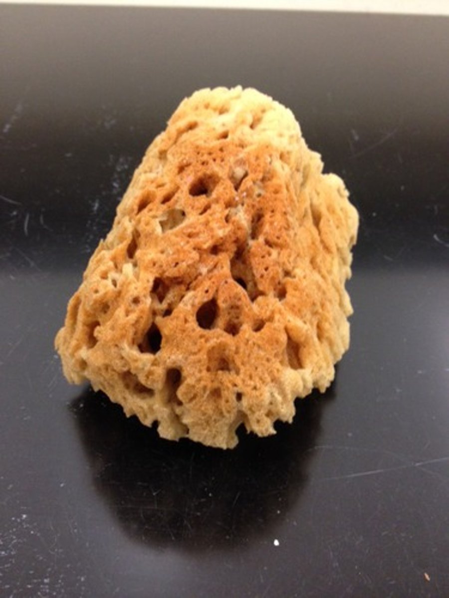
What class of sponge does this belong to?
Calcarea
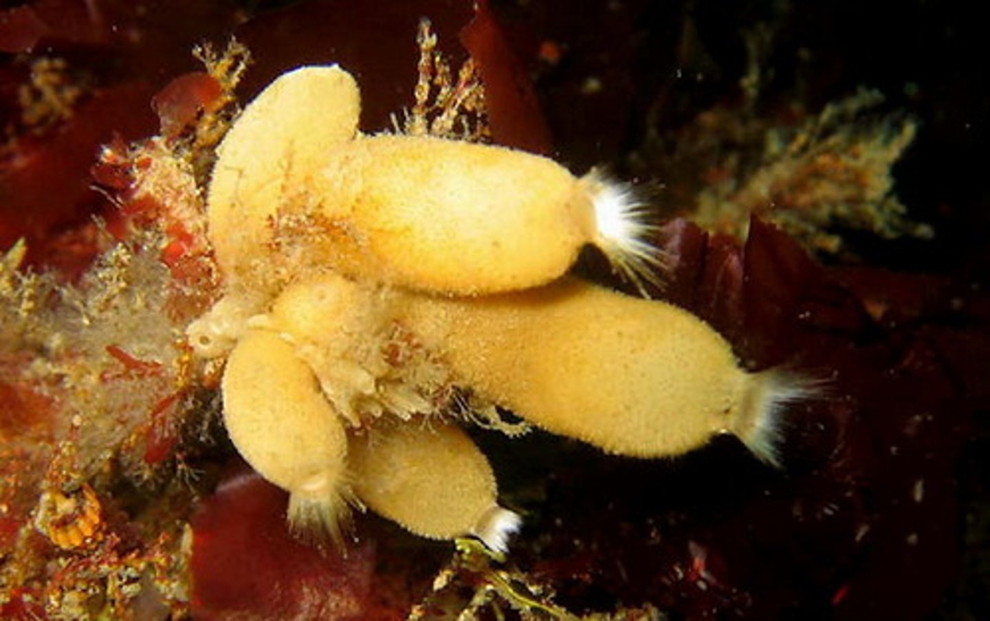
What class of sponge does this belong to?
Calcarea
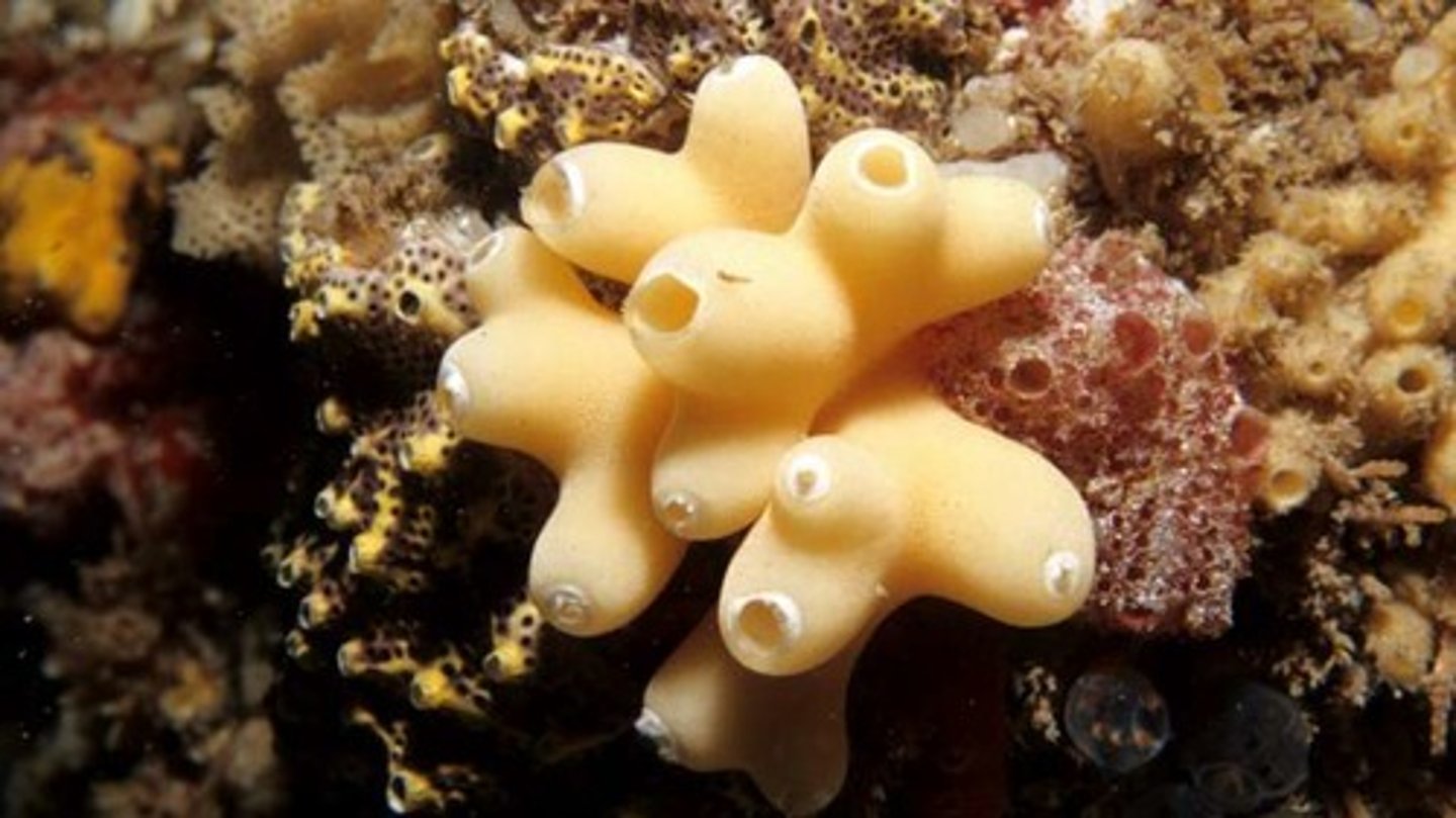
What class of sponge does this belong to?
Calcarea
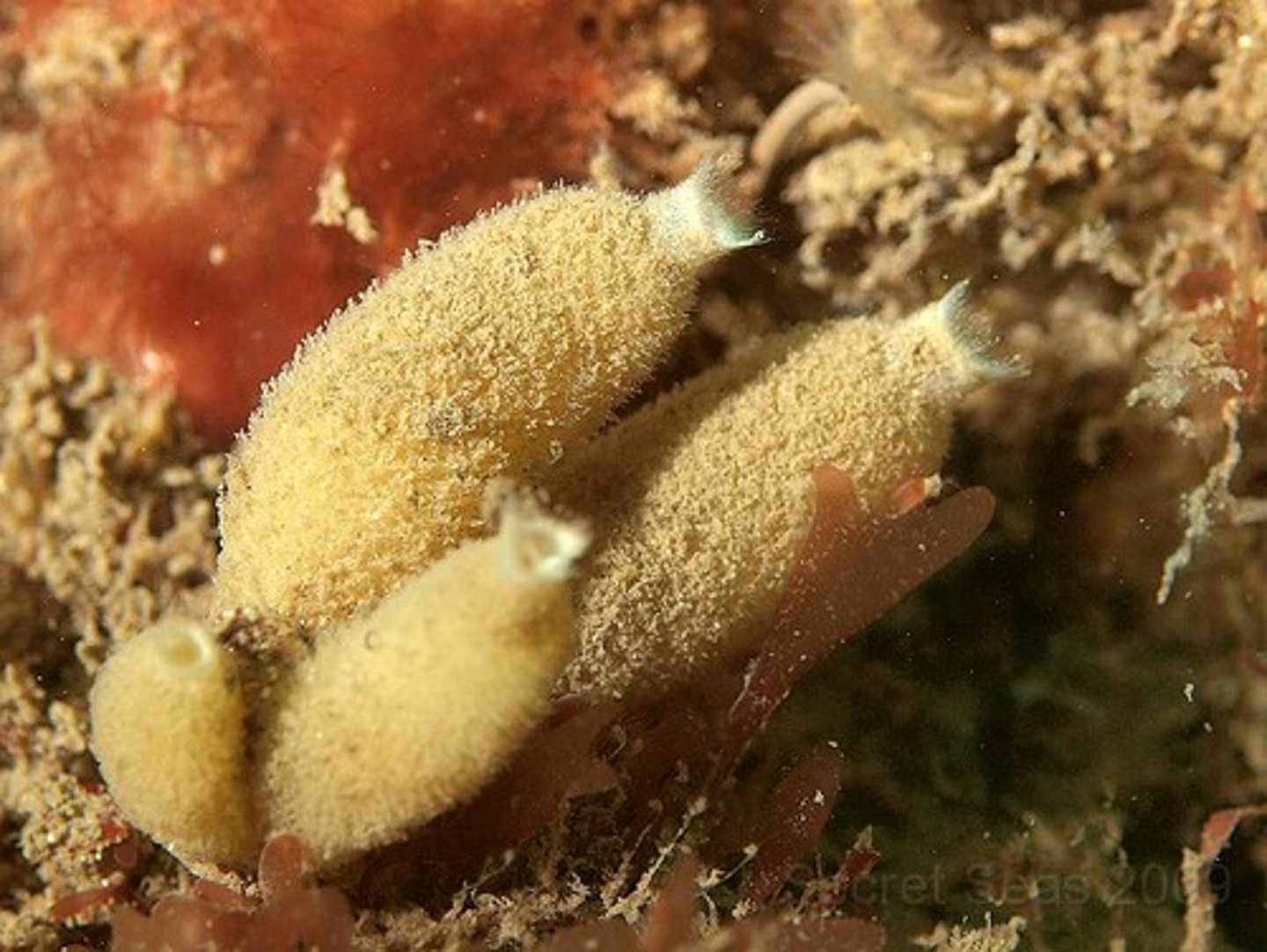
What class of sponge does this belong to?
Desmospongiae