septal defects
 Call Kai
 Learn
Learn Practice Test
 Spaced Repetition
 Match
Match Flashcards
 Knowt Play
1/85

There's no tags or description
Looks like no tags are added yet.
Name | Mastery | Learn | Test | Matching | Spaced | Call with Kai | Chat |
|---|
No analytics yet
Send a link to your students to track their progress
86 Terms
sternal scar indicates
open heart surgery that required cardiopulm bypass
lateral scar at 4th/5th ribs indicate
shunt replacement, coarc repair, PDA ligation
what is the order of identification if you suspect CHD?
establish situs
identify chambers and great vessels that connect to them
then begin looking for defects
what is situs solitus?
ao stomach spleen on left side
liver and IVC on right side
RA is identified by
right atrial appendige (triangular)
lots of pectinate muscle
rv is identified by
coarsely trabeculations (more than LV)
la is identified by
LAA (finger shaped pouch)
less pectinate muscle
lv is identified by
smooth walls that are thicker than the rv
differentiate between ao and pulm
ao has 2 branches and pulm has 3
what kind of murmurs for VSD?
holosystolic
what are small vs large VSDs?
small : less than 1/3 diameter of ao annulus → restrictive flow
large : greater than ½ size of ao annulus → non restrictive flow
what are rv and lv pressures when there is non restrictive flow
relatively equal
normal qp/qs vaules should be
around 1:1
signficant shunt defect is Qp/Qs of
2 or higher
what is Qp and how do you get it
pulmonary cardiac output
pulmonic SV : CSA x VTI x 100
csa : rvot diameter at base of pulm leaflets
CSA =.785 x D²
vti : pw doppler tracing of rvot flow
pulm CO : SV x HR
what is Qs and how do you get it
systemic cardiac output
systemic SV : CSA x VTI x 100
csa : lvot diameter at base of pulm leaflets
csa = .785 xD²
vti : pw doppler tracing of lvot flow
systemic CO : SV x HR
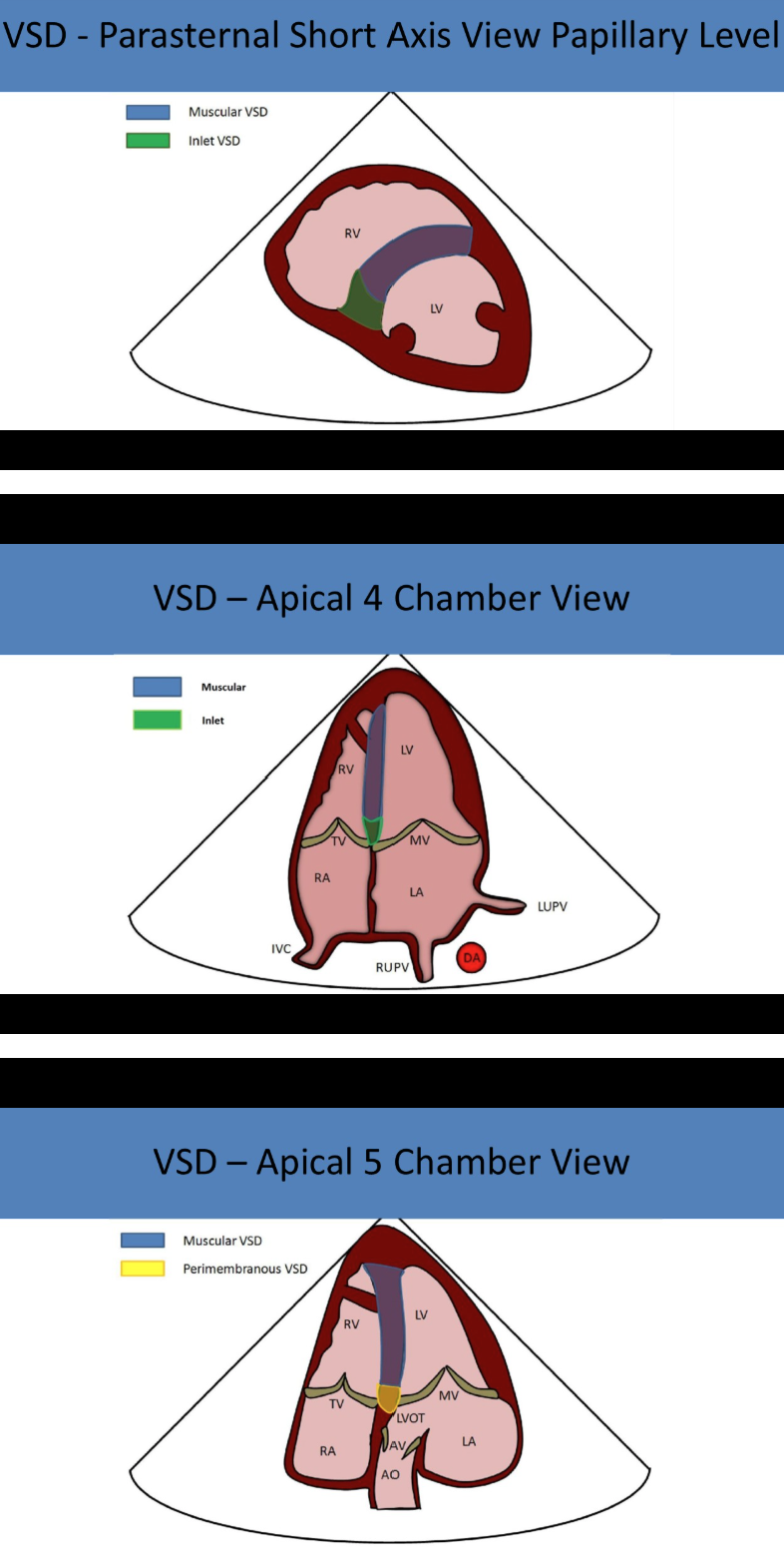
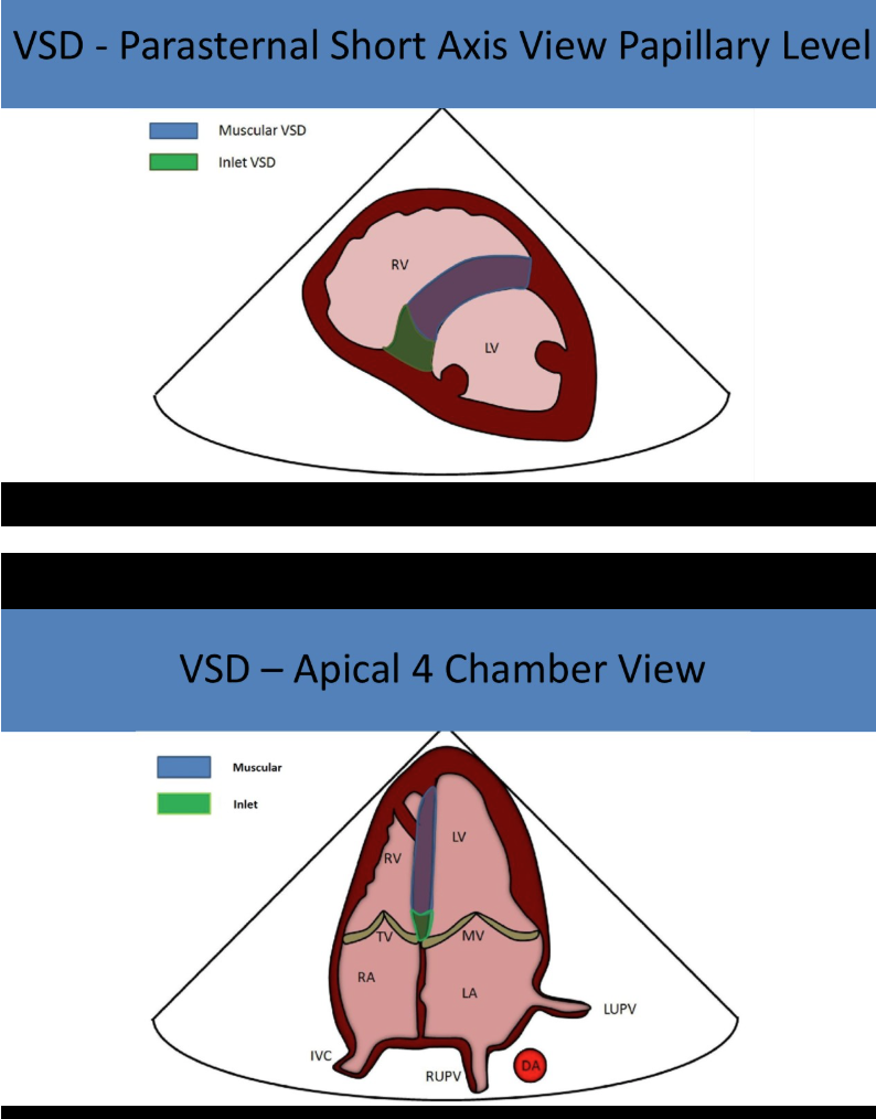
what are the types of vsds?
perimembranous
muscular/trabecular
outlet/supracristal/subpulmonic/doubly committed
inlet
what is the most common vsd?
perimembranous
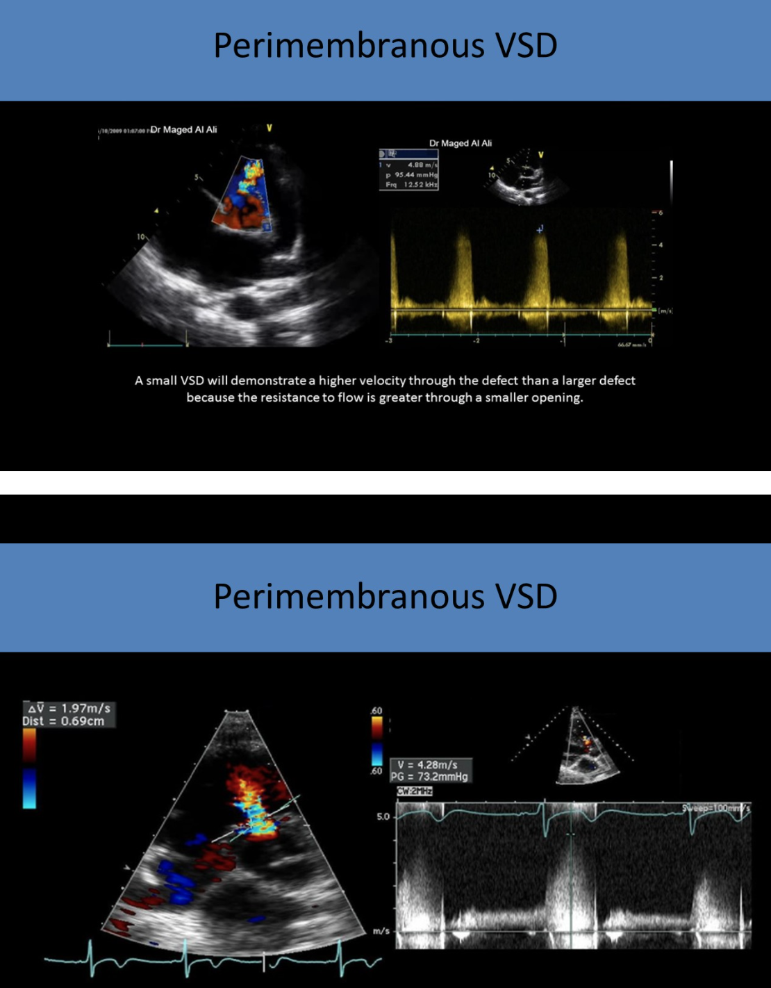
where are perimembranous VSDs found?
in LVOT near aortic valve
inferior to right coronary cusp and adjacent to septal leaflet of tricuspid valve
perimembranous VSDs are best evaluated in
PLAX
(any view where ao is in view)
perimembranous VSDs are commonly associated with
aortic insufficiency
ventricular septal aneurysm
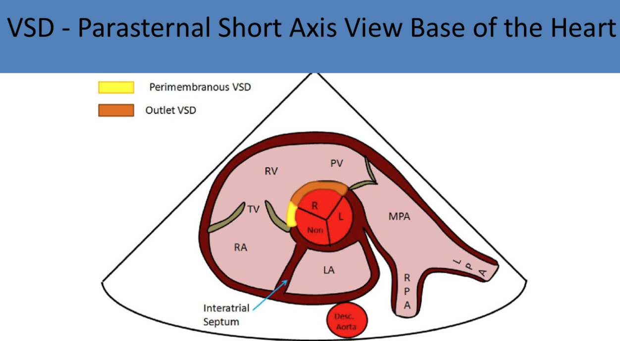
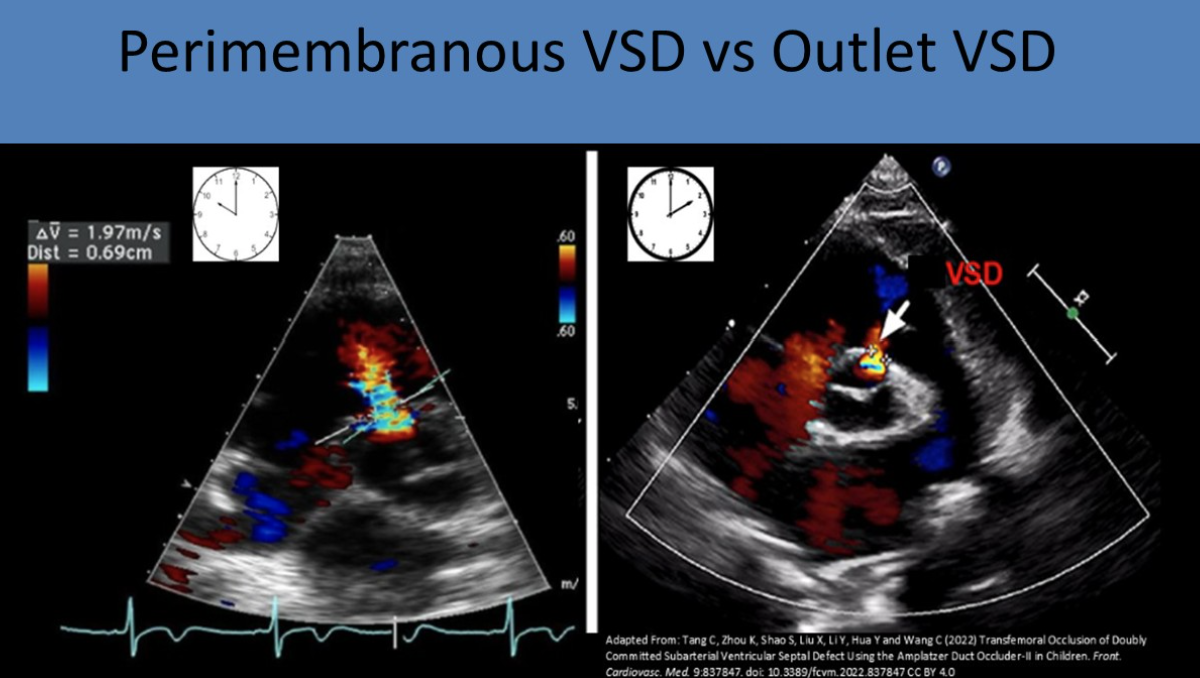
where are perimembrnous VSDs seen in PSAX?
10-12 oclock position
what is the second most common type of VSD?
muscular/trabecular
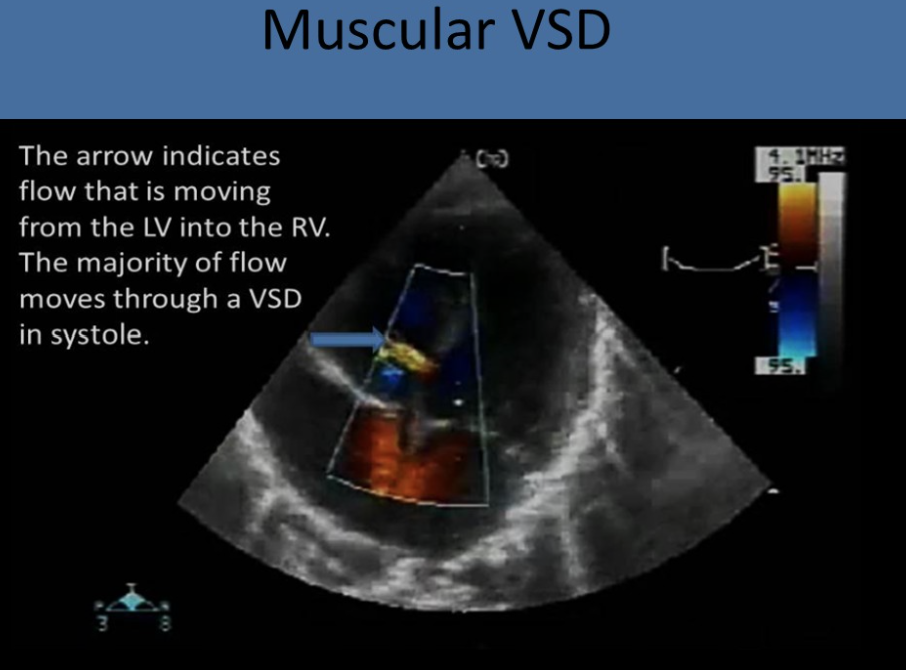
muscular VSDs are best evaluated in
subcostal views
where is the outlet VSD located?
in RVOT near pulmonic valve
inferior to left coronary leaflet of ao valve and adjacent to pulmonic valve
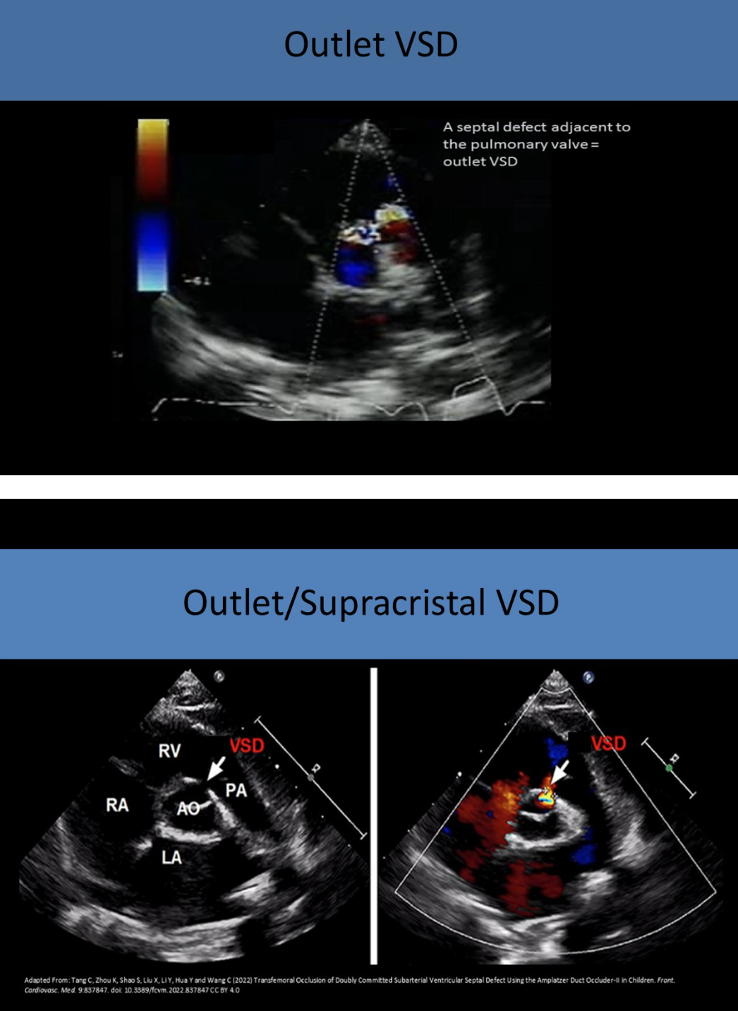
outlet VSDs are aka (3)
supracristal
subpulmonic
doubly committed
where are outlet VSDs best seen?
high parasternal short
PLAX
where are outlets VSDs seen in PSAX?
12-2 oclock
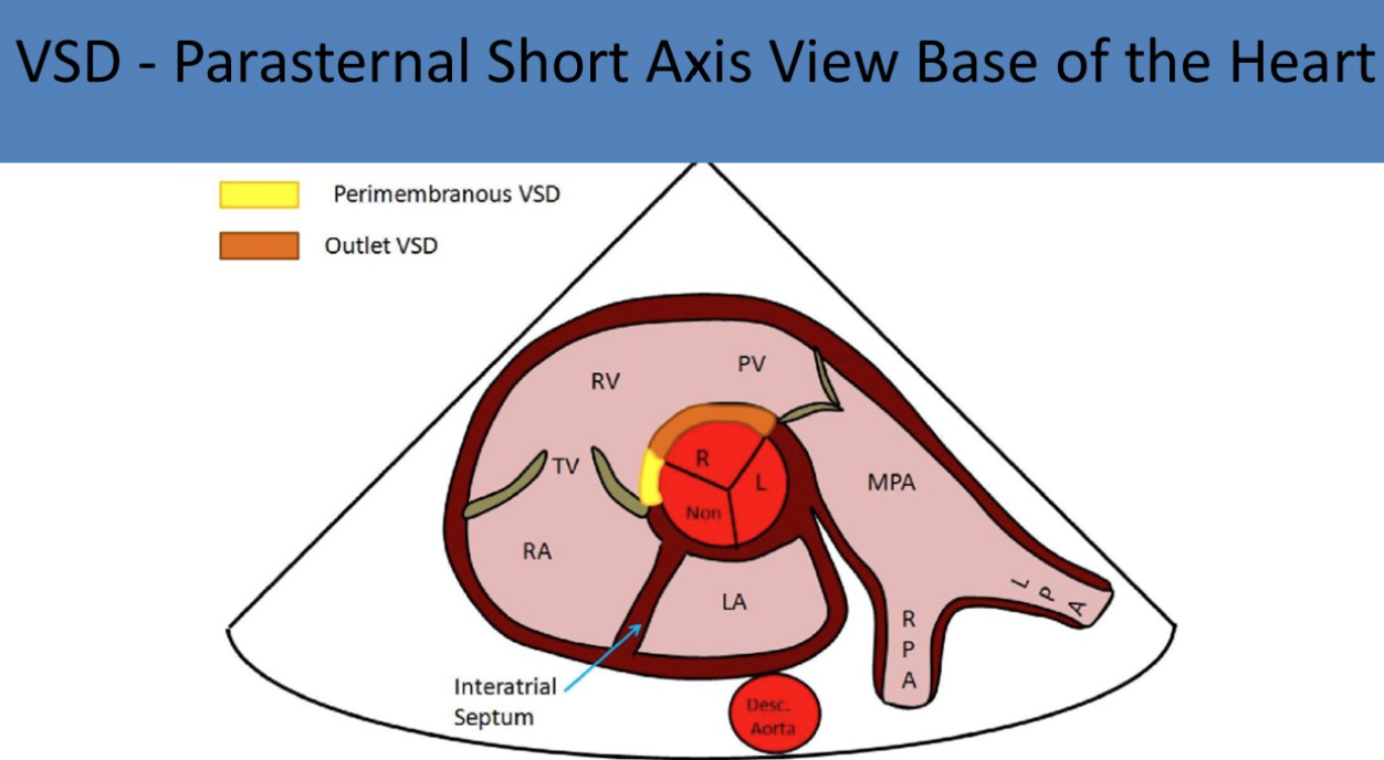
what is commonly associated with outlet VSDs?
60-70% have right ao cusp prolapse
ao insufficiency
where are inlet VSDs located?
in posterior septum, near posterior leaflet of tricuspid valve
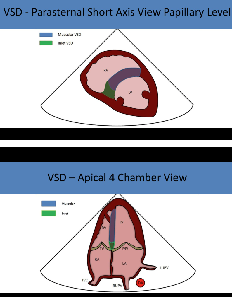
inlet VSDs are associated with
AVSD
inlet VSDs are best evaluated in
high parasternal short
PLAX
A4ch
large VSDs can lead to
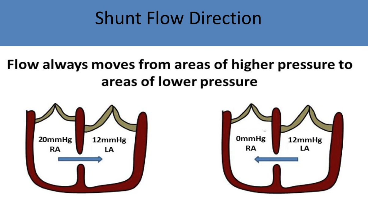
eisenmenger syndrome with right to left shunting
shunt flow is normally
left to right
the higher the gradient the - the defect
smaller
how do you calculate RVSP with VSD?
systolic BP - VSD gradient = RVSP
what is the second most common form of CHD identified in adults?
ASD
what is the most common form of CHD identified in adults?
bicuspid AV
large ASDs will cause
right heart volume overload and reduced ventricular compliance
what are the types of ASDs?
septum primum
septum secundum
sinus venosus
coronary sinus defects
common atrium
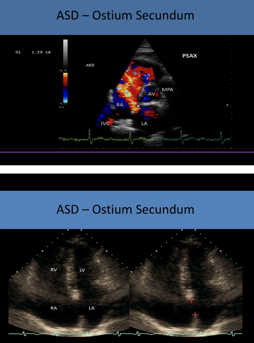
what is the most common type of ASD?
septum secundum
where are septum secundum asds located?
mid septal area
septum secundum asds are associated with
MVP
what is the second most common ASD?
septum primum
where are septum primum asds located?
close to av valves
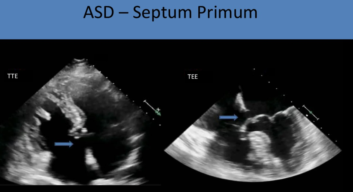
septum primum asds are associated with
cleft mitral valve
where are sinus venosus ASDs located?
near SVC entrance
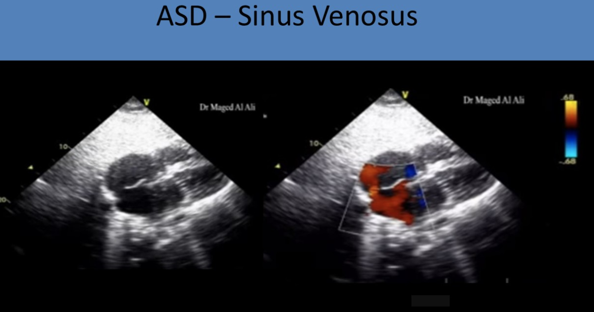
what are sinus venosus ASDs associated with
persistent LSVC
common atriums are associated with
ellis van creveld syndrome : bone growth disorder
what are the signs and symptoms of asds?
murmur
dyspnea
orthopnea
jugular vein distention
peripheral edema
what views are optimal for evaluating atrial septum?
subcostal
normal ASD shunt flow should be
left to right atrium with small flow reversal at early systole
if pressures in both atria are normal what will the avg peak velocity/peak pressure gradient be?
avg peak velocity : 1 m/s
peak pressure gradient : 5 mmHg
(because normal difference in pressure between right and left atrium is 5mmHg)
bidirectional flow in ASD indicates
increased right atrial pressure equivalent to left atrial pressure
ASDs are associated with
interatrial septal aneurysms
defect of septumprimum layer
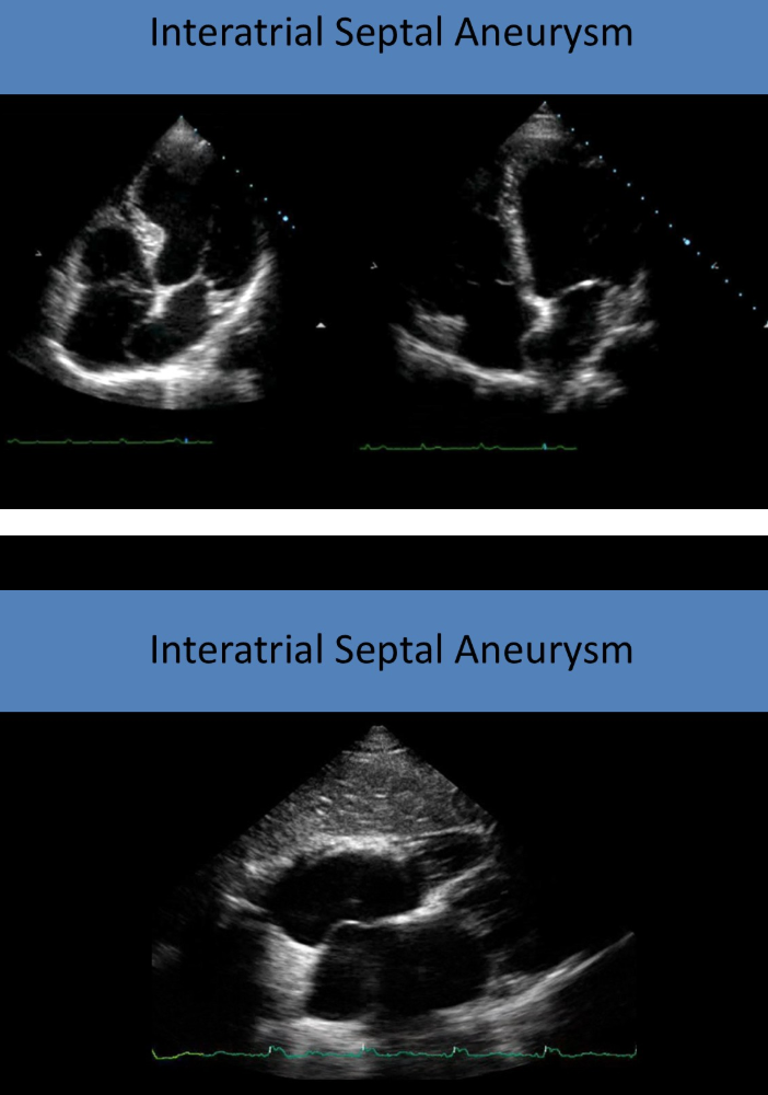
ASD will cause - heart dilation bc -
right bc of shunting of flow across septum to right atrium
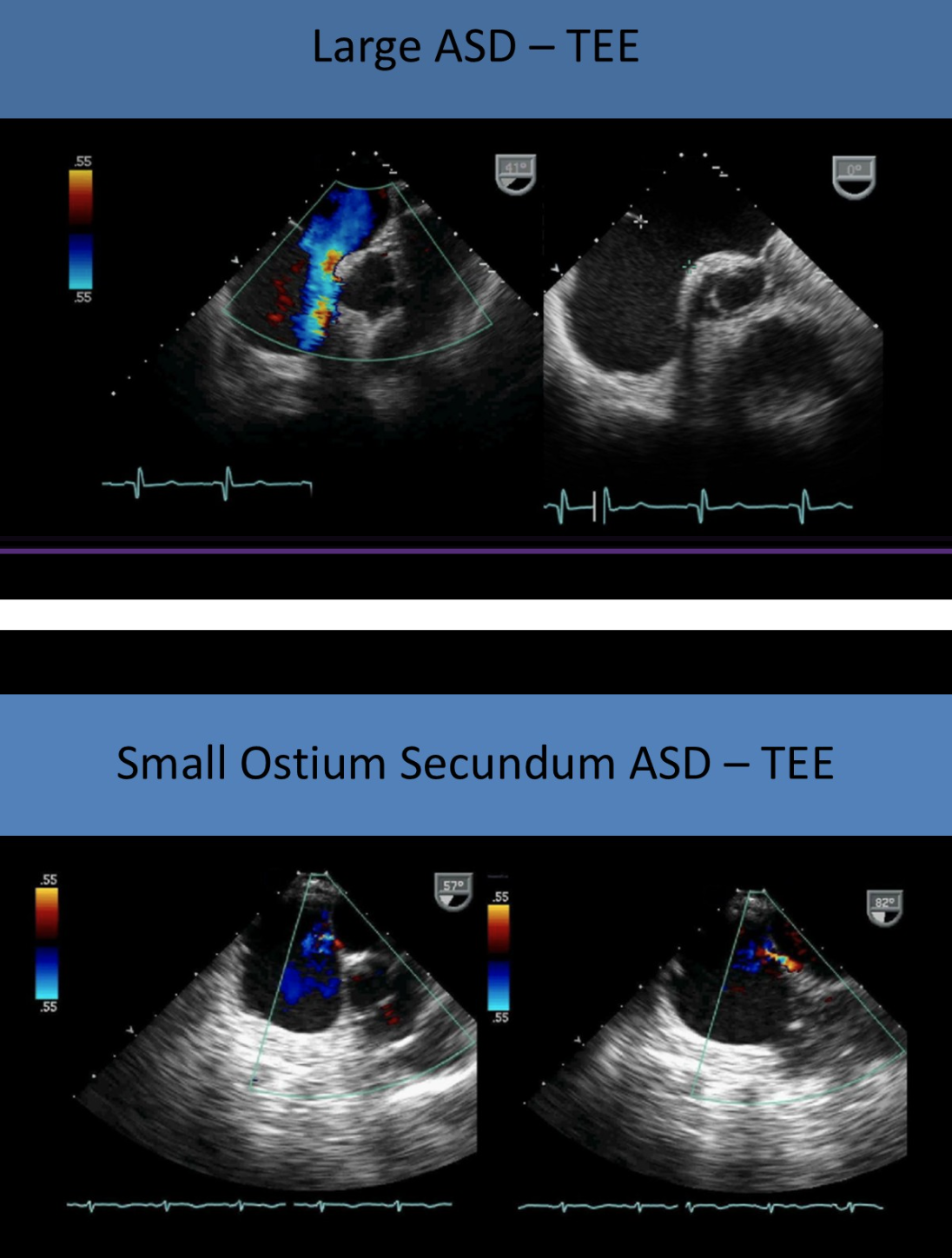
saline bubble study is positive if
there is presence of bubbles in LA and ventricle
how can secundum ASDs be treated?
percutaneous transcatheter device closure
called amplatzer
implanted through balloon catheter
how are septum primum/sinus venosus defects treated?
surgical intervention
what is endocardial cushion defect? aka
incomplete development of endocardial cushions
aka atrioventricular septal defect (AVSD) or AV canal defect
endocardial cushion defects are associated with
trisomy 21
partial endocardial cushion defects have
septum primum ASD
cleft mitral valve
complete endocardial cushion defects have
septum primum ASD
inlet VSD
common av valve with 5 leaflets
how do common av valves appear
as a single linear structure between ventricles and atria
(lack of normal offest of TV toward apex)
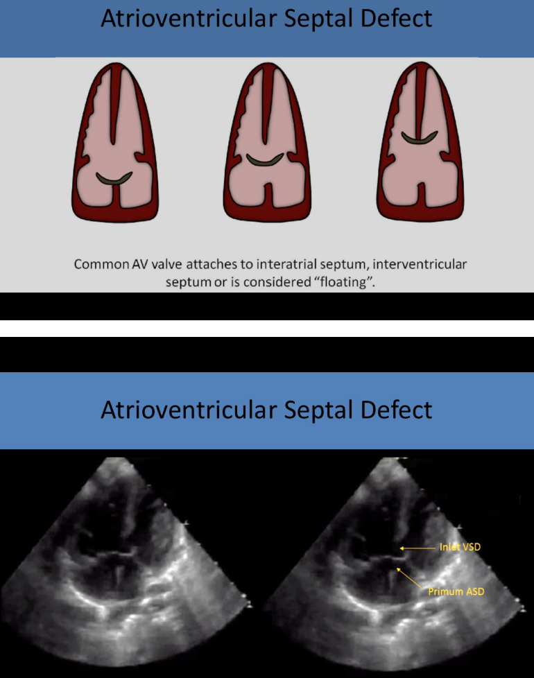
what will you see with AVSD?
right heart volume overload
dilated RV w paradoxical ventricular septal motion
in the embryonic development of the cardiac vessels there are - total arches that form and - of these arches develop into normal structures
a 6 6
b 12 6
c 4 8
d 8 4
b
which of the following is primary difference between a standard adult echo and an adult echo performed for congenital heart defects
a pt history is reviewed for prior surgery
b protocol includes a complete evaluation of abd organs
c 10-20 second clips are stored of sweeps through cardiac anatomy
d image display is inverted during the exam
c
a lateral scar on a patients chest is most suggestive of what type of surgery
a cardiac bypass
b pericardiocentesis
c valve replacement
d cardiac shunt replacement
d
which of the following describes how to document situs on an echo pt
a parasternal approach with transducer notch at 10
b apical approach w transducer at 9
c subcostal approach with transducer at 3
d suprasternal approach w transducer notch at 2
c
what is the best way to differentiate the pulm artery from the aorta on an echo
a location
b size
c number of valve leaflets
d number of branches
d
the size of a VSD is compared to the size of what cardiac structure to estimate severity
a ao
b pulm
c foramen ovale
d la
a
the qp/qs ration is used to compare
a peak systolic velocities in pulm art and ao
b pulm and systemic cardiac output
c end diastolic velocity in pulm artery and ao
d right and left ventricle diameters
b
in psax view, perimembranous VSD are identified
a near apex of heart
b at 12-2 position in septum
c at 10-12 position in septum
d inferior to left coronary leaflet of ao valve
c
which type of VSD is located in the posterior septum, near the posterior leaflet of tricuspid valve
a outlet
b perimembranous
c inlet
d muscular
c
which type of VSD is most commonly associated with right ao cusp prolapse
a outlet
b perimembranous
c inlet
d muscular
a
describe hemodynamic changes associated with small and large VSDs
small
dilated LV with hyperkinesis and LA dilated = left heart overload
large
can lead to eisenmenger syndrome w right to left shunting
when evaluating flow through VSD, smaller defects will demonstrate
a low peak velocity flow
b bidirectional shunting
c right to left shunting
d high peak velocity flow
d
which of the following is used to correct a VSD?
a stitches
b piece of pericardium
c occlusive devise
d all of the above
d
list the 5 asds and associated defects
septum primum : cleft MV
septum secundum : MV prolapse
sinus venosus : PAPVR
coronary sinus defects : persistent LSVC
common atrium : ellis van crevald syndrome
which of the following is a common symptom of ASD?
a diastolic murmur
b jugular vein distention
c systemic HTN
d premature ventricular contractions
b
what echo view is preferred to evalute the integrity of the atrial septum?
a parasternal
b apical
c subcostal
d suprasternal
c
which of the following findings is associated with an ASD in an adult
A qp/qs >1.5 : 1
b d shaped lv
c t artifact
d all
d
what type of defect can be repaired with amplatzer?
a sept primum
b sept secundum
c sinus venosus
d all
b