A&P 1 Lab Exam 1
 Call Kai
 Learn
Learn Practice Test
 Spaced Repetition
 Match
Match Flashcards
 Knowt Play
 Podcast
1/101

There's no tags or description
Looks like no tags are added yet.
Name | Mastery | Learn | Test | Matching | Spaced | Call with Kai | Chat |
|---|
No analytics yet
Send a link to your students to track their progress
102 Terms
how would you describe the location of a bruise on the front right lower leg?
anterior, distal to knee
What directional term would be used to indicate sunburn on a patient's back?
posterior
What’s another name for the frontal plane
coronal plane
what’s another name for sagittal or midsagittal plane
Median plane
Which directional term is used to describe the relationship of the skin to the skeletal muscles?
superficial
the knee is proximal to the thigh
False
Which statement is correct
Which of the following statements regarding the umbilicus and the chin is true?
The umbilicus is caudal to the chin.
The lungs are what to the heart
lateral
the arm is what to the forearm
proximal
The bridge of the norse is what to the left eye
medial
the stomach is what to the spine
anterior
the fingers are what to the wrist
distal
The anatomical position is characterized by all of the following EXCEPT ________.
palms turned posteriorly
Which of the following describes a parasagittal plane?
any sagittal plane except in the midline
What is a vertical section through the body, dividing it into anterior and posterior regions called?
frontal
The elbow is proximal to the shoulder
false
Which of the following is NOT correct for anatomical position?
Palms facing inward towards the thighs
Which of the following is located in a median position?
mouth
The knee is proximal to the ankle. Which of these other terms could also be used to correctly describe the relationship between the knee and the ankle?
The knee is superior to the ankle.
The cranial bones almost entirely surround the brain and protect it. Which of the following terms best describes the relationship between the cranial bones and the brain?
The cranial bones are superficial to the brain.
Which of the following is a term that can be used instead of
dorsal?
posterior

Which of the following is a correct pairing of a body cavity with its contents?
The cavity C contains the lungs

The ventral cavity includes which of the indicated subdivisions in the figure?
C,D, and E
The dorsal body cavity is divided into which of the following subdivisions?
the vertebral/spinal and cranial cavities
In which quadrant of the abdominopelvic cavity is the stomach located?
left upper quadrant
The thoracic cavity contains the ________. It is found ________ to the vertebral cavity.
heart and lungs; anterior
Which of the following epithelial tissue types is NOT correctly matched to its function?
stratified squamous epithelium; absorption
Pseudostratified epithelia are well adapted to the digestive system because the cilia increase the surface area for absorption.
False
What function do transitional epithelia have?
stretching
You observe a tissue under a microscope. There appears to be a lumen on one side of the tissue. Lining this lumen is a single layer of epithelial cells that seem to have long, fingerlike projections. Beneath the projections, the cells seem to have the shape of shoeboxes and are packed tightly together. What type of tissue are you looking at?
simple columnar epithelium
Which of the following is a modification of the simple columnar epithelium that allows for efficient absorption along portions of the digestive tract?
dense microvilli
A ciliated version of pseudostratified columnar epithelium containing mucus-secreting goblet cells ________.
lines most of the respiratory tract
Which of the following epithelia forms the walls of the air sacs across which gas exchange occurs in the lungs?
simple squamous
________ epithelium appears to have two or three layers of cells, but all the cells are in contact with the basement membrane.
Pseudostratified columnar
A many-layered epithelium with cuboidal basal cells and flat cells at its surface would be classified as ________.
stratified squamous
Select the correct statement regarding epithelia.
Stratified epithelia are present where protection from abrasion is important.
Simple cuboidal epithelia are usually found in areas where secretion and absorption occur.
true

identify the highlighted structure
cilia

Identify the highlighted type of epithelial tissue
pseudostratified ciliated columnar
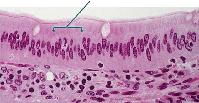
identify the epithelium indicated by the arrows
simple columnar

Identify the highlighted epithelium
simple cuboidal
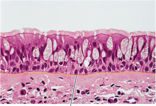
What structure is highlighted
basement membrane

What structures are highlighted
microvilli

What structures are highlighted
nuclei

Which epithelial type is highlighted
simple columnar epithelium

Which epithelial type is highlighted
stratified squamous epithelium

Which epithelial type is highlighted
transitional epithelium
Squamous epithelial cells _____.
are thin and flat, like fish scales
Simple epithelia have ________.
one type of epithelial cell, arranged in a single layer
Which of the following best describes pseudostratified epithelium?
Single layer of cells with various heights
What are the visible characteristics of ciliated pseudostratified columnar epithelium?
The nuclei are found in multiple planes, giving the illusion of multiple cell layers.
Cilia project from the apical surface into the lumen.
Goblet cells are columnar cells that can be seen interspersed within the tissue.
Identify the functions of ciliated pseudostratified columnar epithelium.
Secretion
Movement
Where is ciliated pseudostratified columnar epithelium found in the body?
Lining the trachea, bronchi, and nasal cavities
What body system contains pseudostratified columnar epithelium?
Respiratory system
A single layer of tall rectangular cells best describes which of the following?
Simple columnar epithelium
Describe the apical surface.
Secretions from goblet cells are released from the apical surface of a cell.
The apical surface is sometimes covered with cilia.
The apical surface is closest to or on the luminal surface.
Identify the primary functions of simple columnar epithelium
secretion and absorption
Where is simple columnar epithelium found in the body?
Lining the small and large intestine
Lining the stomach
Lining the uterine (fallopian) tubes
Identify the one characteristic that does NOT describe a goblet cell.
Component of a mucous gland
What is the function of cilia?
Movement of something across the cell's surface
Identify the best description for the shape of squamous cells.
Plate-like
Which of these are characteristics of simple squamous epithelium?
Simple squamous cells attach to form a sheet-like arrangement.
Simple squamous epithelium is found next to free space called the lumen.
Identify the primary function(s) of simple squamous epithelium cells.
Filtration
Diffusion
Where is simple squamous epithelium found in the body?
Kidneys (glomerular capsule)
Air sacs of the lungs
Lining of blood vessels
Having multiple layers of flat cells against a free surface describes which of the following?
Stratified squamous epithelium
Where is the basement membrane located?
At the boundary between connective tissue and epithelium
Which of these are visible characteristics of stratified squamous epithelium?
Stratified squamous epithelium is always adjacent to a free space or lumen.
The squamous cells closer to the lumen are flatter while the squamous cells closer to the connective tissue are fuller and rounder.
There are so many layers of cells that it would be difficult to count them precisely.
What is the function of stratified squamous epithelium?
protection
Where is stratified squamous epithelium found in the body?
Lining of the vagina
Epidermis
Lining of the esophagus
Which description is best for transitional epithelium?
Cells that are more cuboidal in a relaxed state but more squamous when stretched.
What are the visible characteristics of transitional epithelium?
Rounded apical surfaces that resemble a tombstone shape.
Neighbors a hollow cavity called the lumen.
Multiple layers of cells.
Identify the function of transitional epithelium.
Flexibility
Where is transitional epithelium found in the body?
Lining of the ureters
Lining of the proximal portion of the urethra
Lining of the urinary bladder
What is the only body system that contains transitional epithelium?
Urinary system
Epithelial tissue is typically localized to the following region(s) of the body.
on the surfaces of organs, on the walls of cavities, on the outer surface of the body, and in glands of the body
Based on its function, where would you likely find simple squamous epithelium?
in places where materials need to pass through a thin sheet of cells
What is the function of ciliated epithelium?
to move fluids across the tissue surface
Which of the following is an example of pseudostratified epithelium?
lining of the trachea
Why are adipose tissue, blood, and bone all considered to be connective tissues?
They have a common origin: the embryonic tissue mesenchyme.
Connective tissues are made of different combinations of ground substance and fibers. As you compare slides of connective tissues, which would most likely be the strongest (toughest) type of connective tissue based on the composition of ground substance and fibers?
connective tissues high in collagen fibers
Which is the most atypical connective tissue since it neither connects structures nor provides structural support?
blood
Which tissue is correctly paired with its primary immature cell type
bone; osteoblast
Which of the following statements is true?
Dense connective tissue is dominated by collagen fibers
The reason that intervertebral discs exhibit a large amount of tensile strength, which allows them to absorb shock, is because they possess ________.
collagen fibers
What tissue has lacunae, calcium salts, and blood vessels?
bone (osseous tissue)
The primary blast cell for bone is the ________.
osteoblast
How is hyaline cartilage different from elastic cartilage or fibrocartilage?
Fibers are not normally visible.
Which of the following is found in hyaline cartilage?
chondrocyte
The shape of the external ear is maintained by ________.
elastic cartilage
Select the correct statement regarding adipose tissue.
Its primary function is nutrient storage.
Tendons and ligaments are composed mainly of dense irregular connective tissue.
False

Which tissue is highlighted?
elastic cartilage

Which component of the connective tissue in this field of view is highlighted?
extracellular matrix

What structure connects the highlighted muscle cells to one another?
intercalated discs
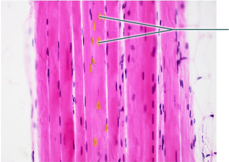
Which structures are highlighted?
nuclei

Which structure is highlighted?
skeletal muscle fiber
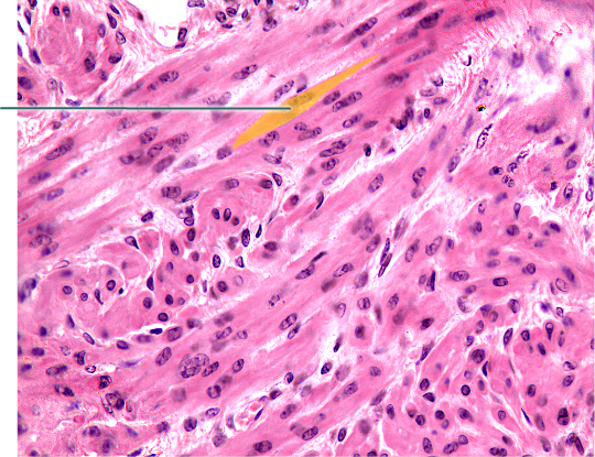
What is a characteristic of the highlighted muscle tissue?
uni-nucleated
Under a microscope you observe a tissue that appears to have long fibers that appear striated. The nuclei are pushed off to the side of the fibers. The tissue looks very vascular. What type of tissue are you observing?
skeletal muscle tissue
Smooth muscle __________.
is under involuntary control