Principles of Biomedical Science Chapter 2.2 Study Guide
 Call Kai
 Learn
Learn Practice Test
 Spaced Repetition
 Match
Match Flashcards
 Knowt Play
1/88

Name | Mastery | Learn | Test | Matching | Spaced | Call with Kai | Chat |
|---|
No analytics yet
Send a link to your students to track their progress
89 Terms
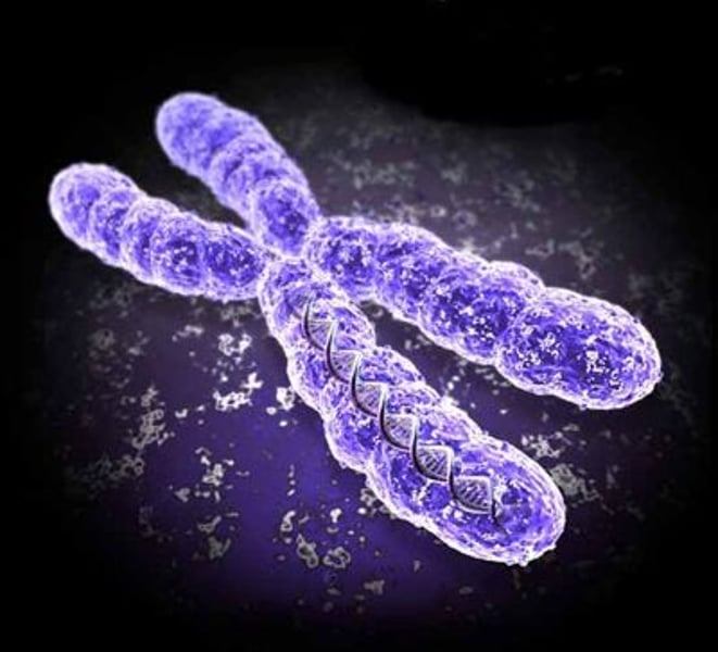
Chromosomes
thread-like structures found in the nucleus of cells that store genetic material in the form of DNA
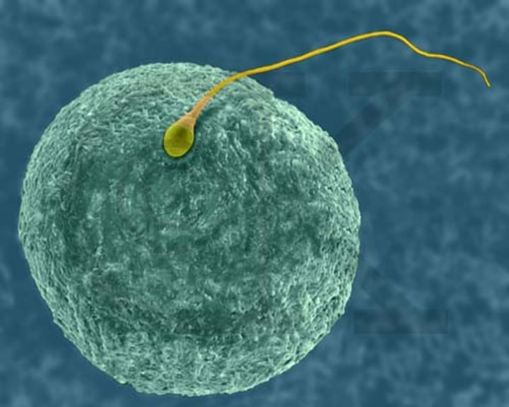
Fertilization
when an egg and a sperm join and chromosomes combine to create a unique set of genetic info
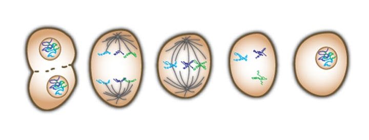
Mitosis
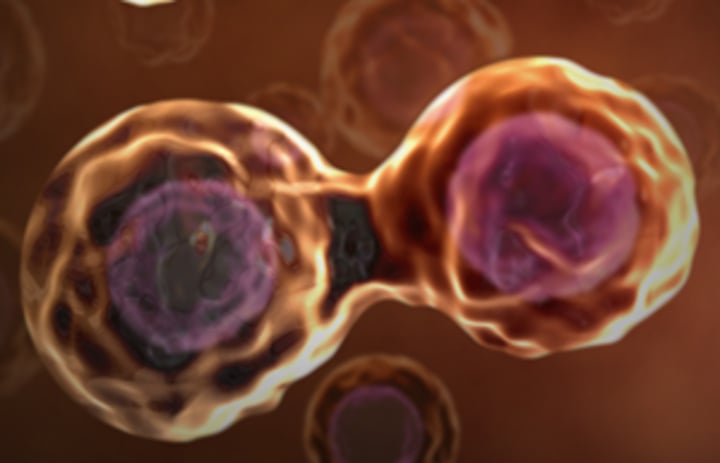
the process of cell division where a single cell divides into two daughter cells
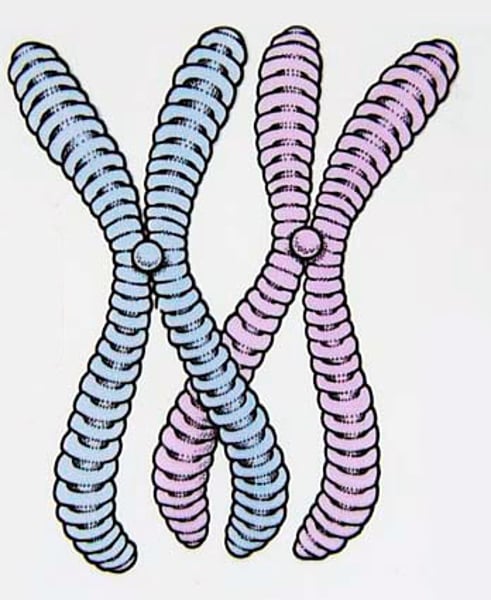
Homologous Chromosomes
pairs of chromosomes that contain the same genetic materials, but different versions of them
Interphase
DNA replicates so that there are two copies of each chromosome; each copy is referred to as a sister chromatid
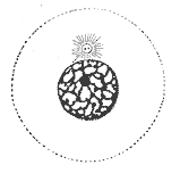
Prophase
chromosomes condense and become visible under a light microscope, and they pair up with their sister chromatids; the mitotic spindle forms and the nuclear envelope disappears
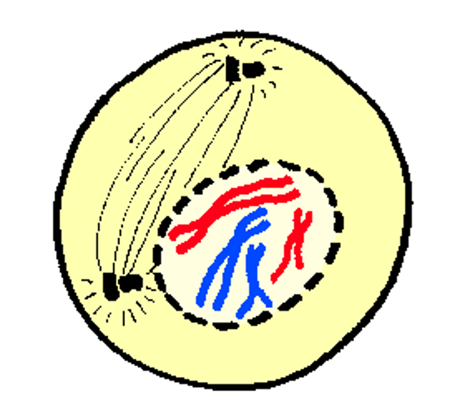
Metaphase
chromosomes line up at the center of the cell; fibers attach to each sister chromatid
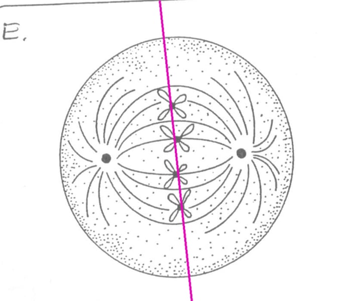
Anaphase
each chromosomes separates and the sister chromatids are pulled to the opposite poles of the cell
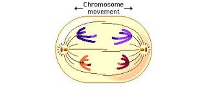
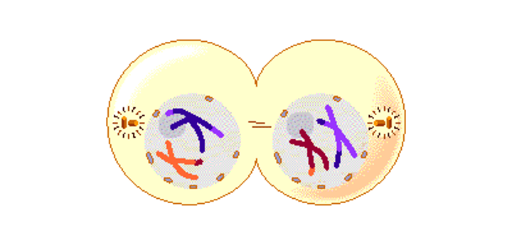
Telophase + Cytokinesis
the cell splits into two; each new "daughter cell" looks identical to the original cell
Tumors
a lump or mass of cells caused by uncontrolled cell-division
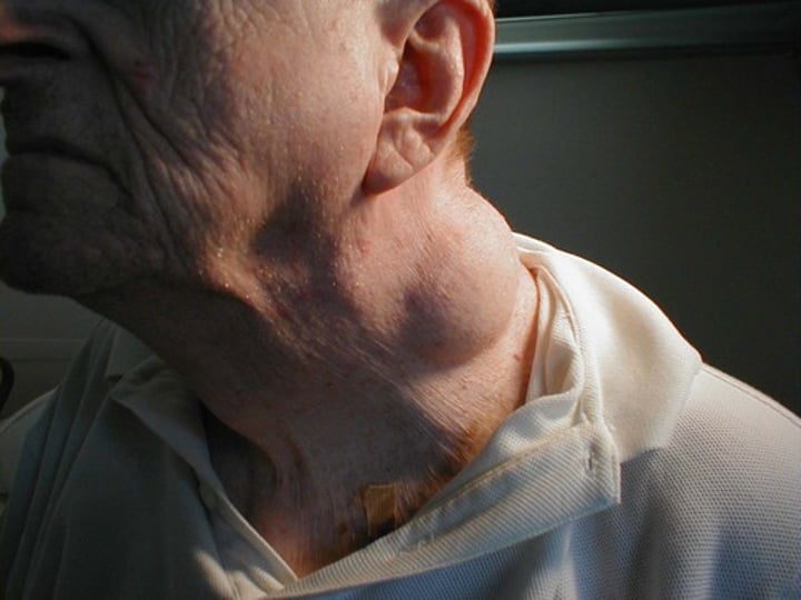
Benign
tumors that are generally considered harmless; will not spread to other parts of the body or invade other tissues; are not cancer
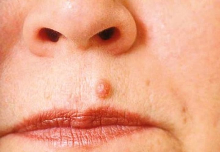
Malignant
also known as cancerous tumors; harmful and can invade other tissues or spread to other parts of the body if not treated; this spread is called metastasis
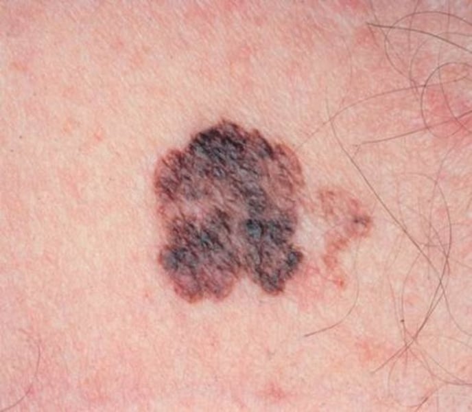
Cancer
a disease caused when cells divide uncontrollably and spread into other tissues; uncontrolled mitosis
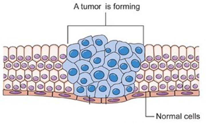
Metastasis
the spread of cancerous cells to other tissues or parts of the body
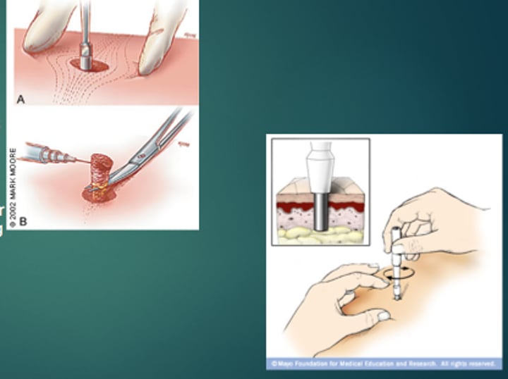
Biopsy
the removal of cells or tissues from a patient's body using a needle, scalpel, or other tools to study them more closely using a microscope
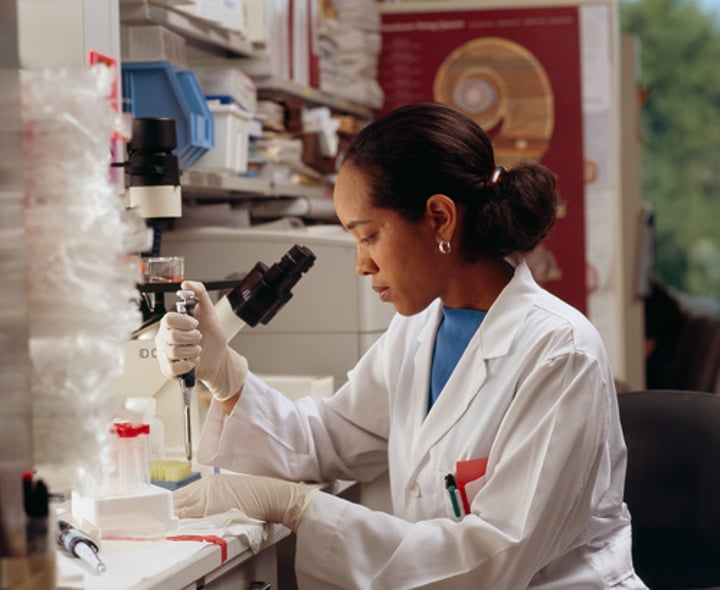
Cytopathologist
examines cells under a microscope to look for signs of cancer
Normal Cell Characteristics
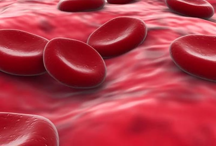
one small nucleus, same size and shape as other cells of the same tissue, divide in an organized manner and die after a limited number of cell divisions, contain distinct, specialized features common to the particular cell type, arranged in an organized manner with well-defined tissue boundaries
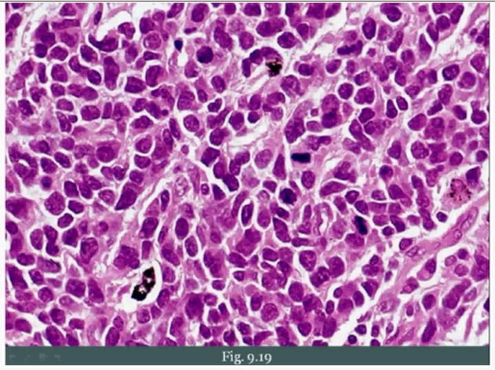
Cancerous Cell Characteristics
large, variably shaped nuclei, vary in size and shape, proliferate indefinitely resulting in a large number of irregularly shaped, dividing cells, lose specialized cell features, preventing them from performing functions properly, disorganized arrangement with poorly-defined tissue boundaries
Are Kai Rossie's bumps harmful?
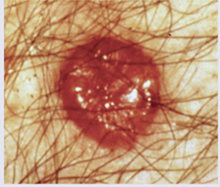
No, because they are benign since they are consistent in size and shape and have single regularly-shaped nuclei; according to the cytopathologist, they are also well-organized
Proteins that drive cellular functions
responsible for digestion, biosynthesis, and controlling cell growth/division (cyclin-dependent kinases (Cdks) control the cell cycle)
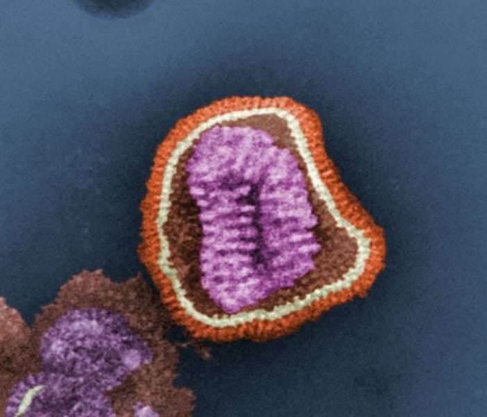
Proteins that provide defense
such as antibodies, combat foreign invaders to the body (such as bacteria, viruses, fungi, pollen, etc)
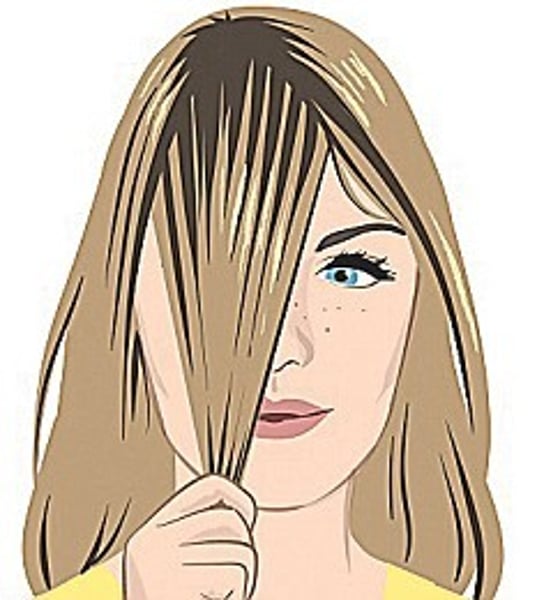
Proteins that build structures
build structures and include silk fibers (spider webs), collagen and elastin (animal connective tissue), and keratin (feathers and hair)
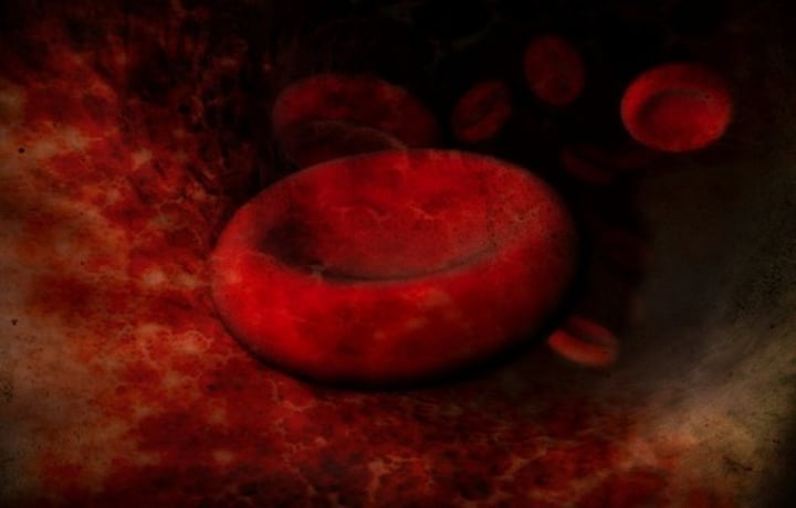
Proteins that transport materials
carry essential nutrients/gasses through the body (hemoglobin transports oxygen for cellular respiration); carry cellular waste for disposal
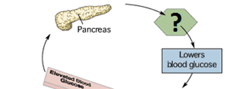
Proteins that coordinate cellular activities
hormonal proteins help maintain homeostasis (insulin helps regulate blood glucose levels); sensory proteins are used to interpret our environment (taste, smell, and sound)
Proteins for movement
motor and contractile proteins, such as actin and myosin, found in muscles and used during cell division
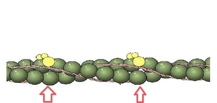
NF1 Gene
found on Chromosome 17 and carries the instruction to create a protein called Neurofibromin; coordinates cellular activities
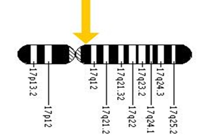
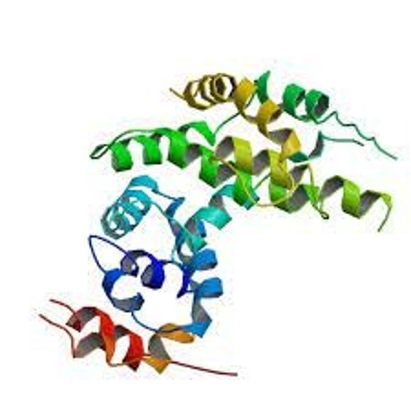
Neurofibromin
regulates mitosis and works to prevent cells from growing and dividing in and uncontrolled way
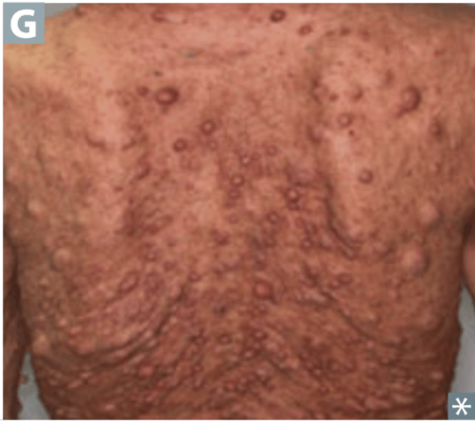
Effects of NF1
cells divide out of control, leading to tumors
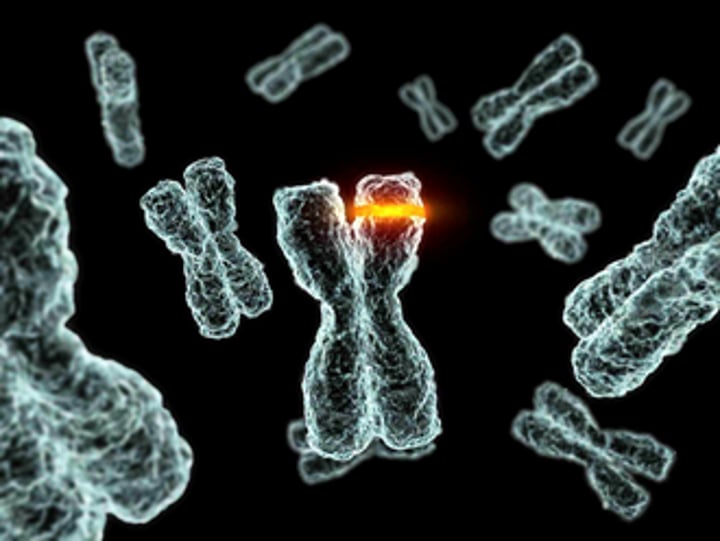
Point Mutation
only a single base is affected
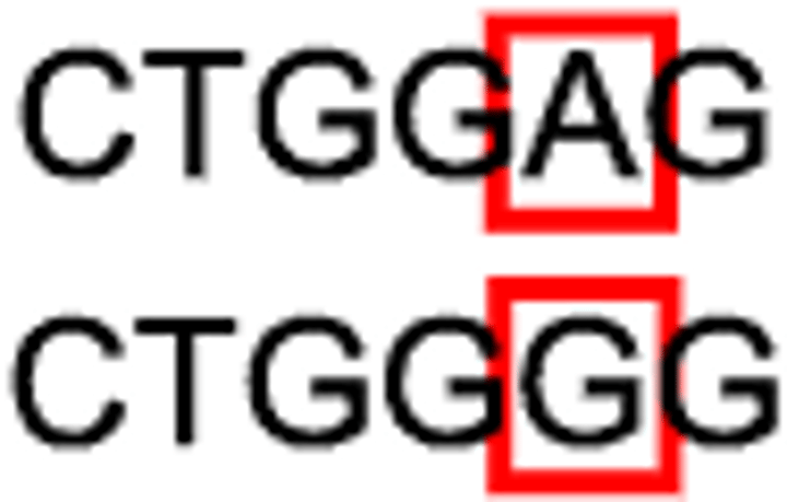
Substitution Genetic Mutation
exchange a single base for another (only one amino acid may be affected)
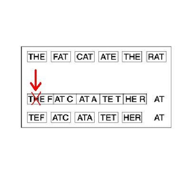
Frameshift Mutation
when the reading frame shifts
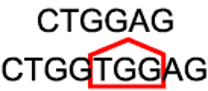
Insertion Genetic Mutation
adds on more base and affects other codons following it
Deletion Genetic Mutation
removes one or more bases
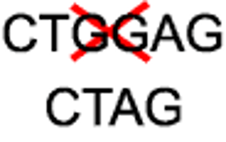
What type of genetic mutation causes Sickle Cell?
Substitution
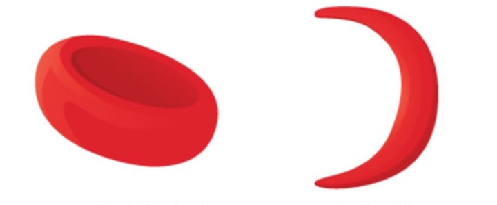
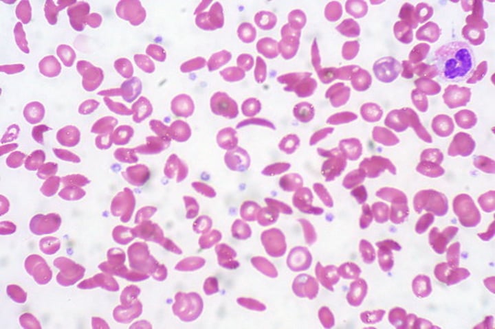
What causes Sickle Cell?
happens when the HBB gene (chromosome 11) is not working well; Thymine is replaced by Adenine, so it shows up as a "U" instead of an "A" when it is transcribed into mRNA
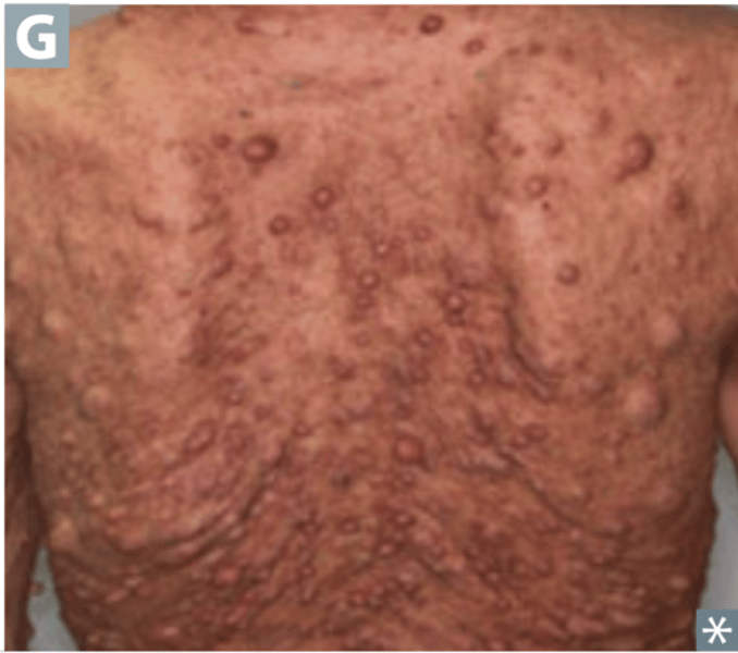
What does Kai have?
Neurofibromatosis Type 1 (NF1)
What caused Kai's NF1?
a frameshift mutation (deletion of CG and insertion of ATAC), leading to a dysfunctional neurofibromin protein
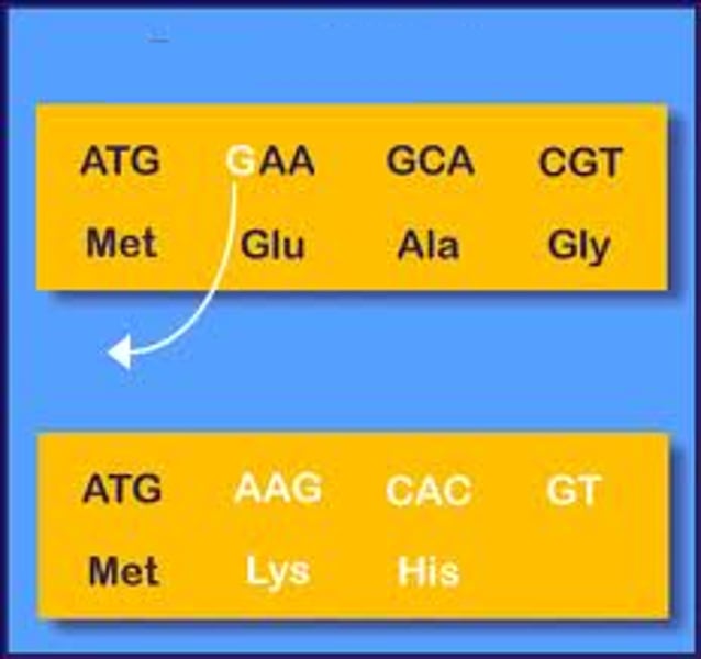
Causes of NF1
hyperactivity (ADHD), numerous tumors (if not treated), trouble healing broken bones, high blood pressure
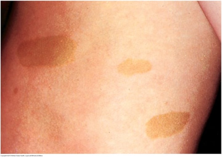
Signs of NF1
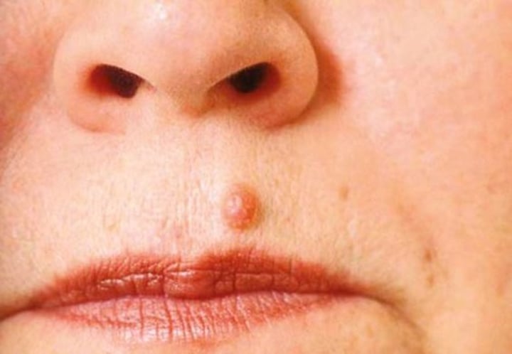
family history, six or more café-au-lait spots on skin, pea sized bumps on/under skin, larger areas on/under skin that appear swollen, seizures, tumors of the eye, bone deformities
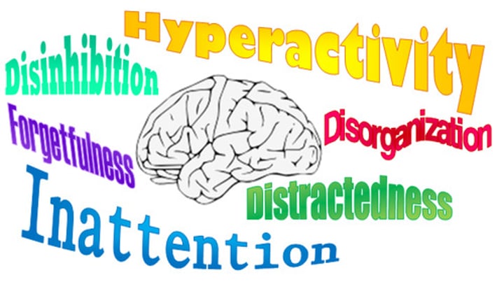
Symptoms of NF1
pain (from affected nerves), ADHD
Possible Treatments for NF1
no specific treatment; tumors causing pain/loss of function may be removed (those that have grown quickly should be removed as they may become malignant), drugs for children with severe tumors, special schooling for some children with learning disorders
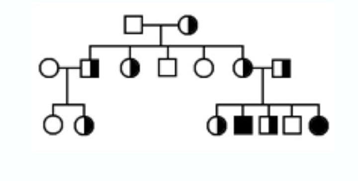
Is the gene represented in the pedigree dominant or recessive? Autosomal or sex-linked?
Autosomal recessive
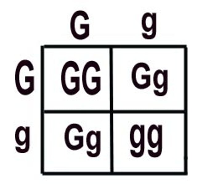
What is the chance the parents will have a heterozygous child?
50%
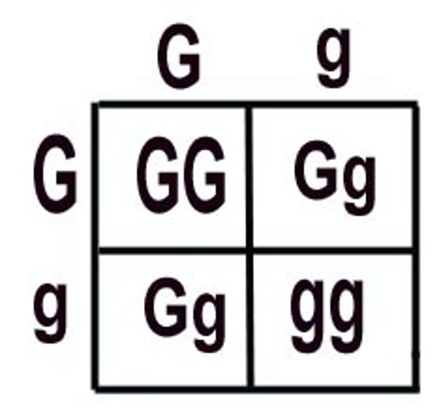
What is the chance the parents will have a homozygous dominant child?
25%
What is the chance the parents will have a homozygous recessive child?
50%
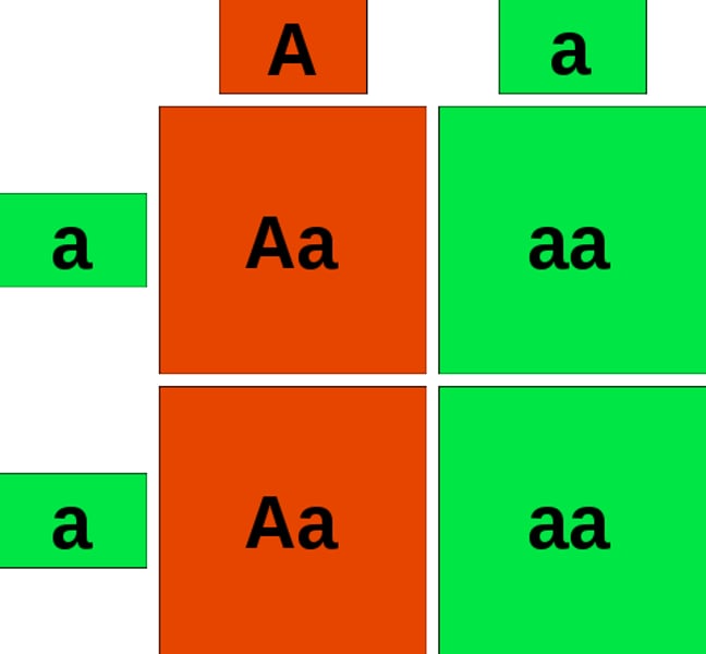
What is the chance the parents will have a homozygous dominant child?
0%
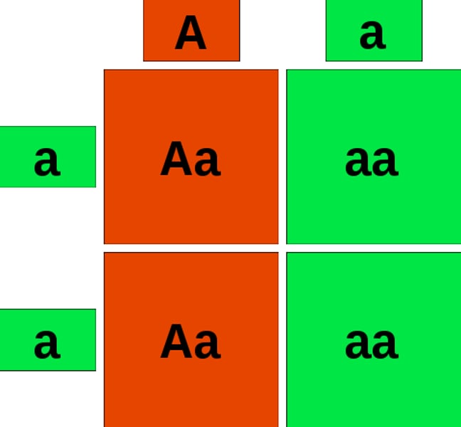
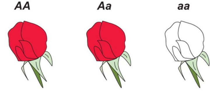
Is NF1 a dominant or recessive trait?
Dominant
Is NF1 an autosomal or sex-linked trait?
Autosomal
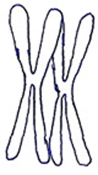
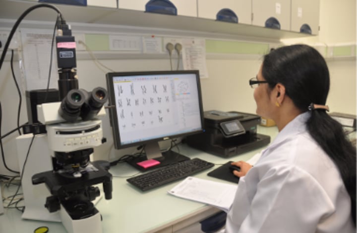
Cytogeneticist
laboratory technicians who harvest cells from biological samples and prepare the cells to view the chromosomes under a microscope
Karyotype
an image of the chromosome pairs of a cell arranged by size and shape
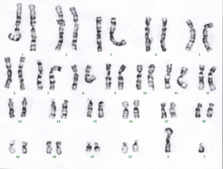
How many chromosomes in total are in a karyotype? How many pairs?
46 chromosomes, 23 pairs
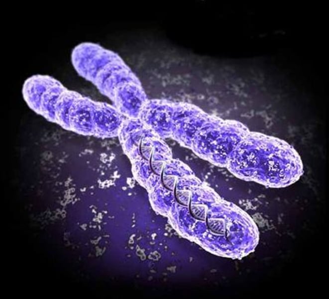
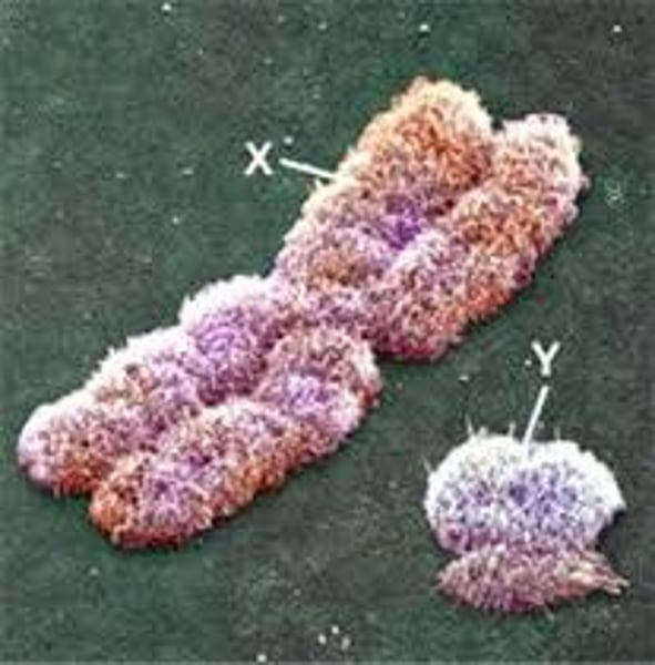
Which chromosome pair determines the sex of a person?
23rd
What are HeLa cells?
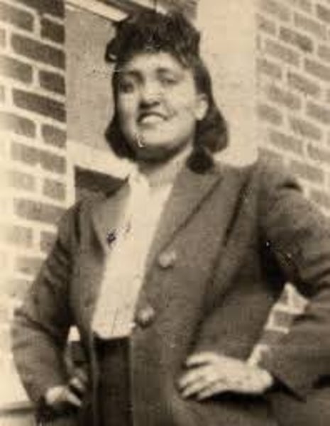
cells from Henrietta Lacks, whose cells were taken from her cervix without her knowledge or consent; her cells contributed to great scientific research
What happened to Henrietta Lacks?
she died in 1951 due to cervical cancer
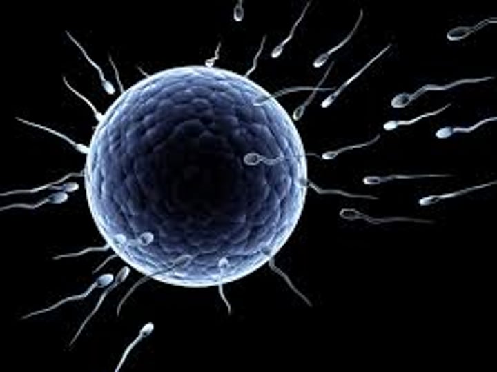
What kinds of cells does Meiosis produce?
gametes (egg and sperm)
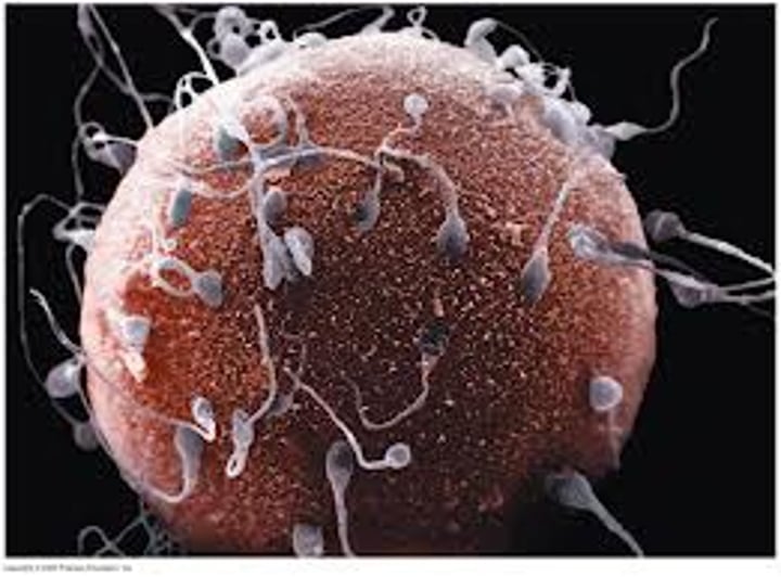
How many chromosomes are in each gamete?
23
How many chromosomes are in each normal cell?
46
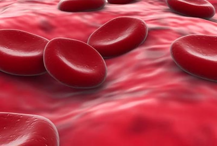
Culture Media
a sterile solution that helps cells grow
Arrest
halt cells in Metaphase
Chromosome Spread - Step 1
add cell sample to culture media
Chromosome Spread - Step 2
culture, or grow, cells in a lab for up to two weeks
Chromosome Spread - Step 3
arresy cells in metaphase by treating them with a chemical that stops cell division at that point
Chromosome Spread - Step 4
swell and drop cells onto microscope slides - cells were burst and spread out their chromosomes
Chromosome Spread - Step 5
Stain with Giemsa dye and observe the chromosomes under a microscope
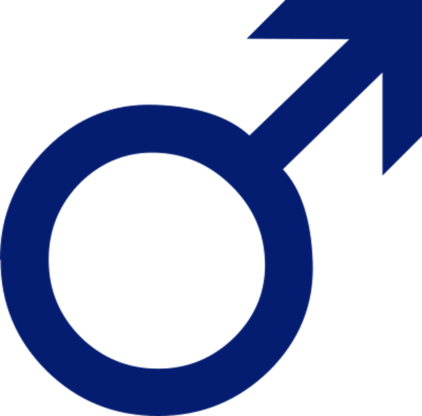
What sex chromosomes does a male have?
XY
What sex chromosomes does a female have?
XX
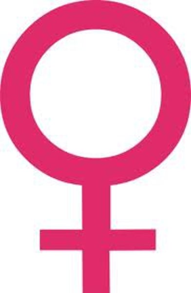
What kind of person is represented by the picture?
unaffected male
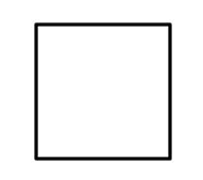
What kind of person is represented by the picture?
unaffected female
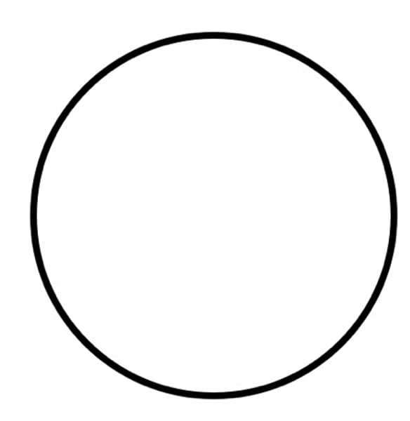
What kind of person is represented by the picture?
affected female
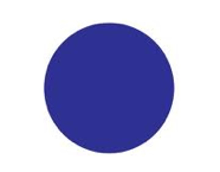
What kind of person is represented by the picture?
affected male

What kind of person is represented by the picture?
male carrier
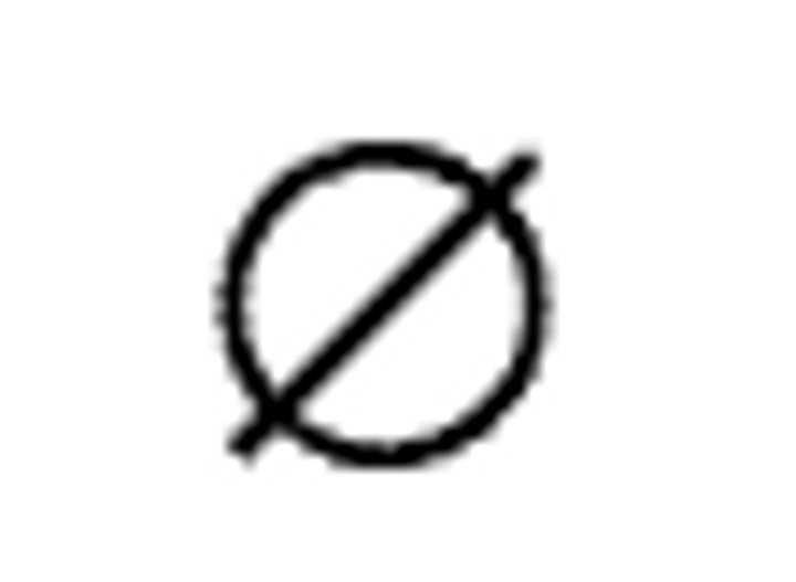
What kind of person is represented by the picture?
deceased female
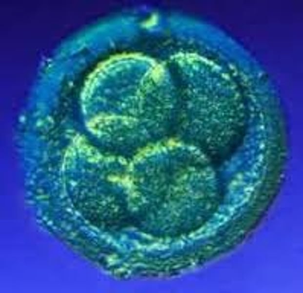
Zygote
a diploid cell made by the fusion of two gametes
Prophase I
homologous chromosomes pair up
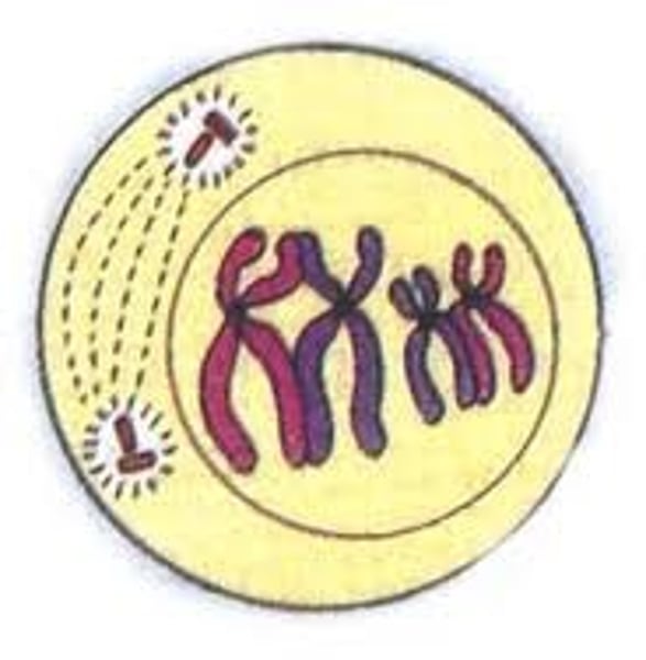
Metaphase I
homologous chromosomes line up at the center of the cell and fibers attach to each homologous chromosomes
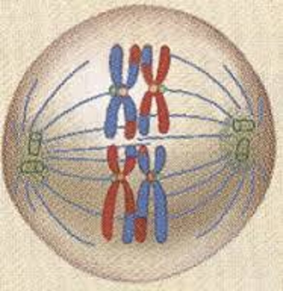
Anaphase I
each chromosome separates and the homologous chromosomes are pulled to opposite poles of the cell
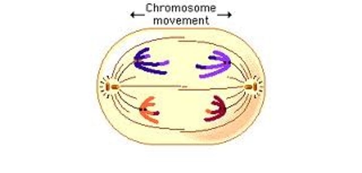
Telophase I + Cytokinesis
the cell splits into two identical daughter cells that are genetically different
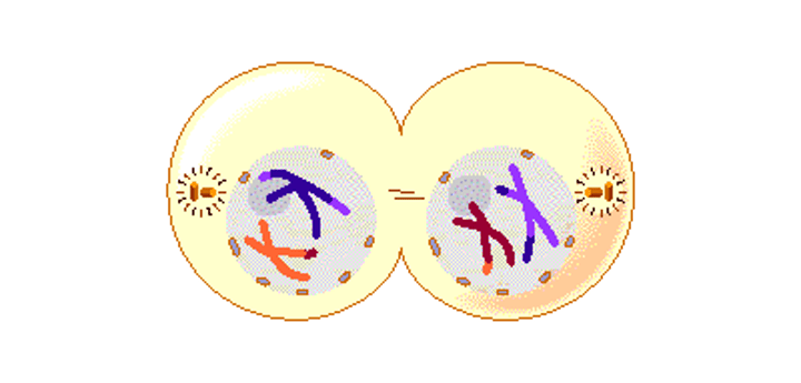
Prophase II
homologous chromosomes pair up in each cell
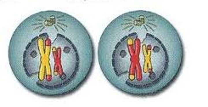
Metaphase II
chromosomes line up in the center of both cells and fibers attach to each chromosome
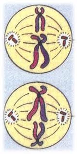
Anaphase II
each chromosome separates and the sister chromatids are pulled to the opposite poles of the cell
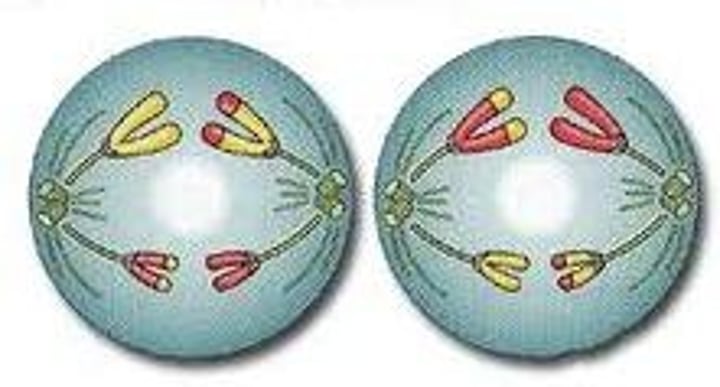
Telophase II + Cytokinesis II
a nucleus forms around each set of chromosomes - cytokinesis takes place, producing four haploid daughter cells
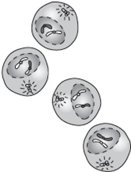
Nondisjunction
an accident during meiosis or mitosis in which members of a pair of homologous chromosomes or sister chromatids to move apart properly
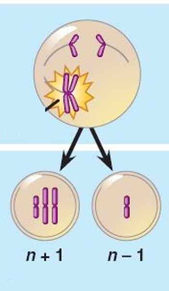
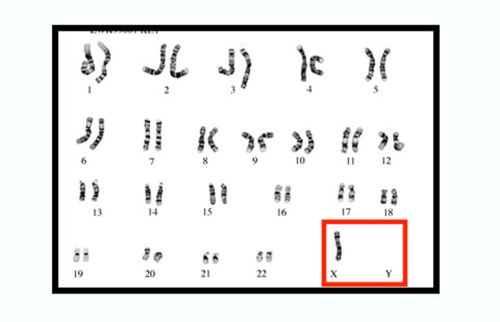
Monosomy
one missing chromosome
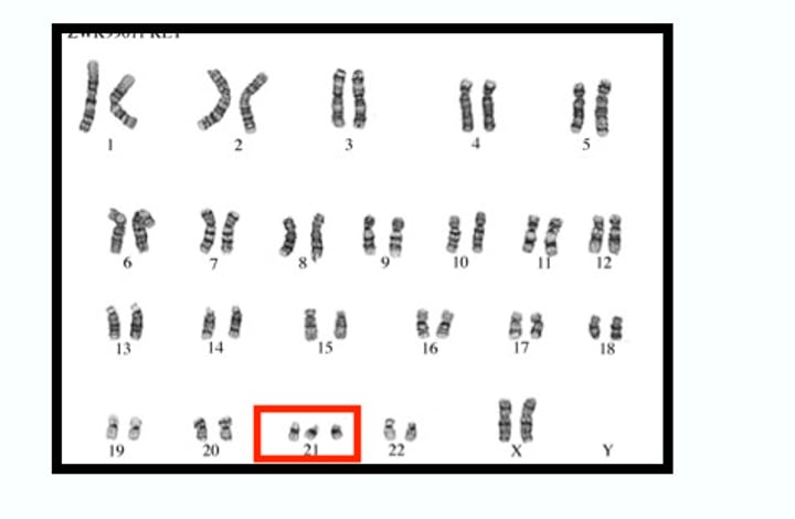
Trisomy
one extra chromosome
Deletion Chromosomal Mutation
occurs when a segment of a chromosome is removed or lost
Insertion Chromosomal Mutation
occurs when a apart of a chromosome is removed and added to another chromosome
Inversion Chromosomal Mutation
occurs when part of a chromosome is placed in the wrong orientation compared to the rest of the chromosomes
Duplication Chromosomal Mutation
occurs when a segment of the chromosome is replicated and inserted next to the original copy
Translocation Chromosomal Mutation
occurs when two chromosomes swap segments