Veterinary Systems Lab Exam 1
 Call Kai
 Learn
Learn Practice Test
 Spaced Repetition
 Match
Match Flashcards
 Knowt Play
1/368

There's no tags or description
Looks like no tags are added yet.
Name | Mastery | Learn | Test | Matching | Spaced | Call with Kai | Chat |
|---|
No analytics yet
Send a link to your students to track their progress
369 Terms
Mitotic Figures
What feature is indicated by the arrows?
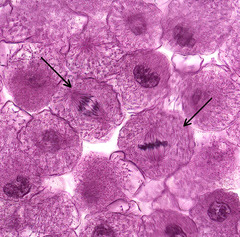
Prophase
What phase of mitosis is indicative of the cell at the arrow?

Metaphase
What phase of mitosis is indicative of the cell at the arrow?
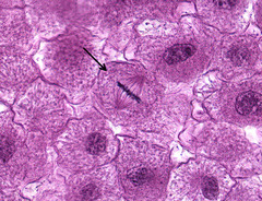
Anaphase
What phase of mitosis is indicative of the cell at the arrow?
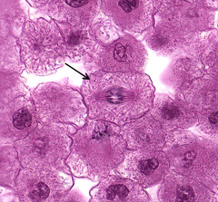
Telophase
What phase of mitosis is indicative of the cell at the arrow?
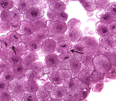
Longitudinal
What plane of section is the starred area?

Cross (Transverse)
What plane of section is the starred area?
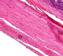
Longitudinal
What plane of section is the vessel at the arrow
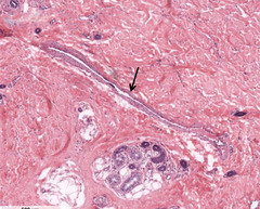
Heterochromatic
How would you describe the nucleous of the indicated cells compared to others?
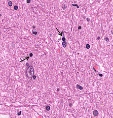
Euchromatic
How would you describe the nucleous of the indicated cells compared to others?
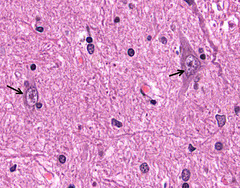
Eosinophilic (Acidophilic)
How would you describe the staining of the indicated cells cytoplasm?

Basophilic
How would you describe the staining of the starred tissue?
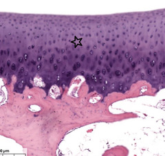
Nucleolus
What feature of the cells are indicated by the arrow?
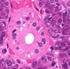
Rough Endoplasmic Reticulum (RER)
What organelle causes basophilic cytoplasm in this cheif cell?
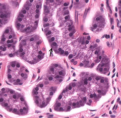
Mitochondria
What organelle causes the cytoplasm of this parietal cell to stain eosinophilic?
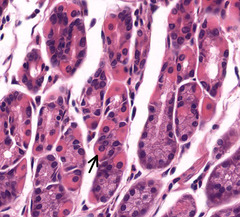
Secretory Vescicles
What are the basophilically staining structures in these cells?

Secretory Vescicles
What are the eosinophilically staining structures in these cells?

Golgi Apparatus
What causes this halo effect near the nucleus of this cell?

Nucleus
What is the starred cell feature?

Plasma Cell
What type of cells are indicated?
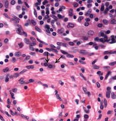
Macrophage
What type of cells are indicated?

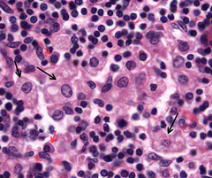
Macrophages - black arrow
Plasma Cells - red arrow
What type of cells are indicated?
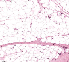
White adipocyte
What type of cells are majority on this image?
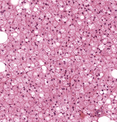
Brown adipose cells
What type of cells are majority on this image?
Nucleus of white adipocyte
What is the indicated structure in the cell?

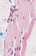
Cross section - skeletal muscle
What plane of section are the indicated cells? What type of cells are these?
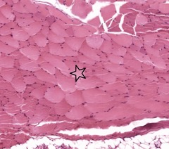
Melanin
What is the brown staining substance in the skin?
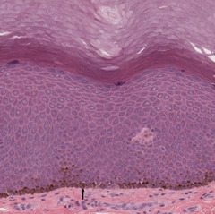
A= anaphase
B= prophase
What mitotic phase is each indicated cell in?
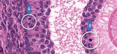
Cross sectional
Plane of section?

Longitudinal
Plane of section?

Secretory vessels in cells of glands
What are the Eosinophilic structures in the cells?
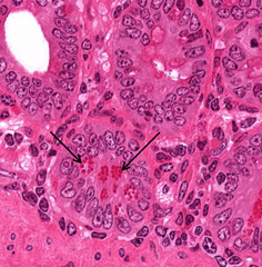
Mesenchyme
What type of connective tissue is pictured?
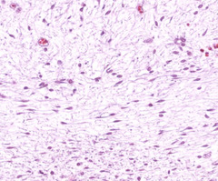
Mesenchyme cells
What are the indicated cells?

Fibroblasts
What are the indicated cells?

Dense Irregular connective tissue
What type of connective tissue is pictured?

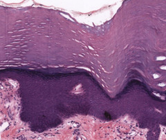
Keratinized simple squamous epithelium
How would you classify the epithelium?
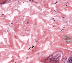
Collagen Fibers
The majority of this connective tissue is staining eosinophilically due to...?
Fibroblasts
What cell is indicated by the area in this connective tissue?
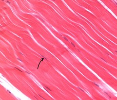
Dense Regular Collagenous Connective Tissue
What type of connective tissue is indicated by the star?
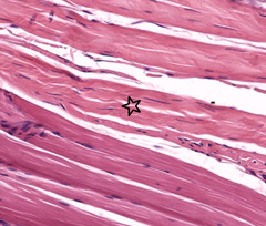
Skeletal Muscle
What is indicated by the star?

Loose connective tissue
What type of connective tissue is visible?

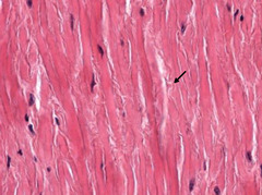
Elastic fibers in dense regular CT
What is indicated by the arrow
Dense regular elastic CT
Blue= collagen fibers
Red = elastic fibers
Name type of connective tissue?

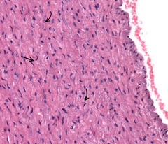
Elastic sheets
What is indicated by the arrows in this layer of the aorta?
Brown adipocytes
What kind of cells are these?

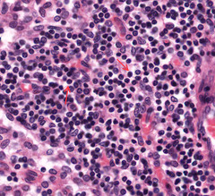
Lymphocytes
Most of the cells and those indicated on this slide are ....?
Red = Plasma cells
Black = Macrophage
What cells are marked by the indicated arrows?

Star = RBCs
Arrow = Neutrophil
What types of cells are indicated by the star and arrow?

Black = Plasma Cell
Red = Macrophage
Blue = Eosinophil
Green = collagen fibers
What is indicated by the arrows?

Fibroblast
What is indicated by the arrow?
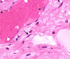
Black = Mast cell
Red = Eosinophil
Blue = fibroblast
Green = Lymphocyte
What is indicated by the arrows?

Elastic Cartilage
What type of tissue is this?

Hyaline Cartilage
What type of tissue is this?
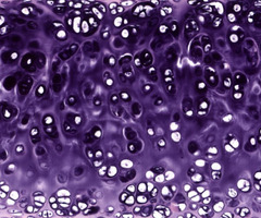
Hyaline Cartilage
What type of tissue is this?
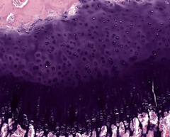
Fibrocartilage
What type of tissue is this?
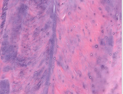
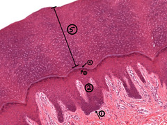
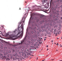
Simple Squamous Keratinized Epithelium
1. Stratum Basale - single layer of germinal cells resting on the basement membrane which is attached to the dermis.
2. Stratum Spinosum - keratinocytes attached to each other by desmosomes on spiny processes.
3. Stratum Granulosum - keratinocytes with numerous basophilic granules in their cytoplasm.
4. Stratum Lucidum - highly refractive zone only seen in very thick skin.
5. Stratum Corneum - thick layer of dead cells
Classify type of epithelium and indicate the layer at each number?
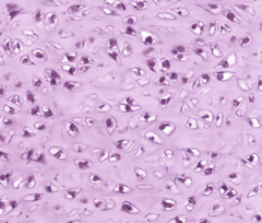
Chrondocytes housed in lacuna
What are the cells and where are they housed in?
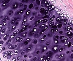
Chondrocytes
What are the cells?
Elastic Cartilage
Red = Ground Substance
Green = Chondrocyte
Blue = Elastic Fibers
Yellow = Lacuna
What kind of tissue? What are the different arrows indicating?

Red = Perichondrium
Blue = Fibrous layer of Perichondrium
Yellow = Chondrogenic Layer of Perichondrium
Green = Cartilage
Black = Chondroblast
What is indicated by the different markings?

Ground substance = proteoglycans/GAGs
What causes the Basophilic staining in cartilage?

A = appositional growth
B= Interstitial growth
In which area would appositional growth occur? What about interstitial growth?

Perichondrium
What is the outer covering around cartilage called as indicated by "A"?
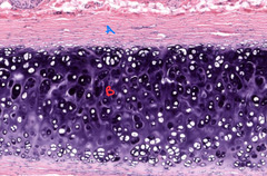
Isogenous Groups
Interstitial growth occurring
What is special about the indicated chondrocytes?

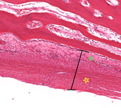
Black = Periosteum
Yellow = Fibrous layer of periosteum
Green = Osteoprogenic layer of periosteum
What is indicated by the different labels?
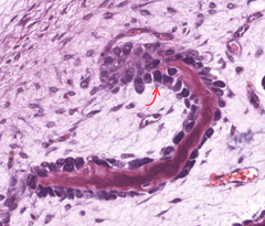
Active osteoblasts
What is indicated by the arrow?
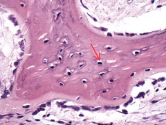
Osteocyte
What is indicated by the arrow?
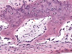
Inactive osteoblast
What is indicated by the arrow?
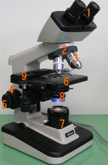
1. Eyepiece lens
2. Revolving nosepiece/turret
3. Objectives lens
4. Course focus knob
5. Fine focus knob
6. Stage
7. Light Source
8. Condenser
9. Knobs controlling x/y stage movement
Identify parts of microscope
Vacuoles
What term is used to describe the clear spaces inside these cells?

C
Where would you find hemidesmosomes?
Mesenchymal cell
Identify the cell at the arrow in this embryo.

Simple cuboidal
Classify the epithelium in these starred tubules.

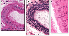
B
Which epithelium is stratified?
Sebaceous gland
Identify the circled skin structure?

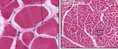
Skeletal muscle
Identify the specific tissue
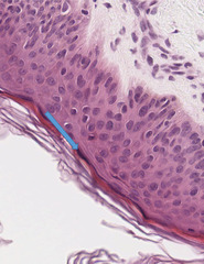
Stratum granulosum
Identify the indicated layer
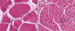
Skeletal muscle
Identify the specific tissue?
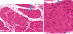
Cardiac muscle
Identify the specific tissue in the box?

Smooth muscle
Identify the tissue?
B
Identify the dense regular connective tissue?

Fibroblast
Identify the connective tissue cell?
Mast cell
Identify the connective tissue cell?
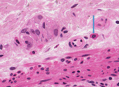
Loose connective tissue
Identify the connective tissue in the circled location?
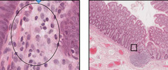
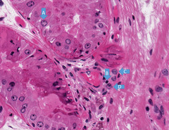
A. Epithelial cell
B. Lymphocyte
C. Macrophage
D. Plasma cell
Identify the cells
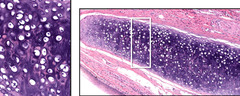
Elastic cartilage
Identify the tissue found in the epiglottis?
Collagen fibers
What causes the pink staining of the extra cellular matrix of this fibrocartilage?
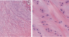
Cell processes of an osteocyte; tissue fluid
What normally occupies the space at the arrow?

Hyaline cartilage at the growth plate
Identify the tissue at the arrow.

Purkinje fibers
Identify the specific cells

Lymph vessel
Identify this vascular structure

Endothelial cell
Name the cell to which this nucleus belongs.
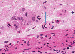
Arteriole
Identify the structure in which the star is placed.

Hyaline cartilage
Identify the tissue at the arrow on the right.

Nissl substance/RER
Identify the Basophilic material in the cytoplasm of this cell
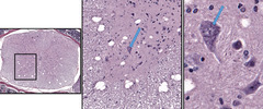
Astrocyte
Identify the glial cell.
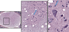
Subarachnoid
In what space is this blood vessel located?

C
In what region is the location of the preganglionic autonomic neuron cell bodies?
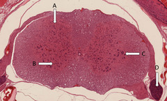
Nerve
Identify the circled structure

Transitional
Classify the epithelium
