Molec Cell Exam 1
 Call Kai
 Learn
Learn Practice Test
 Spaced Repetition
 Match
Match Flashcards
 Knowt Play
 Podcast
1/87

Earn XP
Description and Tags
Q and A podcast
Name | Mastery | Learn | Test | Matching | Spaced | Call with Kai | Chat |
|---|
No analytics yet
Send a link to your students to track their progress
88 Terms
DNA Synthesis
DNA replication: One strand of DNA serves as a template strand for the synthesis of a complementary strand in the 5’ → 3’ direction
DNA Polymerase
Adds new necleotides to the 3’ end of the growing strand (complementary to the template strand)
DNA Helicase
Unwinds the DNA double helix by breaking hydrogen bonds (forms a replication fork)

Single Strand Binding Proteins
Stabilize single stranded DNA to prevent it from re-joining

Topoisomerases
Relieves supercoiling and tension ahead of the replication fork by cutting and re-joining DNA strands

DNA Primase
Synthesize a short RNA primer to provide a free 3’OH group required for DNA polymerase to start
DNA Ligase
seals nicks and joins Okaizaki fragments (short length of DNA) together into a continuous strand
Directionality of DNA Synthesis
DNA strands are Antiparallel (5’→3’, 3’→5’)
Leading strand: synthesized continuously in the same direction that the replication fork opens
Lagging strand: synthesized discontinuously in short segments (akazaki fragments) moving away from the advancing fork → later glued together by DNA Ligase
Replication Fork
Y shaped DNA junction at the site where the DNA is being replicated
DNA polymerase proofreading
DNA polymerates double checks the newly synthesized DNA using 3’→5’ exonuclease activity (removing wrong nucleotides) → DNA polymeraze then replaces it with the correct nucleotide by resuming the 5’→3’ polymerase activity
Consequences of defective proofreading
Hypermutability: loss of exonuclease activity → severe mutator → rapid increase in number of mutation rates
Genomic instability: uncorrected replication errors will disrupt normal gene function and cellular processes
Disease predisposition: inherited mutation that disable proofreading can cause conditions such as Polymerase Proofreading-Associated Polyposis (PPAP) or increase risk to cancer
Replication errors
Origin: direct mechanic and enzymatic errors by DNA polymerase during cell division
Mechanism: the enzyme (DNA polymerase) inserts incorrect nucleotide creating insertion or deletion loops
Fix/ Faliure: caught by proofreading or MMR (if missed it becomes permanent in daughter strand)
Spontaneous Mutations
Origin: Intrinsic, natural chemical reactions inside the cell occurring independently of external radiation or chemicals.
Mechanism: driven by normal cell conditions (thermal fluctuations, water attacks leading to hydrolysis)
Depurination: loss of a purine base
Deamination: loss of an amino group from a base (C→U, reading U as T)
Fix/ Faliure: Processed by BEP (if left unfixed damaged template mispairs or halts polymerases during the following replication cycle
Mismatch Repair (MMR)
corrects mispaired/non-complementary bases left behing by copying mistakes
after proofreading
Only occurs during and after DNA replication
Steps of MMR
1) Mismatch Detection
2) Newly synthesized strand is identified :which is the daughter strand with mistake
in prokaryote the Parent DNA strand is methylated, daughter strand remains unmethylated
3)Recruitment and insition: MutS recruits MutL complexes
these encode MMR proteins
4)Section of DNA is removed: Helicase and exonuclease target and remove single stranded segment with incorrect nuclease→leaves gap
5)DNA is re-synthesized: DNA polymerase fills gap DNA Ligase seals
Deamination
loss of an amino group from a nucleotide base by a hydrolic reaction
Causing changes such as:
C to U: U is read as T and will pair with A (C→G to T→A)
5-Methylcytosine to T: hotspot for spontanous mutation
A to Hypoxanthine: is read as G and will pair with C ( A→T to G→C)
G to Xanthine: will pair with C, but stalls replication
Depurination
loss of A or G bases (by spontaneous hydrolysis→ loss of glycosidic bond on base)
leads to deletions or mutations
UV induced mutation
a cyclobutane ring forms between adjacent T bases → T’s can not pair with A’s
leads to distorted strand
Repaired by NER/photolyase
Base Excision Repair (BER)
General repair mechanism for nucleotide damage (chemically altered or modified single base)
damaged base is replaced before the next replication cycle
Operates throughout the entire cell cycle
BER steps
1) Recognition and removal of damaged base: DNA glycosylase identifies and removes the base
2) Addition of new repaired base by DNA polymerase
3) DNA Ligase seals the nick in the backbone
Nucleotide Excision Repair (NER)
fixes thymine dimers and multiple base damages (Bulky damages)
Defects can lead to Xeroderma Pigmentation
Transposons
segments of DNA (motile) that can change its location inside the genome
LINEs: Long Interspersed Nuclear Elements (~6000-8000BP), Autonomous (move independently)= reverse transcriptase and endonuclease
SINEs: Short Intersperesed Nuclear Elements (~100-400BP), Non-autonomous (Needs a partner LINE to move) = non coding sequences
Bacterial Transposons
May carry antibiotic resistance genes
Occures by two pathways
Replicative transposons: copy-paste
Non-replicative transposons: cut-paste
Replicative transposons
the enzyme transposase cleaves the insertion sequence at the end of the inverted repeat of the donor site
cleaves a random target site
insertion sequence: cu from donor site pasted in target site
result: insertion sequence moves from one site in the genome to another
Replicative Transposon
a copy of the insertion sequence is made by local DNA replication and pasted into target site
result: copy of insertion sequence stays intact and appears in target site
Retrotransposition
done by reverse transcriptase synthesizing a DNA copy of transcribed retrotransposon
Only in eukaryotes
can generate many copies of original mobile element
in some plants retrosposons account for most of the DNA in the genome
reshape genome structure, alter gene expression, and contribute to genetic diversity
DNA Virus infection cycle
1) Enter host cell (nucleus)
2) Replicate viral genome
3) Protein synthesis (transcription, translation)
4)Assemble progeny
5) Release
RNA virus infection cycle
1) enter host cell (cytoplasm)
2) synthesize proteins (translation)
3)Assemble RNA replicase complex
4) Synthesize complementary RNA
5) goes two ways:
Retroviruses
Use reverse transcriptase
Organization of Eukaryotic chromosomes
DNA double bonded helix→ Nucleosomes→ Chromatin fiber → higher order loops and topological domains → condensed metaphase chromosomes
Nucleosomes
DNA wrapped around 8 Histones (repeating unit of chromatin)
Higher order chromatin structures
compact meters of DNA into a microscopic nucleus while precisely controlling gene activity, DNA repair, and replication.
Histone and chromatin associated proteins
organize long DNA molecules into compact structures inside the cell nucleus while controlling gene access
The four core histone proteins
H2A, H2B, H3, and H4
They bind together to form a protein disc called a histone octamer
role of Histone H1
It acts as a linker histone
It binds the entry/exit sites of DNA on the nucleosome (locks the strand into place to help fold the 10-nm fiber into a more compact 30-nm chromatin fiber)
chromatin-associated remodelers
use ATP energy to slide, evict, or modify nucleosomes
This shifts DNA packaging to dynamically open or close specific areas of the genome
Euchromatin
Loosely packed
Accessible
Transcriptionally active (genes are on).
Gene rich
Heterochromatin
Highly condensed
inactive/ Silenced (genes are off)
inaccessible
Repeat Rich
post-translational modifications (PTMs)
use:
Phosphorylation: kinases add phosphate groups to amino acids creating a physical block or an electrical shift that activates or halts an enzyme
Cleavage: targeted cutting triggers a sudden release of biological activity in proteins (by insuline protein)
Alters gene access
chemical groups added to histone tails (acetylation/methylation) change the physical tightness of the DNA-histone grip and serve as binding docking sites for gene regulatory proteins
three essential functional elements required for eukaryotic chromosome maintenance and inheritance
Centromeres, telomeres, and origins of replication
structural hallmark of a eukaryotic centromere
Repetitive DNA packed with specialized CENP-A histone variants instead of standard H3, which epigenetically marks the region for chromosome segregation.
primary role of the centromere during cell division
consists of repetitive G-rich sequences (like TTAGGG) that form a single-stranded overhang
this overhang loops back into a T-loop stabilized by the shelterin protein complex
telomeres solve
end-replication problem
act as non-coding buffer zone (prevents loss of genetic data during shortening of DNA during replication)
Eukaryotic chromosome origins of replication
enriched with A and T bases
A-T base pairs share only two hydrogen bonds (compared to three in G-C pairs), making it energetically easier for replication machinery to pull the strands apart
role of the Origin Recognition Complex (ORC)
binds directly to origins of replication during the cell cycle to license and initiate bidirectional DNA duplication
Chromatin condensation
tightly packs DNA into Hetorochromatin (physical tightening of DNA)
Causes gene innactivation
Histone modification
chemical tags on Histone Tails
DNA Methylation
Chemical tags directly on DNA bases
Adding methyl groups to specific amino acids (like H3K9 or H3K27) recruits silencing proteins
Methyl groups (usually added to cytosine bases in CpG islands) physically block transcription factors from binding to the promoter region
Silencing
Histone Deacetylation
Removing acetyl groups increases the positive charge of histones, making them bind DNA more tightly
How do DNA methylation and histone modifications work together to lock a gene silent
Methylated DNA attracts Methyl-CpG-binding domain proteins (MBDs)
These proteins recruit Histone Deacetylases (HDACs), which strip acetyl groups and force the chromatin to condense
Xist (X-inactive specific transcript)
makes a long non-coding RNA molecule (lncRNA) that physically coats the X chromosome from which it is transcribed, triggering silence
X-chromosome inactivation
Female mammals shut down one of their two X chromosomes to match the single X chromosome dose found in males
Epigenetic inheritance
The chromosome which is silenced is tagged (the tags are part of epigenetics)
DNA Methylation : cells add methyl groups to DNA (small chemical tags) to insure inactivity
Histone modifications: histones receive inhibitory marks, and active histones are removed
Miotic Memory: daughter cells of inactive chromosome will have tags to ensure inactivation
Promoters
Act as the primary binding site for RNA polymerase and general transcription factors to initiate transcription
Found just upstream (at the 5' end) of the main coding region
Includes:
core promoter: positions the enzyme correctly (often containing a TATA box)
proximal promoter: help modulate the basal rate of transcription
Regulatory sequences
Controls when, where, and at what level a gene is expressed
upstream, downstream, or inside introns
Includes:
enhancers: boost transcription when specific activator proteins bind
silencers: repress transcription when repressor proteins bind
Exons
sequences retained in the mature messenger RNA (mRNA) after splicing
expressed/ kept sequences
Segments interspersed throughout the transcribed portion of the gene
many exons contain the protein-coding sequence (CDS) translated into amino acids
contain the untranslated regions at the ends of the transcript
Introns
Transcribed into initial pre-mRNA but completely removed during RNA splicing before the mRNA is translated
Found in intervening non-coding sequences that alternate with exons in the DNA and pre-mRNA
Not expressed/ cut out
Contain vital signals like splice donor and acceptor sites, and sometimes harbor auxiliary regulatory motifs or alternative splice variants
Unregulated regions (UTR’s)
Regulate translation efficiency, mRNA stability, and subcellular localization, though they are not translated into protein
How and when gene expression happens
Found at both ends of the processed mRNA transcript, originating from terminal exons
Include:
5′ UTR: Sits before the start codon, assists in ribosome binding and translation start
3′ UTR: Sits after the stop codon, contains polyadenylation signals and binding sites for microRNAs that control mRNA degradation
Genome size and density comparison
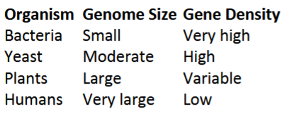
Humans have:
Large introns
Extensive repetitive DNA
Regulatory sequences spread over large regions
Tandem repeats
repeats adjacent to each other (together)
found in centromeres and telomeres
simple repeats
Transposable elements (interspersed repeats)
can move or copy themselves
make up a large fraction of eukaryotic genome
include:
retrosposons (copy and paste, LINEs, SINEs): class I
DNA transposons (cut and paste): class II
Repeated sequences
influence chromosome structure
Drive genome evolution
Affect gene regulation and stability
Polypeptides
linear polymer of amino acids
synthesized by ribosomes
Opposes directionality:
synthesis occurs from N terminus to C terminus
Connected by covalent peptide bonds
Polypeptide synthesis
Condensation/ Dehydration reaction: two amino acids are joined by removing a water molecule
A protein’s amino acid sequence
consists of:
Primary structure: amino acid sequence (bases) (covalent peptide bonds)
Secondary structure: alpha-helices and beta sheets (hydrogen bonds)
Tertiary Structure: 3D fold driven by side chain interactions (side chains)
Quartery structure: assembly of multiple peptide subunits (hydrogen bonds, ionic bonds, hydrophobic interactions, and occasionally disulfide bonds or covalent links)
Molecular chaperone
help protein fold correctly
Misfolding consequences
aggregation: loss of function or toxic gain of function
linked to neurodegenerative disease
Protein Folding
spontaneous process where polypeptide adopts its native energetically favorable confirmation
protein misfolding consequences
disrupts proper three-dimensional conformation
lead to a loss of function or gain of toxic properties ( linked to disease)
Prion disease
Infectious misfolded proteins template the conversion of healthy proteins into a toxic, aggregated state via a self-propagating cascade
touches normal protein and acts like a mold or template to unfold it
can lead to Transmissible spongiform encephalopathies (TSE): demaged brain tissue
Disulfide bonds
form between thiol groups of cystein residues
Stabilized tertiary + quarternary structures of proteins
Occurs in Oxidizing/ reducing environments ( ER, Cytoplasm)
Denaturation
loss of 3D structure due to disruption of non-covalent bonds by heat, pH extremes, or chemicals
can be reversible
Bad environment→ protein loses shape→protein loses function
Breaks 2-4 structure of proteins
Reducing Agents
reduce/ break covalent disulfide bonds, convertthem into free thiols (beta-mercaptoethanol)
breaks 3-4 structure of proteins
Ubiquination
Ubiquitin cahin is attached to tag a protein for degradation
polyubiquitin chains targett proteins
proteosome degrades ubiquitilated protein into peptides (ATP dependent)
Elastin fibers
a rubberlike elastic meshwork present in the extracellular matrix of some cell types
allow tissues such as skin, arteries and lungs to stretch and recoil without tearing
Fibrillar collagens
major structural proteins of connective tissues
built of triple helices of procollagen polypeptides
Macromolecules
Large, complex molecules vital for life, built from smaller building blocks
Monomers
single subunit of polymers
Dehydration/ condensation synthesis
building polymers by covalently bonding monomers
releases a water molecule
Hydrolysis
breaking down polymers by adding a water molecule
four major classes of macromolecules
1) Carbohydrates
2) Lipids
3)Proteins
4)Nucleic Acids
Carbohydrates
subunit is monosaccharides (simple sugars)
are carbon rings or chains
provide short term energy and support
Lipids
glycerol + fatty acids
For long term energy (triglycerine), creating cell membrane (phospholipids), and structural support (steroids→cell signaling)
include:
Saturated fatty acids: no double bond, pack tightly
Unsaturated fatty acids: double bonds, create kinks
Proteins
monomers are amino acids
made from amino, carboxyl, and r-groups
provide enzymes, structure, transport and defense
linked by peptide bonds
Nucleic acids
monomers are nucleotides
made from phosphate, pentose sugan, and nitrogen base
store and transmit genetic info
Chemical Interactions
covalent bonds
ionic bonds
hydrogen bonds
van der waals interactions
Sickle Cell Disease
Changing a single amino acid in hemoglobin alters its structural folding
causes the proteins to clump into rigid fibers, distorting red blood cells into a sickle shape and reducing oxygen transport