Exam 2: Bridging structural and cell biology with cryo-electron microscopy
1/71
There's no tags or description
Looks like no tags are added yet.
Name | Mastery | Learn | Test | Matching | Spaced |
|---|
No study sessions yet.
72 Terms
What is single-molecule fluorescence spectroscopy?
allows real-time observation of conformational changes in individual DNA, RNA, and proteins during their function
What is the importance of single-molecule fluorescence spectroscopy?
Measures real-time structural dynamics of individual molecules.
Helps correlate molecular structure with function.
Reveals molecular heterogeneity—identical molecules can behave differently under nearly identical conditions.
Previous skepticism regarding heterogeneity has been addressed with technical advancements and stronger biological connections
What was found using single-molecule fluorescence spectroscopy?
Hairpin ribozyme and holiday junction dynamics
tRNA Dynamics in ribosome
Helicase protein movement on ssDNA
Rotational steps of F0F1-ATP synthase
What is the mechanism of hairpin ribozyme and holiday junctions?
hairpin ribozyme: docking/undocking reactions, cleavage or ligation reactions
holiday junctions: stacking conformer dynamics and single-step branch migration
Memory effect - molecules switch between two distinct internal clocks
WHta is the mechanism for tRNA in ribosomes
transient intermediates involved in tRNA accommodation were observed for first time
What is the mechanism for helicase protein movement on ssDNA
ATP-powered helicase protein moves along single-stranded DNA (ssDNA).
When encountering obstacles, the protein performs acrobatic movements to return and restart the process.
what is the mechanism for rotational steps of F0F1-ATP synthase
First direct measurement of discrete rotational steps during ATP synthesis
Direction of rotation (clockwise vs. counterclockwise).
Asymmetry in dwell times at three rotor angles.
What is Single-Molecule Fluorescence Resonance Energy Transfer (FRET)?
powerful tool for measuring relative distance changes between two fluorophores (donor & acceptor) attached to specific sites on a macromolecule, able to see fluorescence intensity variations, able to see lifetime and spectral properties of fluorophores
What are the advantages of using FRET?
Provides precise insights into conformational changes at a molecular level.
Captures dynamic structural transitions.
What is the hairpin ribozyme?
RNA enzyme that catalyzes site-specific self-cleavage and ligation of viral RNA
What is the structure of a hairpin ribozyme?
modular architecture, consisting of a four-way junction and two adjacent loops that interact to form the catalytic site. The docking of these loops, which is stabilized by divalent metal ions, is significantly enhanced by the junction's natural fluctuations
Is the cleavage reaction of a hairpin ribozyme irreversible?
No, it is able to be retained
What are the docking/undocking dynamics of a hairpin ribozyme?
at 1mM magnesium, most ribozymes were found in a stably docked state (high FRET), interrupted by rapid docking/undocking bursts
When a noncleavable mutant showed steady FRET signals, what was the hypothesis for a hairpin ribozyme?
Stable docking = ligated ribozyme
Rapid fluctuations = cleaved ribozyme
What was the experimental evidence that supported that stable docking = ligated ribozyme
and rapid fluctuations = cleaved ribozyme?
Upon adding magnesium ions, ribozymes transitioned from undocked to stably docked states. Rapid fluctuations appeared later, suggesting these were triggered by cleavage
Short cleavage substrate ribozymes were exposed to magnesium, leading to cleavage and quick dissociation of the product. The resulting impaired ribozyme structure could not refold (low FRET signal). When a buffer containing a high concentration of long (7 bp) cleavage product was introduced, the ribozyme underwent: Low FRET → Rapid fluctuations → Stable high FRET. Since the ribozyme was in a cleaved state immediately after binding, the rapidly fluctuating signal was assigned to the cleaved ribozyme
How was real-time tracking of cleavage and ligation reactions achieve? What did this enable?
achieved by monitoring docking/undocking kinetics
enabled single-molecule enzymology, allowing researchers to study mutations and solution conditions without interference from conformational complexity
“Why is undocking accelerated after cleavage?” What was the hypothesis?
Rapid undocking may facilitate presentation of the cleavage product for the next step in viral RNA replication, rather than promoting religation.
“Why is undocking accelerated after cleavage?” What were the unexpected findings?
When the cleavage product had a nonnatural terminus, this acceleration was not observed
modifications to the ribozyme can impact both folding and catalysis
What is the structure of a holiday junction/what is it?
four-way DNA structure formed during homologous recombination, a key process for maintaining genome stability and diversity.
How did the design the experiment using Holiday junctions to find if homologous junction behaves like nonhomologous junction?
designed a monomigratable junction, allowing a single step of branch migration, and used single-molecule fluorescence resonance energy transfer (FRET) to observe its dynamics
How does the Holiday junction behave when it is in the presence and absence of metal ions?
Absence: junction adopts an open structure with four arms forming a square
Presence: junction folds into a stacked X-structure, which exists in two conformations(isol(low FRET) and isoll(high FRET) whihc exchange thorough open intermediate, needed for branch migration
What is the role of open structure for Holiday Junctions?
open structure facilitates spontaneous branch migration, while the stacked X-structure inhibits it
what is monomigratable junction design?
The designed junction could migrate only one step between two defined branch points, U and M.
The arms of the junction were labeled with two fluorophores, which produced low FRET (isoI) and high FRET (isoII) signals
How does single-molecule analysis work?
Four-state fluctuations were observed, grouped into two distinct phases:
Slow-fluctuating phase (assigned to branch point U)
Fast-fluctuating phase (assigned to branch point M)
Molecules switched between these phases, which was interpreted as branch migration
How is Holiday junctions like a pendulum?
Oscillates between two states
How is branch migration similar to two pendulums?
acts as a spring linking two pendulums:
If one pendulum starts swinging, energy eventually transfers to the second one.
If the coupling is weak, each pendulum swings multiple times before energy transfers
Why is migrability a weak perturbation for holiday junctions?
Retains behavior of nonhomologous junctions within each branch point
What are the factors that effect branch migration?
GC vs. AT base pairs: GC pairs (stronger bonds) slowed migration more than AT pairs.
Magnesium concentration: Affected the overall rate of fluctuations but not the relative populations of isoI and isoII states
When Holiday junctions were being analyzed, what did branch points exhibit? What does this suggest?
Different branch points exhibited up to 30-fold variations in their lifetimes (Figure 3C).
This suggests that spontaneous migration can be rapid within local regions, but some branch points act as strong barriers
What did the study find for Holliday junction dynamics?
provides new insights into junction dynamics, which could aid in understanding how enzymes recognize and process Holliday junctions
protein-free Holliday junctions may rapidly migrate to stable branch points, potentially influencing recombination outcomes
In bacteria what do ribosomes consist of?
two subunits, 30S and 50S. The mRNA binds to the 30S subunit, allowing tRNA to recognize codons via anticodon base pairing
In order to analyze tRNA, how the experiment set up?
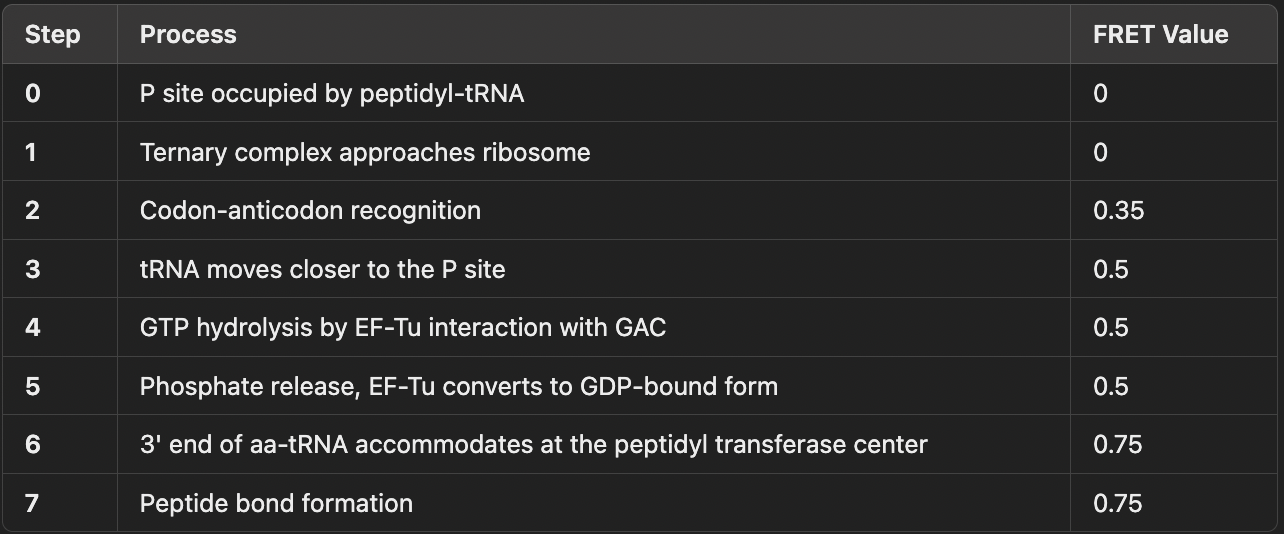
The P site was occupied by a donor-labeled tRNA, bound to a biotinylated mRNA attached to a PEG-coated surface.
A ternary complex (aminoacyl-tRNA, EF-Tu, and GTP) was introduced while FRET was recorded during its accommodation at the A site.
Because the timing of ternary complex binding varied, a post-synchronization technique aligned traces at the first moment when FRET exceeded 0.25
What FRET transitions did tRNA show during tRNA accommodation?
Low FRET (0.3–0.5) → High FRET (0.6+) as the ternary complex accommodated into the A site.
What was found during tRNA accommodation, excluding the FRET transitions?
Antibiotic tetracycline prevented full accommodation, stalling at a short-lived 0.35 FRET state.
Replacing GTP with GDPNP stalled the process at 0.5 FRET for ~8s, indicating more stable interactions before GTP hydrolysis.
Cleavage of the sarcin-ricin loop (GTPase activating center, GAC) also stalled the reaction at 0.5 FRET, confirming its role in tRNA selection
What happened to the near-cognate tRNA at the end of the tRNA experiment?
Near-cognate tRNAs (one-base mismatch) tended to dissociate from the ribosome at low and intermediate FRET states, failing to reach full accommodation
What was concluded about near-cognate tRNA and translation fidelity at the end of the tRNA experiment?
The probability ratios for cognate vs. near-cognate tRNAs at each FRET transition confirmed that two proofreading steps exist in tRNA selection.
The fidelity of tRNA selection was estimated at 1 miscoding error per 5000 translation events
What was the stepwaise model of tRNA selectrion based on FRET transitions?
After A site tRNA accommodation, what did fluctuate between? What do each of these numbers mean?
After A site tRNA accommodation, FRET fluctuated between 0.74 and 0.45.
0.74 FRET (Classical State):
tRNAs remain in A and P sites, both anticodon and 3’ ends properly positioned.
0.45 FRET (Hybrid State):
Anticodons stay in A and P sites, but 3’ ends shift to P and E sites.
in the tRNA experiment, how did peptide bond formation effect the lifetime folding? How does this effect ribosome translocation?
Peptide bond formation reduced the classical state lifetime 6-fold, increasing time spent in the hybrid state, which primes the ribosome for translocation
What was the overal concepts found in the tRNA experiments?
quantitative fidelity estimates, confirmed the two-step proofreading model, and demonstrated dynamic transitions between classical and hybrid states
What does helicase do?
ATP-powered translocation along single-stranded DNA (ssDNA) or RNA, unzipping enzymes
What does the Escherichia coli Rep helicase do?
moves in the 3' to 5' direction as a monomer but cannot unwind DNA alone
How was Escherichia coli Rep helicase studied? What did it find?
using single-molecule FRET with Cy3-labeled Rep and Cy5-labeled DNA
repetitive shuttling" behavior, where Rep continuously translocates and then snaps back in a sawtooth-like pattern rather than simply dissociating at the DNA junction
What was the experimental set up for Single-Molecule FRET Observation of Rep Translocation?
30-tailed partial duplex DNA with an acceptor-labeled (Cy5) junction was immobilized on a polymer-coated surface.
Cy3-labeled Rep helicase was introduced with ATP, and translocation events were recorded with 15 ms time resolution
What was found through FRET signal interpretation?
Upon Rep binding to DNA, a donor (Cy3) signal increase was observed.
Gradual FRET increase (donor decrease, acceptor increase) indicated 3' to 5' translocation toward the junction.
Surprising Result: Instead of dissociating at the junction, FRET abruptly dropped back to its initial level.
This cycle of gradual FRET rise and sudden drop repeated multiple times, forming a sawtooth-like pattern
What was the conclusion to Single-Molecule FRET Observation of Rep Translocation?
The gradual FRET rise corresponds to ssDNA translocation.
The sudden FRET drop represents snapback, where Rep returns to the 3' end and restarts translocation.
This newly observed behavior was termed "repetitive shuttling."
How is repetitive shuttling dependent on ssDNA Tail Length?
Time between successive snapbacks (Δt) increased with longer ssDNA tails.
This confirmed that the FRET increase directly corresponds to translocation, while snapback resets the process.
Rep does not maintain contact with the 3' end during translocation, as shown by FRET data when Cy5-labeled Rep moved away from a Cy5-labeled 3' end
What was the hypothesis for why there was a snapback during repetitive shuttling?
encountering a physical blockade (e.g., duplex junction) induces Rep to rebind the 3' end, triggering snapback
What was the supportive evidence found for why there was a snapback during repetitive shuttling?
Conformational analysis of the ssDNA tail during repetitive shuttling revealed the formation of a transient DNA loop, synchronized with the shuttling cycles.
Since ssDNA is highly flexible, it can rapidly form loops, supporting this looping mechanism for repetitive shuttling
What are the two crystal conformations of Rep?
Open form (low FRET).
Closed form (high FRET), which is favored when bound to partial duplex DNA
How did they test if encountering a blockade may induce Rep to adopt the closed form?
Rep translocation assay was performed using dual-labeled Rep to distinguish between open (low FRET) and closed (high FRET) conformations
What was found during the Rep translocation assay?
Rep gradually closes as it moves toward the duplex junction.
Upon reaching the blockade, Rep abruptly opens, coinciding with snapback and translocation restart
What was concluded after Rep translocation assay?
blockade-induced closing increases Rep’s affinity for the 3' end, facilitating snapback
What is Rep’s role in DNA replication?
DNA damage can stall replication, requiring restart after repair. Rep is known to be involved in replication restart
How was Rep’s role in replication restart confirmed?
observed repetitive shuttling of Rep on ssDNA adjacent to stalled replication forks, reinforcing its role in replication restart mechanisms
How is Rep different from other helicases?
Unlike the traditional helicase function of DNA unwinding, Rep’s repetitive shuttling represents a new class of helicase activity that plays a role in:
Replication restart.
DNA-protein homeostasis (clearing unwanted proteins from ssDNA)
What is ATP synthesized by?
F-type ATP synthases in mitochondria, bacteria, and chloroplasts
What facilitates changes in ATP formation and release?
enzyme utilizes a proton gradient to induce conformational changes, altering substrate (ADP and P_i) binding affinities
What is the structure of F0F1-ATP synthase
Consists of two main components: F_0 and F_1.
F_0 (hydrophobic, membrane-embedded): Contains subunits a, b_2, and c_10 (in E. coli).
F_1 (hydrophilic, catalytic domain): Made up of three α, three β, γ, δ, and ε subunits.
β subunits contain catalytic sites for ADP and P_i, forming part of the stator.
The ε subunit is responsible for switching the enzyme between active and inactive states
The stator includes the α, δ, and αβ dimer subunits, keeping the complex stable.
The rotor is made of γ, ε, and c_10 subunits and rotates as protons pass through F_0, driving ATP synthesis
What was the purpose of studying F1-ATPase? What was the experimental setup?
F_1-ATPase (ATP synthase without F_0) was tested to determine if it functions as a rotary motor
Setup:
α_3β_3γ subunits were immobilized on a glass surface.
An actin filament was attached to the γ subunit to visualize rotation.
Rotation was observed in three discrete 120º steps
When studying F1-ATPase, what were the findings?
Confirmed ATP hydrolysis drives γ subunit rotation.
The experiment refined our understanding of this stepwise rotary motor.
Later studies improved time resolution and used advanced imaging techniques like FRET (Borsch et al., 2002; Yasuda et al., 2003).
Observing rotation during ATP synthesis was much more challenging, as the full complex, including F_0, must be studied in a membrane under a proton gradient
What were the objectives of studying gama subunit rotation?
Observe γ subunit rotation during ATP synthesis.
Determine if the rotor moves in the same or opposite direction during hydrolysis and synthesis.
Investigate whether γ subunit movement is stepwise or continuous
Describe the setup of the experiment that studied gama subunit of F1-ATPase rotation
ATP synthase mutants were labeled with donor and acceptor fluorophores at engineered cysteines.
The γ subunit was labeled with the donor, while a bifunctional acceptor crosslinked the β subunits.
The enzyme was reconstituted into liposomes and observed using a confocal microscope
What was found through the observation of gama subunit of F1-ATPase?
The large size of liposomes significantly slowed diffusion, allowing longer data collection (~hundreds of milliseconds per liposome).
Photon bursts showed varying intensity, but FRET efficiency remained constant when the donor-acceptor distance was unchanged.
Rapid pH changes were used to establish a proton gradient that drove ATP synthesis for several minutes
How was did the gama subunit in F1-ATPase react during ATP hydrolysis during the reactions?
1 mM ATP was added to the reconstituted F_0F_1-ATP synthase.
Three distinct FRET states (termed 1, 2, and 3) were observed.
Transitions between FRET states occurred in less than 1 ms.
In over 70% of traces, the switching sequence followed: 1 → 3 → 2 → 1
How was did the gama subunit in F1-ATPase react during ATP synthesis during the reactions?
ATP synthesis was induced by adding ADP, P_i, and a proton gradient.
Again, three distinct FRET states were observed, but switching occurred in the reverse order:
1 → 2 → 3 → 1 (in over 80% of traces).
This confirmed that ATP synthesis and hydrolysis drive γ subunit rotation in opposite directions
What happens to the gama subunit of F1-ATPase when there was a forced reverse rotation of the rotor?
Led to ATP synthesis
How was dwell time of FRET states used to confirm the structural model of gama subunit of F1-ATPase
dwell times of the FRET states correlated with ATP turnover rates from bulk experiments:
Hydrolysis dwell time: 19 ms.
Synthesis dwell time: 51 ms.
This confirmed that FRET state transitions align with catalytic events.
Donor-acceptor distances from FRET measurements matched structural model prediction
What was the findings of the study that examined movement of epsilon subunit?
synthesis and hydrolysis drive opposite γ subunit rotation.
Discovered asymmetry between the three FRET states in terms of:
Dwell times.
State populations
asymmetry was not observed in earlier F_1-ATPase studies
What are possible explanations for the asymmetry of F1-ATPase gamma subunit?
In F_1-ATPase, the peripheral stalk is absent, allowing symmetrical function.
In the full F_0F_1 complex, the peripheral stalk may introduce asymmetry by interacting with one of the three αβ units.
Additionally, FRET values changed significantly in the inactive enzyme, suggesting that the ε subunit undergoes a conformational change during activation
What are the rotary motor functinos of F0F1-ATPase?
ATP hydrolysis powers rotation in one direction.
ATP synthesis drives rotation in the opposite direction
Gamma subunit rotates in discrete 120 degree steps
How does rotation during hydrolysis and synthesis compare to FRET state transitions? How does it relate to structural models?
it follows opposuty FRET state transitions
FRET-based observations align with structural models and bulk ATP turnover measurements.