Hepatocellular Carcinoma
 Call Kai
 Learn
Learn Practice Test
 Spaced Repetition
 Match
Match Flashcards
 Knowt Play
1/14

There's no tags or description
Looks like no tags are added yet.
Name | Mastery | Learn | Test | Matching | Spaced | Call with Kai | Chat |
|---|
No analytics yet
Send a link to your students to track their progress
15 Terms
HCC is the most common
primary form of liver cancer
HCC is not encountered as often as
metastatic liver disease
HCC is most commonly seen in
men, and frequently accompanied by cirrhosis or chronic hepatitis
the malignant mass associated with HCC is referred to as
a hepatoma
other causes of HCC
hemochromatosis, von gierke disease, and wilson disease
hepatomas can invade the
portal veins or hepatic veins
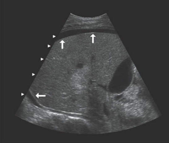
this older hematoma has become sonolucent. fresh hematomas may appear isoechoic to normal liver tissue
clinically, patients with HCC will have possible
abnormal liver function tests, signs of cirrhosis, history of chronic hepatitis, unexplained weight loss, hepatomegaly, fever, abdominal swelling with ascites, and perhaps a palpable mass
a tumor marker for HCC is
serum alpha-fetoprotein (AFP)
in the fetus, AFP is produced in
large amounts by the liver
in the adult (AFP)
low levels of AFP exist
most patients with HCC will have
elevated AFP
elevation of AFP occurs because
AFP is produced in excess by the malignant hepatocytes that make up the tumor
clinical findings of HCC
elevated AFP
abnormal liver function tests (possibly)
cirrhosis
chronic hepatitis
unexplained weight loss
hepatomegaly
fever
palpable mass
abdominal swelling with ascites
sonographic findings of HCC
solitary, hypoechoic mass
heterogenous masses scattered throughout the liver
mass with a hypoechoic halo and central echogenic portion (“target” or “bull’s-eye” lesion)
possible ascites