Lecture 23: Overview of Infectious Agents Related to Pericarditis, Myocarditis, and Endocarditis | Quizlet
 Call Kai
 Learn
Learn Practice Test
 Spaced Repetition
 Match
Match Flashcards
 Knowt Play
1/104

There's no tags or description
Looks like no tags are added yet.
Name | Mastery | Learn | Test | Matching | Spaced | Call with Kai | Chat |
|---|
No analytics yet
Send a link to your students to track their progress
105 Terms
Fibrous Pericardium
the outer layer of the thin, fibrous sac around the heart
Serous Pericardium
the inner layer of the thin, fibrous sac around the heart
Pericardium
acts as a mechanical protection for heart and big vessels and reduces friction between heart and surrounding structures
Acute Pericarditis
-inflammatory disorder of the pericardium
-severe, sharp, and positional chest pain
-aggravated by supine position, inspiration, and cough
-improved by sitting up and leaning forward
-may radiate to neck, shoulder, and scapular ridge
Acute Pericarditis
-friction rub
-new widespread ST elevation or PR depression
Acute Pericarditis
At least 2 of the 4 should be present:
1. typical chest pain
2. pericardial friction rub
3. suggestive ECG changes
4. new or worsening pericardial effusion
Myocarditis
-inflammatory disease of cardiac muscle
-acute: symptoms of heart failure developing within < 3 months
-Chronic: symptoms of heart failure developing > 3 months
Myocarditis
-majority of pts are young and healthy
-no specific PE findings
-excessive fatigue, chest pain unexplained sinus tachycardia, S3, S4 or summation gallop, cardiomegaly, cardiogenic shick, respiratory distress, hepatomegaly
Myocarditis
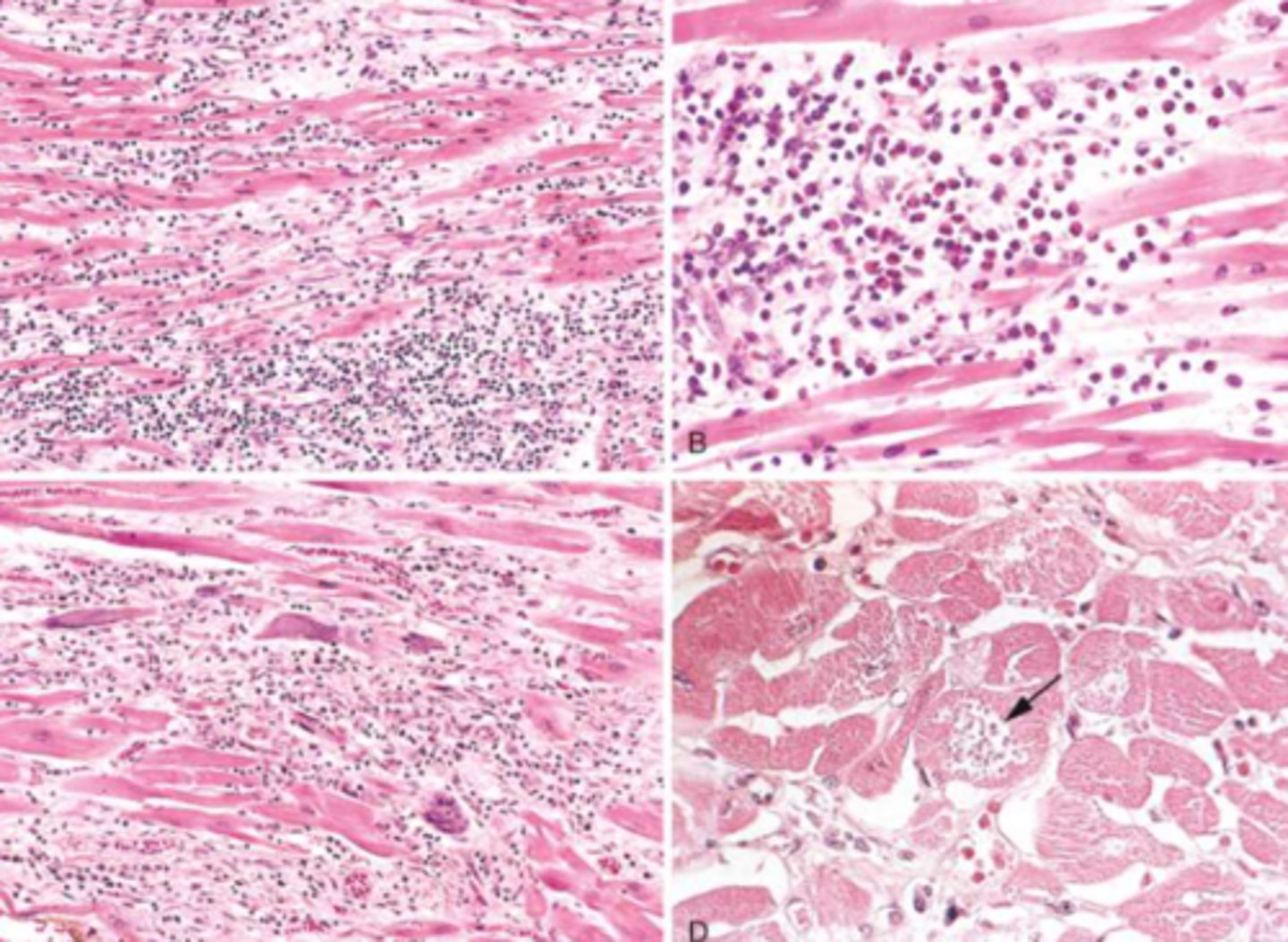
Endocarditis
-inflammation of endocardium
Endocarditis
-usually caused by a bacterial infection
-risk factors: > 60, male, IV drug use, poor dental health, heart disease
Endocarditis
-micro-traume or chronic disease causes damage to endothelium, and then that damaged endothelium becomes colonized by bacteria
Endocarditis
• Janeway lesions
• Osler nodes
• Roth spots
Endocarditis
• Fever, fatigue, flu-like symptoms
• Petechiae or splinter hemorrhages
• Cardiac murmurs are common
• Complications can include glomerulonephritis, sepsis, arrhythmias, systemic embolization
staphylococci, streptococci, and enterococci
three most common causes of IE worldwide
1. Viridans streptococci
2. S. aureus
leading and secondary cause of community acquired Native Valve Endocarditis
1. S. aureus
2. Viridans strep or CoNS
leading and secondary cause of healthcare-assoc Native Valve Endocarditis
1. S. aureus
2. Viridans strep or Candida
leading and secondary cause of IV Drug use Native Valve Endocarditis
1. S. aureus or CoNS
2. Gram negative bacilli or Candida
leading and secondary cause of early onset Prosthetic Valve Endocarditis
1. Streptococci, S. aureus
2 CoNS or Enterococci
leading and secondary cause of late onset Prosthetic Valve Endocarditis
12 months
Late onset prosthetic valve endocarditis occurs greater than __________________ after surgery
S. aureus
most common overall cause of IE in the US
S. aureus
-generally nasopharynx colonization
-may be assoc with staff, equipment, etc if it is a healthcared associated infection
S. aureus
-catalase AND coagulase positive
S. aureus
• Attachment factors, especially fibronectin-binding proteins
• Coagulase production
• Production of clumping factor
• Biofilm formation
• Exotoxins
Low; High
Incidence of MRSA is ___________ in community-acquired NVE, and __________ in healthcare-associated IE
Staphylococcus epidermidis
-Catalase positive
-Coagulase negative
-common cause of PVE
Staphylococcus epidermidis
• Contamination from skin, during manufacturing (mechanical valve), removal (bioprosthetic valve), or implantation
Staphylococcus epidermidis
• Attachment factors, especially fibronectin-binding proteins
• Biofilm formation
• Lower virulence allows establishment of infection (time for biofilm formation) without rapidly activating host immune responses
Streptococcus spp.
-very common cause of IE
-viridans most common
Streptococcus spp.
Identification:
• Gram positive cocci in chains, chain length varies by species
• Catalase negative
• Hemolysis pattern on blood agar in CO2
Quellung Reaction
when identifying the causative agent, what test is specific for S. pneumoniae
α
_____-hemolytic:
-pneumoniae
-viridans
β
_____-hemolytic:
-pyogenes
-agalactiae
γ
_____-hemolytic:
-enterococcus
Viridans Streptococci
-Group D streptococci
S. mitis and S. salicaris
-members of viridans streptococci spp.
-colonize oral cavity (dental plaque) and cause endocarditis
-transmitted thru dental work or in ppl with periodontal disease
S. gallolyticus
-members of viridans streptococci spp.
-colonizes intestinal tract and causes endocarditis
-transmitted after GI trauma, surgeyr, or in colon cancer
S. mitis and S. salicaris
-group D streptococci
-strong biofilm formation, dextran production
S. gallolyticus
-group D streptococci
-capsule and biofilm
-adherence to proteins that bind colon cancer cells
-enzymes that break down GI epithelium and help bacteria invade tissue
Enterococcus spp.
-colonize GI tract
-facilitates healthcare associated infection
-increasing resistance to vancomyocin
-disease spread generally caused by spread of endogenous bacteria to other mucosal sites when normal flora are killed by antibiotic treatment
Enterococcus spp.
• Adherence to host cells
• Polysaccharide capsule
• Biofilm formation
• Natural resistance to penicillin G and cephalosporins enhances when antibiotic treatment eliminates other bacterial flora
Enterococcus spp.
identification:
Gram positive diplococci (looks like S. pneumoniae), combined with biochemical tests (growth at 45°C and in 6.5% NaCl)
Enterococcus spp.
Patient risk factors:
• Patients frequently or recently hospitalized for prolonged periods
• Use of urinary or intravascular catheters
• Prolonged antibiotic treatments, especially with cephalosporins
S. pneumoniae
-colonizes oropharynx and nasopharynx
-disease usually caused by spread of endogenous bacteria, sometimes spread by respiratory droplets
S. pneumoniae
• Adhesins
• Polysaccharide capsule
• Pneumolysin and secretory IgA protease enhance bacterial dissemination and cause
cellular damage and tissue destruction
S. pneumoniae
Identification:
Gram-positive diplococci (looks like enterococci) combined with biochemical
tests (Quellung reaction)
Culture-negative Endocarditis (Intermittent or Occasional)
-cultures require enriched media and CO2 to grow
-cultures may be negative early and then grown in 5-7 days
-most often due to HACEK organisms
Culture-negative Endocarditis (Constant)
-cultures do not grow on most commonly used bacterial media and culture conditions
-most often are intracellular pathogens
HACEK Bacteria
• Group of Gram-negative coccobacilli associated with endocarditis
• Haemophilis spp.
• Aggregatibacter spp.
• Cardiobacter hominis
• Eikenella corrodens
• Kingella kingae
HACEK Bacteria
• Fastidious
• Slow growing
• May require CO2 for growth
• Colonize the oropharynx
• May cause periodontitis, then allowing bacteremia
HACEK
______________-related endocarditis is generally sub-acute and occurs in pre-existing heart disease
H. parainfluenza
the Haemophilus species that most commonly causes subacute endocarditis
Aggregobactor spp.
-a HACEK bacteria
-bacteria easily clump due to expression of surface proteins
Cardiobactor hominis
-a HACEK bacteria
• Oxidase-positive, catalase-negative, indole producing
• Does not grow on selective media (e.g. MacConkey's) that is used for most Gram-negative bacilli
• Most strains resistant to erythromycin
Eikenella corrodens
-a HACEK bacteria
-more common cause of subacute endocarditis
-commonly associated wtih human bites
• Grows on chocolate agar with ~50% of strains producing a pitting in the agar
• Produces a bleach-like odor during growth
• Many strains resistant to oxacillin, 1st gen cephalosporins, clindamycin, erythromycin, and aminoglycosides
Kingella kingae
-a HACEK bacteria
-commonly associated with osteoarticular infections in young children
-colonization of oropharynx and/or upper respiratory tract
-slow growth
key virulence factors common to all HACEK bacteria
Coxiella burnettii
• Causes Q fever
• High fever and severe headaches, flu-like illness
• Most common cause of culture-negative IE
Coxiella burnettii
• Zoonotic spread
-consider in pts who have contact with animals or live in endemic areas
• Cannot be cultured in lab (intracellular pathogen) and acidic environment of phagosome required for bacterial metabolism inactivates most antibiotics
Small cell variant (Phase I)
which variant of Coxiella burnettii is the infectious form?
Large cell variant (Phase II)
Which variant of Coxiella burnettii is the metabolically active form?
Bartonella spp.
• Gram-negative rod that causes intracellular infections in endothelium, RBC, and/or macrophages
• Zoonotic infections transmitted directly from
infected animals or via insect vectors
• Disease in immunocompetent patients associated
with recurrent fevers and/or angioproliferative
disorders
Bartonella henselae
cat scratch fever
Bartonella quintana
trench fever
Bartonella quintana
what Bartonella spp. is assoc with homelessness, alcoholism, and body lice?
Bartonella henselae
what Bartonella spp. is associated with contact with cats, and previous valvular disease?
Tropheryma whipplei
Whipple's Disease
Tropheryma whipplei
• Gram-positive bacilli that establishes intracellular infection in macrophages
• GI infection; PAS+ foamy macs in intestinal lumen
• Major associated symptoms are Cardiac, Arthralgias, and Neurologic
Tropheryma whipplei
-causes subacute, culture negative IE
-prominent fibrosis, slight inflammation with foamy macrophages
-presenting cardiac symptoms but no indication of infection
• Use 16S rRNA PCR of explanted heart valves
Tropheryma whipplei
which agent is assoc with foamy macs
Brucella spp.
-Brucellosis
-undulant fever
-mediterranean fever
-malta fever
Brucella spp.
-fever, fatigue, malaise, arthralgias, night sweats with moldy odor
-Zoonotic infection transmitted by ingestion of food products or contact with tissue or fluids
• Intracellular pathogen, infects monocytes, macrophages, DCs
• Detect in a clinical specimen using PCR
Brucella spp.
• Unpasteurized dairy products are most common sources of infection
• Can be an occupational disease
• Infected animals include cattle, sheep, goats, camels, pigs, etc.
• Most common zoonosis globally
Legionella pneumophila
• Aerobic, fastidious Gram-negative bacilli; facultative intracellular pathogen
• Causes pneumonia (Legionnaires' disease) and Pontiac fever
• Usually infection source is a contaminated water supply
Pseudomonas aeruginosa
• Aerobic, oxidase positive Gram-negative bacilli
• Grape-like odor, green pigment
• IE strongly associated with injection drug use
Candida
-most common cause of fungal IE
-Albicans: ferments glucose, maltose, and sucrose
-does not ferment lactose
Aspergillus
-fungal cause of IE
-primarily in pts with prosthetic valves
Corynebacterium diphtheriae
• Gram-positive, anaerobic, club-shaped rod with blue and red granules
• Toxin-producing strains cause diphtheria (Elek's test)
• Myocarditis due to toxin production and dissemination; develops 1-2 weeks after onset of pharyngeal symptoms
Borrelia burgdorferi
• Spirochete, culture is difficult
• Causes Lyme disease (tick-borne illness)
• Stage 2 can cause cardiac dysfunction
• Diagnose with serologic testing
Enteroviruses
most common implicated virsues in viral myocarditis in north america and Europe
HIV, hepatitis C virus, influenza A and B virus
Viruses that indirectly trigger myocarditis by activating the immune system
MERS CoV, SARS-CoV, SARS-CoV-2
Viruses with ACE2 tropism that can potentially mediate direct cardiac injury
Parvovirus B19
-Vasculotropic viruses likely to have lifelong
persistence
-viral myocarditis
Enteroviruses
coxsackieviruses, rhinoviruses, polioviruses, and echoviruses
Enterovirus
• Small (15-30 nm), non-enveloped, +ssRNA genome, high mutation rate
• Relatively acid stable (maintain infectivity over wide pH range), resistant to ether and
alcohol
• Tropism for respiratory or GI tract
Coxsackievirus and Adenovirus Receptor (CAR)
what viral receptors are present in intercalated discs that link myocardial cells
Coxsackievirus and Echovirus
-myocarditis
-more common and more severe in infants
• Sudden onset heart failure in infants
• Cyanosis
• Tachycardia
• Cardiomegaly
• Hepatomegaly
Adenovirus
• Non-enveloped, linear dsDNA genome
• Transmitted by aerosols, fecal-oral route,
direct contact, and fomites
• Illness is usually self-limited in
immunocompetent patients
Adenovirus
• Pharyngitis, respiratory illness, conjunctivitis, gastroenteritis
• Infects epithelial cells, persists in lymphoid tissues and can be shed for weeks after infection
• Binds to CAR
Parvovirus B19
• Non-enveloped, linear ssDNA genome
• Transmitted by aerosols, direct contact, and fomites
• Binds to blood group P antigen
• Tropism for RBC precursors
Parvovirus B19
• Causes erythema infectiosum (fifth disease)
• Associated with myocarditis with increasing frequency, especially in adults
Human Herpes Virus 6 (HHV-6)
• Enveloped, dsDNA genome
• Binds to CD46
• Replicates in a wide range of host cells,
integrates into host genome
-most people infected as children
• Causes roseola infantum
• Primary infection in adults may result in mononucleosis-like illness
Mumps
• Hallmark is parotitis
• - sense, ssRNA genome; part of Paramyxoviridae family
• Associated with myocarditis
• May see transient ECG changes (such as ST segment depression)
Rubella
• + sense, ssRNA genome; part of Matonaviridae family
• AKA German measles
• Myocarditis and pericarditis are rare complications
Trypanosoma cruzi
Chagas Disease
Trypanosoma cruzi
-parasite
-• Most common in rural areas of Central and South America, Trinidad, and southern US
-reduviid bug: insect defecates during feeding, host scratches bite, parasite in feces enters the host
• Acute infection: fever, chills, malaise, edema, Romaña sign, chagoma
• Chronic Chagas cardiomyopathy: biventricular heart failure, cardiac arrhythmias, thromboembolism
Trypanosoma cruzi
• Antigenically distinct life cycle stages: alternates between intracellular proliferative form and extracellular nonproliferative infectious form
• Weak TLR activation prevents macrophage activation
• Parasite mucins suppress T cell responses
Candida spp.
• Dimorphic yeast
• Pseudohyphae and budding at 20°C
• Germ tubes at 37°C
• Immunocompromised patients, ICU patients, patients with indwelling devices, IV drug use
Candida spp.
• Produces extracellular proteases, phospholipases, lipases, hydrolytic enzymes, and adhesins
• Biofilm formation
• Endocarditis in PWID, prosthetic valves, valve damage
• Pericarditis in immunosuppressed patients, or after thoracic surgery