Lab Three - Exercise 27: Structure of the Heart
 Call Kai
 Learn
Learn Practice Test
 Spaced Repetition
 Match
Match Flashcards
 Knowt Play
1/96

There's no tags or description
Looks like no tags are added yet.
Name | Mastery | Learn | Test | Matching | Spaced | Call with Kai | Chat |
|---|
No analytics yet
Send a link to your students to track their progress
97 Terms
arteries
vessels that carry blood from the heart
veins
return blood to the heart
coronary arteries
carry blood from the heart, but they carry the blood to the heart muscle
cardiac veins
take blood from the heart muscle back to the heart for recirculation
mediastinum
contains the heart, the coverings of the heart (the pericardia), and other structures, such as the esophagus and descending aorta
what is the first structure you find when opening the chest cavity, made of tough, outer connective tissue?
fibrous pericardium - encloses the heart
what lining is deep to the fibrous pericardium?
parietal pericardium (outer layer of the serous pericardium)
what is deep to the parietal pericardium?
pericardial cavity; which contains a small amount of serous pericardial fluid
serous pericardial fluid
reduces the friction between the outer surface of the heart and the parietal pericardium
what layer is closest to the heart (inner layer of the serous pericardium)?
visceral pericardium (epicardium)
what is the outermost layer of the heart wall?
epicardium - composed of epithelial and connective tissue
what is the middle layer of the heart wall?
myocardium; thickest of the three layers; mostly made of cardiac muscle
how is cardiac muscle arranged in the heart?
spirally; providing a more efficient wringing motion to the heart
endocardium
inner layer of the heart wall; serous membrane that consists of endothelium (simple squamous epithelium) and connective tissue
where does blood enter the heart?
right atrium - which flows into the right ventricle
From the right ventricle, where does the contraction send the blood?
to the lungs
once being oxygenated in the lungs, where does the blood reenter the heart?
left atrium
where does blood move from the left atrium?
left ventricle; which pumps it to the rest of the body
where are the apex and base of the heart?
pointed end is the apex and the base is where the great vessels leave the heart - the base is actually on top of the apex!
what delineates the right ventricle from the left ventricle?
ventricular sulcus (groove)
what’s found in the ventricular sulcus?
coronary arteries and cardiac veins
which wall of the heart is thicker?
left ventricle
auricles
earlike flaps on the anterior, superior region of the heart
atrioventricular sulcus (groove)
junction of the right atrium and right ventricle
coronary sinus
large venous chamber that carries blood from the cardiac veins to the right atrium
what do the superior and inferior vena cavas do?
return blood to the right atrium
pulmonary veins
carry blood from the lungs to the left atrium
what arteries does the left coronary artery (rooted in the ascending aorta) branch into?
anterior interventricular artery and the circumflex artery
what arteries does the right coronary artery (rooted in the ascending aorta) branch into?
posterior interventricular artery and right marginal artery
when do arteries fill with blood?
ventricular relaxation
where does the great cardiac vein feed into?
cardiac sinus
where does the small cardiac vein feed into?
coronary sinus
where does the coronary sinus empty into?
right atrium
what are the ventricles separated by?
interventricular septum; which forms a wall between the two ventricular chambers
what is the medial wall of the atrium called?
interatrial septum
fossa ovalis
thin, oval depression in the atrial wall
what is the fossa ovalis called in fetal hearts?
foramen ovale
what do pectinate muscles do?
provide additional strength to the atrial wall
tricuspid valve
prevents the return flow of blood from the right ventricle into the right atrium during ventricular contraction
what are the thin, threadlike attachments of the tricuspid valve called?
chordae tindinea; which are attached to larger papillary muscles, which are extensions from the wall of the ventricle
trabeculae carnea
small extensions of the right ventricle that strengthen the ventricle wall
where does blood from the right ventricle flow into?
the pulmonary trunk toward the lungs
pulmonary semilunar valve
appears as three small cusps between the right ventricle and the pulmonary and keeps blood flowing in reverse from the pulmonary trunk into the right ventricle during ventricular relaxation
where does blood from the pulmonary trunk flow to?
pulmonary artieries before entering the lungs
what does blood in the lungs do?
releases carbon dioxide and picks up oxygen
where do the pulmonary veins carry the blood from the lungs?
into the left atrium
where are the pulmonary veins located?
in the superior, posterior portion of the left atrium
where is the bicuspid valve found?
between the left atrium and left ventricle
what is the bicuspid valve also known as?
mitral valve or left atrioventricular valve
what does the bicuspid valve also have?
attached chordae tendineae and papillary muscles
where is the aortic semilunar valve located?
the junction of the left ventricle and the ascending aorta; prevents blood from the aorta into the left ventricle
where does blood from the left ventricle go?
into the aorta and the rest of the body
what are the differences between the human heart and sheep heart?
position of the superior and inferior venae cava
The heart is located between the lungs in an area known as the ______________ .
Mediastinum
What is the name of the layer that is superficial to the pericardial cavity?
Parietal pericardium
What is the innermost layer of the heart called?
Endocardium
Is the apex of the heart superior or inferior to the rest of the heart?
Inferior
What is the name of the depression between the two ventricles on the anterior surface of the heart?
Interventricular groove
Are auricles extensions of the atria or the ventricles?
Atria
What three vessels take blood to the right atrium?
Superior vena cava, inferior vena cava, coronary veins
Where do the great cardiac vein and the small cardiac vein take blood?
Coronary sinus
What blood vessels nourish the heart tissue?
Coronary arteries
What structure separates the left atrium from the right atrium?
Interatrial septum
What is the name of the thin spot between the atria?
1. Since fetuses do not need oxygen, allows blood to flow directly from right atrium to left atrium without going to the lungs
Fossa ovalis
The bicuspid valve is located between what two chambers of the heart?
Left atrium and left ventricle
Name the structure between the atrioventricular valve and the papillary muscle
Chordae tendinae

What is the function of the aortic semilunar valve?
Prevents back flow of blood
What is another name for the tricuspid valve?
Right atrioventricular valve
What cell type makes up most of the myocardium?
Myocytes
What adaptation do you see in the walls of the left ventricle being thicker than those of the right ventricle?
Pushes blood through the body, so it needs to be thicker and stroner
How does cardiac muscle resemble skeletal muscle?
Striations
In terms of function, how is cardiac muscle different from skeletal muscle?
Contracts independently from nervous impulses
Superior vena cava

Right atrium
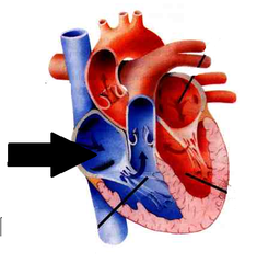
Right ventricle

Aortic arch

Base of heart
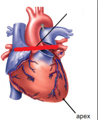
Pulmonary trunk

Left atrium

Left ventricle

Apex

Name the structures surrounding the heart, starting with the heart and moving outward.
Heart ---> visceral pericardium ---> pericardial cavity
---> parietal pericardium ---> fibrous pericardium
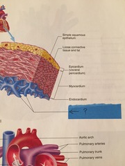
Epicardium (visceral pericardium)

Myocardium

Endocardium

Trabeculae carneae
Aortic semilunar valve

Pulmonary semilunar valve

Tricuspid valve

Pulmonary arteries

Pulmonary veins

Bicuspid valve
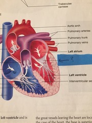
Interventricular septum

Body ---> travels back to heart in veins ---> enters right atrium via superior vena cava, inferior vena cava, or coronary sinus ---> through tricuspid valve to right ventricle ---> through pulmonary semilunar valve out of pulmonary trunk ---> pulmonary arteries take blood to the lungs
Flow of deoxygenated blood
Lungs ---> oxygenated in pulmonary capillaries ---> goes back to heart via pulmonary veins ---> enters left atrium ---> through bicuspid valve to left ventricle ---> through aortic semilunar valve ---> up and out of aorta
Flow of oxygenated blood
Coronary sulcus

Papillary muscles
