Surface Anatomy and Clinical Correlation
 Call Kai
 Learn
Learn Practice Test
 Spaced Repetition
 Match
Match Flashcards
 Knowt Play
1/88

Earn XP
Description and Tags
Name | Mastery | Learn | Test | Matching | Spaced | Call with Kai | Chat |
|---|
No analytics yet
Send a link to your students to track their progress
89 Terms
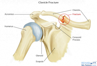
Clavicular Fracture
Occurs at the junction between the lateral and medial curvature of the clavicle
The weakest point of the clavicle
SYMPTOMS:
Muscle guarding wherein the
Commonly Caused:
Falling on outstretched hand: Wrist → radius → ulna (through the interosseous
membrane) → humerus → scapula → clavicle
Greenstick Fracture
Known as the partial break of the clavicle, wherein the bone cracks on one side but not all the way through
Common In Children (i.e shoulder dystocia in newborns) due to their softer bone

Scapular Fracture

Scapular Fracture
Brought upon by high-energy force such as blunt trauma to the scapula
Scaphoid Fracture
Can present as a complication of Prieser’s disease due to the avascular necrosis of the scaphoid
MRI
Best mode of radiographic imaging used for suspected avascular necrosis
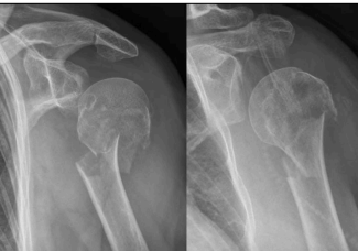
Fracture of the surgical neck of the humerus
Affected structures of the fracture:
Circumflex Humeral Artery can be compromised due to its location in the _______ ____, especially if the proximal region of the humerus
Can lead to avascular necrosis
Axillary Nerve can be compromised since it passes through the posterior region of the _______ ____

Fracture of the Anatomical Neck of the Humerus
Injury to the ________ ___ can cause loss of blood
supply to the humeral head, and can cause rotting/necrosis
● Can also injure the circumflex humeral arteries
○ May result in:
■ Avascular Necrosis
■ Bone death
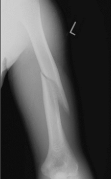
Humeral Shaft Fractures
Can damage structures related to the spiral groove
Radial nerve
Characterized by wrist drop
Triceps may lose function
Profunda brachii artery
torn/ruptured
Transverse Humeral Shaft Fracture

Oblique Humeral Shaft Fracture
Has a sharp edge which has the most potential to lacerate the radial nerve and profunda brachii artery
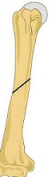
Spiral Humeral Shaft Fracture
Most common humeral shaft fracture

Comminuted Humeral Shaft Fracture
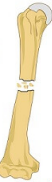
Segmental Humeral Shaft Fracture

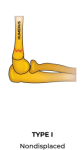
Type I supracondylar fracture
A hairline crack in the supracondylar region of the humerus
Vasculatures affected:
○ Brachial artery
■ Absent pulse suggests possible brachial artery
damage.
■ Loss of strength may indicate the possibility that
the brachial artery is cut off.
○ Median nerve
■ Motor function test: “OK” Sign
● Normal: Patient can form a proper circle.
● Median/AIN damage: Flattened or “pinch” sign
instead of a circle

Type II supracondylar fracture
Fracture which demonstrates a slightly displaced anterior wall in the supracondylar region of the humerus
Vasculatures affected:
○ Brachial artery
■ Absent pulse suggests possible brachial artery
damage.
■ Loss of strength may indicate the possibility that
the brachial artery is cut off.
○ Median nerve
■ Motor function test: “OK” Sign
● Normal: Patient can form a proper circle.
● Median/AIN damage: Flattened or “pinch” sign
instead of a circle

Type III supracondylar fracture
Radial complete displacement( both anterior and posterior wall)
Vasculatures affected:
○ Brachial artery
■ Absent pulse suggests possible brachial artery
damage.
■ Loss of strength may indicate the possibility that
the brachial artery is cut off.
○ Median nerve
■ Motor function test: “OK” Sign
● Normal: Patient can form a proper circle.
● Median/AIN damage: Flattened or “pinch” sign
instead of a circle
Medial epicondylar fracture
Usually affects the ulnar nerve
● Common causes:
○ Direct trauma
○ FOOSH
○ Avulsion fracture
■ Occurs when a very forceful traction of muscles or
ligaments pulls on the bone, breaking it.
■ Avulsion fracture of the ___________ can
be attributed to the pull of the anterior compartment
muscles, most commonly the common flexor
origin as these muscles attach to the medial
epicondyle
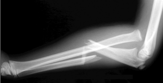
Colles’ fracture, Smith’s fracture and Galeazzi fracture
Types of Radial Head Fractures
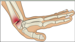
Colles’ fracture
Cause: FOOSH injury while wrist is extended
● A posterior/dorsal displacement fracture in the distal
radial segment towards the lister’s tubercle.
● Results in “Dinner fork” deformity

Smith’s Fracture
Also called “Reverse Colles’” or “Garden Spade”
● Cause: FOOSH injury while wrist is flexed
● An anterior/volar displacement fracture in the distal
radial segment near the wrist.

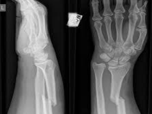
Galeazzi Fracture
Radius fracture with dislocation of the distal radioulnar
joint
Monteggia Fracture
Ulnar fracture with dislocation of the radial head
Lunate Fracture
Just like the scaphoid bone, the _______ is also at risk for
avascular necrosis called Kienbock’s Disease.
○ Rare condition where the _______ loses blood supply,
resulting in bone death.
○ Presents as a complication of the fracture
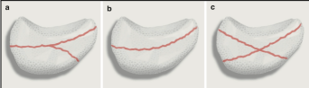
Hamate Fracture
Fractures can occur at the _______ due to its anterior projection.
● Causes:
○ FOOSH
○ Sports equipment handling injury (e.g., by baseball bats
or clubs)
■ May occur if the player hardly hits the incoming ball
as the tension or force is transmitted into the hook of
hamate
● Affects the ulnar artery and ulnar nerve that pass through
the Tunnel of Guyon
■ Pisohamate ligament which is found on the top of
the hook of hamate and pisiform forms the Tunnel
of Guyon or Guyon’s Canal

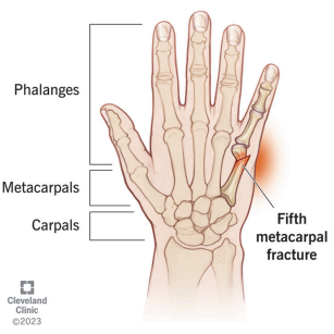
Boxer’s Fracture
Fracture of the neck of the 5th metacarpal
● Cause:
○ Striking an object with a closed fist (common in boxers)
● Symptoms:
○ Pain
○ Swelling
○ Depression of the knuckle

Bennett’s Fracture
Fracture at the base of the 1st metacarpal
● Often involves subluxation (partial dislocation) of the
carpometacarpal (CMC) joint
● Cause:
○ Axial force on a partially flexed/bent thumb (e.g., impact
from punching or a fall)
● Symptoms:
○ Pain, swelling, bruising at the base of the thumb
○ Difficulty or pain with gripping
○ Thumb misalignment
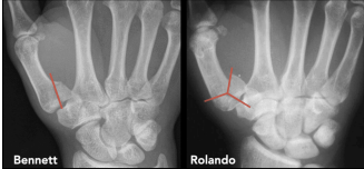
Rolando’s Fracture
Fracture at the base of the 1st metacarpal, with a Y- or
T-shaped pattern
● Multi-fragmented fracture
● Cause:
○ Axial force on a partially flexed/bent thumb (e.g., impact
from punching or a fall)
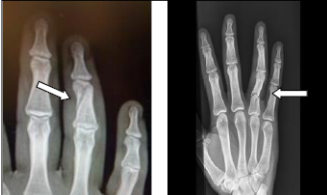
M. Phalangeal Fracture
Causes:
○ Acute trauma (e.g., finger caught in a car door)
○ Crush injuries
○ Forceful twisting or jamming of the fingers
○ Tumors may weaken the bone, making it more
susceptible to fracture

Dislocations
Characterized by total separation of joint components
Anterior Sternoclavicular Joint Dislocation
The separation of the sternum and the sternal/medial end of the clavicle
Common causes: Traumatic events such as collisions in
sports (e.g., rugby)
● The displacement can be anterior or posterior
○ Anterior dislocation: the clavicle moves forward
■ Most common
○ Posterior: the clavicle moves backward
■ a dangerous type of dislocation, since the clavicle can damage or lacerate structures posterior to the sternum (e.g., trachea, esophagus, subclavian artery)

Posterior Sternoclavicular Joint Dislocation
The separation of the sternum and the sternal/medial end of the clavicle
Common causes: Traumatic events such as collisions in
sports (e.g., rugby)
● The displacement can be anterior or posterior
○ Anterior dislocation: the clavicle moves forward
■ Most common
○ Posterior: the clavicle moves backward
■ a dangerous type of dislocation, since the clavicle can damage or lacerate structures posterior to the sternum (e.g., trachea, esophagus, subclavian artery)

Acromioclavicular Joint Dislocation
Displacement of the _________ joint:
○ If the coracoclavicular ligament is still intact, the dislocation is not that severe and undisplaced.
○ If the coracoclavicular ligament is torn, the _________ joint is displaced.
● The acromion and scapula are pulled downwards by the
weight of the upper limb, the clavicle displaced upward,
resulting in a step deformity.

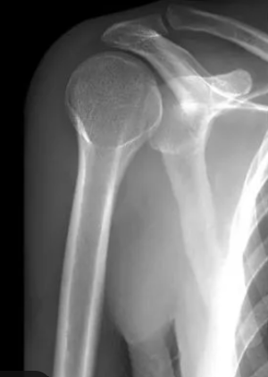
Anterior Shoulder Joint Dislocation
The humeral head will move anterior to the scapula
Test always involve a screening for associated fractures; Patient is positioned to have a flattened shoulder, arm in slight abduction/flexed elbow
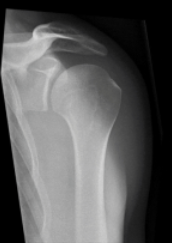
Posterior Shoulder Joint Dislocation
X-ray reveals that the humeral head is aligned with the acromion
Test always involve a screening for associated fractures; Patient is positioned to have a flattened shoulder, arm in slight abduction/flexed elbow

Normal shoulder joint alignment
The glenoid fossa is at the center of the Y line.

Elbow Joint Dislocation
Separation of the radius and ulna from the distal humerus
the Collateral ligament tears possibly with the valgus force
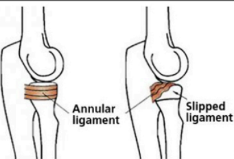
Pulled Lebow/ Nursemaid’s elbow
Common injury in children less than 5 years old when the radial head slips out of the annular ligament.
Characterized by the annular ligament slipping over the radius bone
Symptoms:
Pain the elbow, leading to elbow guarding
Reluctance to use the arm
Wrist Join Dislocation
Rare
Collateral ligaments can be damaged
The radiocarpal joint is displaced
Anterior or Posterior Displacement
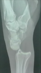
Lunate Bone Dislocation
Lunate bone is dislocated
Lunate is aligned with 3rd metacarpal
Murphy’s Sign: The patient is asked to make a fist. If the 3rd metacarpal is aligned with the 2nd and 4th metacarpal, a lunate dislocation is suspected(+)
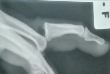
Metacarpophalangeal/Interphalangeal Joint/Finger dislocation
Seen in Murphy’s sign
Can occur anteriorly or posteriorly
Can rupture the collateral ligaments surrounding the joints
Joints must be repositioned in extended position
Collateral ligaments are loose in extended position & taut in flexed position
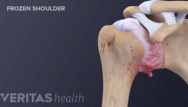
Frozen shoulder/ Adhesive Capsulitis
Commonly occurs in the elderly
Usually idiopathic or occurs after trauma/immobilization
Progression: Capsular inflammation > adhesion > stiffness

Analgesia + early PT intervention
Management for adhesive capsulitis
Stage 1/Pre-freezing of Adhesive Capsulitis
Stage of adhesive capsulitis which occurs 1-3 months and pain is first noticed in the shoulder region
Stage 2/Freezing phase of Adhesive Capsulitis
Stage of adhesive capsulitis which lasts from 10 weeks to 8 months and is known to be the most painful phases. Movement is also restricted
Stage 3/Frozen Phase of Adhesive Capsulitis
Stage of adhesive capsulitis which lasts from 4-12 months and the shoulder is notably stiff
Stage 4/Thawing phase of adhesive capsulitis
Stage of adhesive capsulitis which lasts from 5 months to 2 years and the shoulder joint has become stiff but gradually loosens
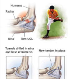
Tommy John Injury
A sprain or tear of the ulnar collateral ligament on the medial side of the elbow
Can also present a tingling sensation along the ulnar nerve distribution in the hand
Can be torn especially during elbow-joint dislocation or other sport injuries
Skier’s Thumb
A sprain or torn ulnar collateral ligament on the medial side of the thumb
Commonly observed in skiers after a bad fall while still holding the ski poles

Rotator Cuff Injury
Tear of the rotator cuff muscles or irritation of the ligaments that are attached to the humerus (holds the shoulder in place)
Causes pain, weakness, limited movement(ex. abduction or lifting above the head)
Can lead to frozen shoulder if not addressed
Common in sports like baseball and tennis
Diagnostic imaging: MRI
Bicipital Tendonitis
The long head of the biceps tendon will pass through the bicipital groove or intertubercular groove
Covered by the bicipital ligament or transverse humeral ligament
Inflammation of the tendon of the long head of the biceps brachii
Continuous use of the biceps brachii will rub its tendon against the ligament surface. This repeated friction irritates the tendon and can lead to tendonitis
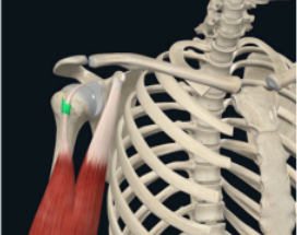
Biceps Brachii Tendon Rupture
Tear of the biceps brachii tendon
May occur at the distal or proximal end
Symptom:
(+) Popeye’s deformity: bulging of biceps brachii
May present with bruising

Golder’s Elbow/ Medial Epicondylitis
Affects the common flexor tendon origin
Common with golfers when they swing the golf club
Excessive use led to inflammation of the common flexor tendon origin

Tennis Elbow/ Lateral Epicondylitis
Affects the common extensor tendon originating from the lateral epicondyle
Posterior muscles in the forearm are affected which can cause small tears and inflammation in the tendon
Common in racket-related sports like tennis, badminton and pickleball
Medial epicondylitis test
Special tests for medial epicondylitis
Cozen’s Tests and Mill’s Test
Special tests Lateral Epicondylitis
De Quervain’s Tenosynovitis
Also called stenosing tenosynovitis
Inflammation of the lareal tendons formed by the abductor Pollicis longus(APOL) and Extensor Pollicis Brevis, which share a common tendon sheath and contribute to the anatomical snuffbox
Common among those who write frequently

Mallet Finger/ Baseball Finger
Tear to the tendon that extends the DIP joint resulting in a flexed position
The extensor digitorum tendon pulls a piece of bone due to sudden force of impact hitting a stretched finger
Avulsion fracture, torn extensor tendon

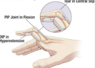
Boutenniere deformity
Bending of the proximal interphalangeal joint posteriorly, affected by the torn central slip extensor tendon
Rupture to the extensor tendon of the proximal interphalangeal joint
Distal joint will extend due to the pull of the tendon
Jersey Finger
Results from an avulsion injury of the flexor digitorum profundus form the insertion at the base of the distal phalanx’
Hyperextension of the DIP joint
Unable to bend the DIP joint due to FDP being unable to act onit
Special test: Sweater finger

Trigger Finger
Delayed finger snapping
Common in elderly where nodules can form on the tendon of the FDP/S
The nodules can get stuck by these flexor sheaths which prevents the smooth glidingg of tendons through the sheath
Management: Steroid injection to reduce the nodule or surgical release of the tendon if conservative treatment fails
Olecranon Bursitis
Inflammation of the olecranon bursa
Commonly known as student’s elbow


Thoracic Outlet Syndrome
Hypertrophy of the muscles obstructs or compresses the subclavian artery and other blood vessels
The pulse is used to determine compression
Compression of subclavian artery will lead to decreased blood flow in upper limb
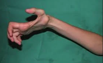
Volkmann’s Ischemic contracture
Severe permanent flexion deformity of the hand and fingers caused by muscle damage from ischemia
The fracture compresses nerves and blood vessels which leads to claw hand deformity and complications of the supracondylar fracture
Lymphatic Edema
Compression of the clavicular and pectoral lymph nodes which can result in reduced lymphatic drainage and swelling of the upper limb

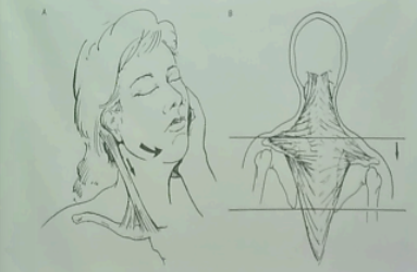
Spinal Accessory nerve
Can be damaged due to acute injuries
Affects sternocleidomastoid and trapezius muscle—leading of lack of head movement and shoulder joint elevation
Winged Scapula
Characterized by a prominent vertebral border of the scapula
Test: ask patient to press palm on wall> prominence of vertebral border of scapula
Caused by long thoracic nerve injury—affecting the serratus anterior which is responsible for pulling near the rib or chest wall
Typically caused by trauma or latrogenic(doctor-induced injuries)
Thoracodorsal Nerve Affectation
Paralyzes latissimus dorsi—affecting the arm extension, adduction and internal rotation
Dorsal Scapular Nerve
Affects the levator scapulae, rhomboid major and rhomboid minor—resulting to difficulty in retracting the scapula
Suprascapular nerve
Affects the supraspinatus and infraspinatus—disables the ability of the arm to abduct (supra—) & externally rotate(infra—)
Leads to atrophy of supra and infra due to lack of movement
Subscapular nerve
Affects the subscapularis and teres major
Difficulty in internal rotation of the shoulder
Plexopathy
Injuries are common and are usually caused by traction, pressure or a stab would
Defined as a form of peripheral neuropathy involving damage or dysfunction of a nerve plexus, which is a complex network of nerves branching from the spinal cord to various parts of the body
Brachial plexopathy
affects the arm, shoulder, and upper chest, often caused by trauma, compression, or inflammation
Damage to the brachial plexus
Erb-Duchenne palsy
Affects C5 and C6 upper ventral rami
Injury to the upper trunk of the brachial plexus
Results from excessive displacement of the head to the opposite side and depression of the shoulder
Common in babies who are difficult to push out during labor
CHARACTERIZED By:
Waiter’s position
Shoulder: adducted(loss of deltoid & supraspinatus function) and medially rotated(unopposed ppectoralis major)
Elbow: Extended(loss of biceps)
Forearm: Pronated(Loss of biceps function)
Fingers and Wrist: Flexed
Klumpke’s Palsy
Traction injury due to excessive arm abduction
When a person falls and clutches an object to save them
Injury to lower trunk of the brachial plexus
Typically involves tearing of the C8 & T1 lower ventral rami
Affected nerves:
Ulnar nerve
Median nerve
Affected muscles
Small muscles of the hand(d/t damage to ulnar and median nerve fibers)
Results in claw hand deformity
Hyperextension of the MCP joints and flexion of the IP joints
Sensory loss along the medial side of the arm
Musculocutaneous nerve
Paralyzed muscles of the anterior fascial compartment due to MC nerve injury
The musculocutaneous nerve rises from the lateral cord of the brachial plexus
The nerve pierces through the coracobrachialis and is covered by the biceps brachii
Its terminal end supplies the lateral skin
Damage results to weak elbow flexion
Weakness of forearm supination
Numbness in the anterolateral surface of the forearm
Common causes are penetrating injury as the biceps brachii covers the nerve
Axillary Nerve
Presentation: Decreased roundness and flattening of shoulder
Common causes: Fractures of the surgical neck of the humerus
Downward displacement of the humeral head in glenohumeral (shoulder) dislocations
Median Nerve
Extends from origin in the brachial plexus until wrist and hand
Rises from the lateral half of the lateral cord and half of the medial cord
No role in brachium but has a role in anterior forearm, thenar eminence, and lumbricals 1-2
Anywhere from its origin can damage the median nerve but differs in its presentation
Proximal Median Nerve Injury
Upper arm, elbow or anterior forearm is affected
All manifestations pertaining to median nerve will be present
Median nerve oppression due to supracondylar fracture
Proximal affectation:
Oppression of the ______ nerve due to a supracondylar fracture
Pronator Teres Syndrome
Compression of the median nerve due to pronator teres
Usually happens for profession that require frequent pronation/supination
Presents with pain on top of the pronator teres
Manifests with hypesthesia
Decrease in sensation of the distribution of median nerve
Presents with papal benediction sign
Only the lateral 2 fingers supplied by ulnar nerve will move
High Median Nerve Injury
Does not supply the FDS and FDP
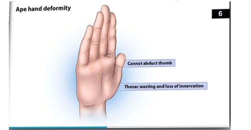
The terminal branch supplies the thenar eminence which will result in its atrophy due to wasting of muscles
Decreased sensation on area being supplied by the median nerve
Presentation: Ape hand deformity, papal benediction
Distal Median Nerve Injury
Lesion at the distal side affects of the FDS and FDP
If the median nerve is compressed, specifically in the carpal tunnel, both FDS and FDP will still be functional
No papal benediction
However, thenar eminence will not be innervated = ape hand deformity
Carpal Tunnel Syndrome
Compression of the median nerve within the carpal tunnel at the anterior wrist
Causes:
Occupation or activities that require prolonged use of the FDP and FDS such as frequent typing and frequent writing
Repetitive use of the flexor tendons can lead to hypertrophy(thickening) of these tendons which can compress the median nerve within the carpal tunnel
Affected Muscles:
Thenar eminence
Lumbricals 1 & 2
Sign: Ape hand deformity
Provocative tests for Carpal Tunnel Syndrome:
Phalen Test
Compressing further the median nerve
Tingling sensation will increase in area
Inverted praying hand position
Tinel Test
Tapping on the area of carpal tunnel
Median nerve is irritated and will present tingling sensation as well
Anterior Interosseus Syndrome
Affects the anterior interosseus nerve
Test:
O sign test
Intact muscle = Negative O sign
Weak FPL and FDP = O sign collapse > Positive O sign
Radial Nerve
Comes from posterior division of trunk
Divides into superficial nerve and the posterior interosseus nerve
Proximal radial nerve injury
At distal humerus
Affects all posterior skin being supplied will be affected
Problem with elbow extension
Triceps is still functional
No sensation at posterior forearm and dorsum of hand
Both proximal and distal injuries present with affectation of posterior forearm muscles
(+) wrist drop
Causes: Improper use of axillary crutch
Radial nerve is compressed
Saturday Night Palsy
Pressing up into the arm
Common in people who fall asleep with one arm over the back of the chair
Spiral Groove Fracture
Results: Claw hand deformity due to atrophy of adductor pollicis, guttering or depression in between the metacarpals occupied by interossei muscles
Clawing due to pull of extensor digitorum
Loss of function of the structures moved by the medial half of the flexor digitorum profundus and the flexor carpi ulnaris
Test: Froment Test