HISTOLOGY/INTEGUMENTARY SYSTEM LAB PRACTICAL
 Call Kai
 Learn
Learn Practice Test
 Spaced Repetition
 Match
Match Flashcards
 Knowt Play
1/37

There's no tags or description
Looks like no tags are added yet.
Name | Mastery | Learn | Test | Matching | Spaced | Call with Kai | Chat |
|---|
No analytics yet
Send a link to your students to track their progress
38 Terms
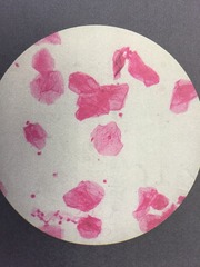
Squamous Epithelium
Flat, "fried egg"
Smear, you can't determine from the appearance if it is simple or stratified.
This is a smear of stratified squamous epithelium.
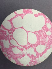
FOUND IN LUNGS, KIDNEYS, SEROUS MEMBRANES
FUNCTIONS IN OSMOSIS AND SECRETION
Simple Squamous Epithelium (lung)
Edge view, alveoli
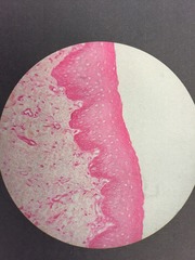
Stratified Squamous Epithelium
Edge view, layers
Focusing on the upper deep pink band
Non-keratinized
FOUND IN ESOPHAGUS AND SKIN
Simple Cuboidal Epithelium (1)
Cube-shaped with centrally located nucleus
FOUND IN KIDNEY TUBULES (LINES DUCTS)
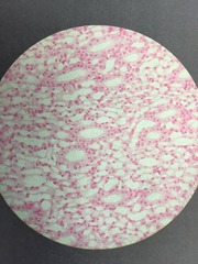
Simple Cuboidal Epithelium (2)
Cube-shaped with centrally located nucleus

Simple Columnar Epithelium
Longer than wide, nucleus close to the basement membrane
FOUND IN LINING OF KIDNEY COLLECTING TUBULES
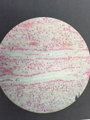
Pseuostratified Ciliated Columnar Epithelium (1)
Cells have surface modification of cilia
Cells not equal in size
All cells in contact with basement membrane, but not all reach surface
FOUND LINING TRACHEA AND BRONCHI
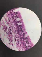
Pseuostratified Ciliated Columnar Epithelium (2)
Cells have surface modification of cilia
Cells not equal in size
All cells in contact with basement membrane, but not all reach surface
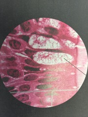
Stratified Ciliated Columnar Epithelium
"Leaves on a tree"
FOUND IN MALE URETHRA AND LARGE EXCRETORY DUCTS OF SOME GLANDS
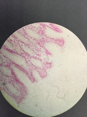
Transitional Epithelium
Stratified epithelium with some cells appearing squamous and stratified cuboidal, and basal cells being cuboidal or columnar.
Cells are flattened while others are flattened side to side, allowing elasticity
LINES THE URETERS, BLADDER, AND PART OF THE URETHRA
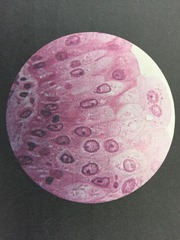
Areolar (Loose) Fibrous Connective Tissue
Matrix of elastic and collagenous fibers
Small amounts of reticular fibers in hyaluronic acid
Elastic fibers thin and dark (thread)
Collagen fibers thick and pink
Fibroblasts large and flat (black dots)
FOUND IN PAPILLARY REGION OF THE DERMIS

Dense Irregular Fibrous Connective Tissue
Lower region of the dermis
FASCIA, RETICULAR REGION OF THE DERMIS
PERIOSTEUM OF BONE
PERIOCHONDRIUM OF CARTILAGE
FIBROUS CAPSULES AROUND KIDNEYS, LIVER, TESTES, LYMPH NODES

Dense Regular Fibrous Connective Tissue
Collagen fibers densely packed in regular parallel rows with fibroblasts between the fibers
TENDONS, MUSCLE TO BONE AND MUSCLE TO MUSCLE
LIGAMENTS, BONE TO BONE
APONEUROSES, MUSCLE TO MUSCLE OR BONE

Adipose Tissue (1)
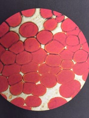
Adipose Tissue (2)
Cytoplasmic membrane (stained purple) between the adipose cells (white)
Nuclei (black dots)

Hyaline Cartilage
Dense matrix
Fibers not visible
Chondrocytes in lacunae
Bare
FOUND IN JOINTS, NOSE, LARYNX, TRACHEA, BRONCHI

Osseous (Bone)
Matrix found in concentric layers (lamellae) around a central canal (haversian canal)
Osteocytes interconnected with haversian canal by tiny cracks (canaliculi)

Blood (1)
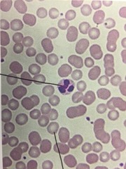
Purple: Leucocyte

Blood (2)
Smooth Muscle (1)
Oriented around (circular) or lengthwise (longitudinal)
Long spindly fibers
Elongated nuclei
INTESTINE AND BLOOD VESSELS

Smooth Muscle (2)

Smooth Muscle (3)

Skeletal Muscle (1)
Striated
Multinucleated
Non-branching
Lack cell boundaries
*Parallel, multinucleated
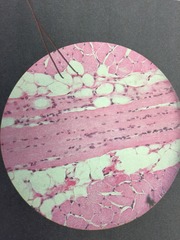
Skeletal Muscle (2)
Striated (lines)
Multinucleated
Non-branching
Lack cell boundaries
*Horizontal, striations

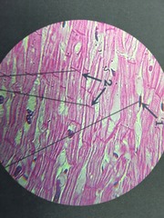
Cardiac Muscle (1)
Striated
Branched
Intercalated discs (heavy bands)

Cardiac Muscle (2)
Intercalated discs stained pink,red
Nucleus is dark dot
Neuron (1)
Axon, thick pink appendage
Dendrite, thin pink appendage
Glial cells, black scattered dots
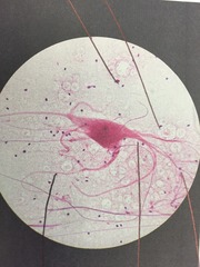
Neuron (2)

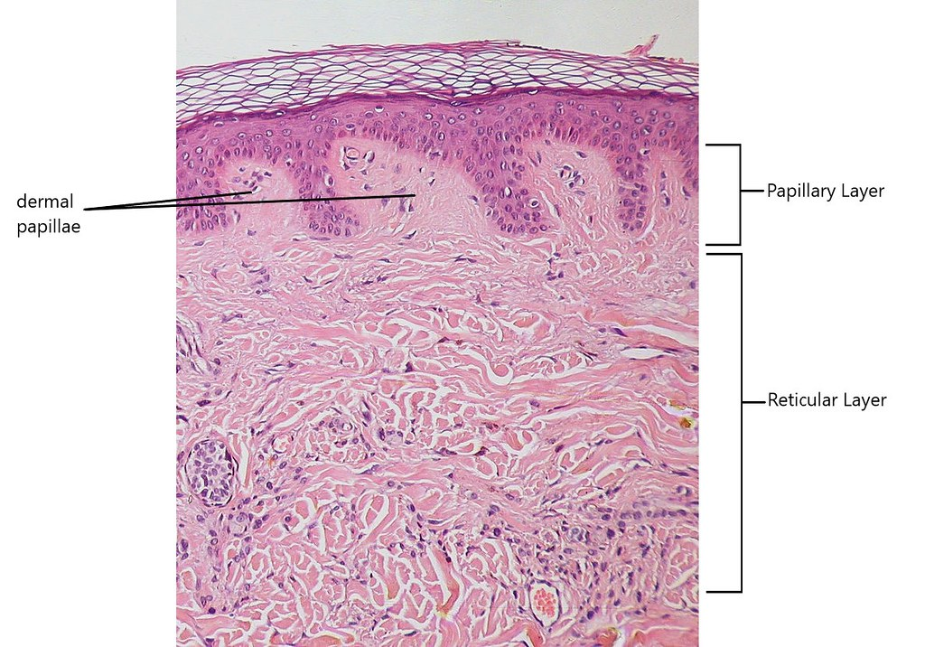
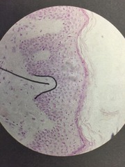
Dermal Papillae
Finger-like projections of the papillary layer
-Made up of areolar connective tissue
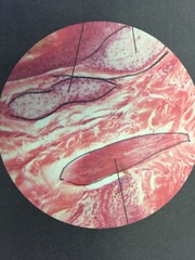
Arrector Pili Muscle
Associated with hair follicles and sebacious oil glands
Smooth muscle tissue
Makes your hair stand up on end
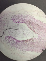
Hair papilla
Invagination
Sudoriferous (Sweat) Glands
Seen as numerous cross sections of tubules

Stratum corneum
Uppermost layer of the epidermis
Dead keratin-filled cell "horny layer"
25-30 layers of dead cells filled with keratin
"Stratified squamous epithelium"
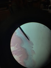
Stratum lucidium
Thin translucent zone
FINGERTIPS, PALMS, AND SOLES (THICK SKIN)
Filled with eleiden (precursor to keratin)
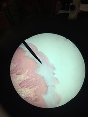
Stratum granulosum
Middle of epidermis
Distinctive feature: dark stained granules of protein keratohyalin
Stratum spinosum
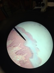
8-10 rows of keratinocytes
Produce spine like cytoplasmic processes that interlock with adjacent cells by desmosomes
Strength and flexibility
Stratum basale
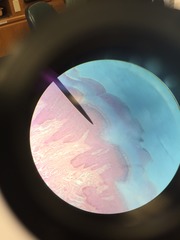
Single layer AT BOTTOM
Youngest layer of epidermis
Columnar or cuboidal cells
High mitotic rate

Reticular Layer