1.02 Synchronous
1/82
There's no tags or description
Looks like no tags are added yet.
Name | Mastery | Learn | Test | Matching | Spaced |
|---|
No study sessions yet.
83 Terms
How many vertebrae are typically found in the human body, and how many are separate bones?
There are typically 33 vertebrae (with individual findings ranging from 32-34) or 26 separate bones
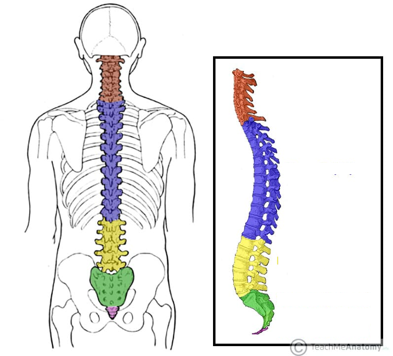
Name the five regions of the vertebral column and the number of vertebrae in each.
Cervical - 7 vertebrae
Thoracic - 12 vertebrae
Lumbar - 5 vertebrae
Sacral - 5 vertebrae
Coccygeal - 2-4 vertebrae
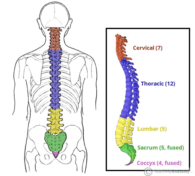
What happens to the vertebrae in the sacral and coccygeal regions?
The 5 sacral vertebrae fuse to make the sacrum, and the 2-4 coccygeal vertebrae fuse to make the coccyx.
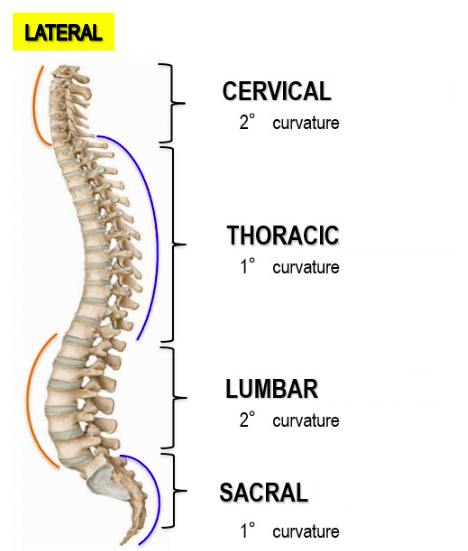
When do primary curvatures develop, and what is their characteristic shape?
Primary curvatures develop during the fetal period; the newborn spine is kyphotic, which is concave anteriorly
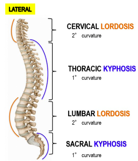
When do secondary curvatures develop?
Secondary curvatures result from the development of the spine in the first few years of life
Describe the shape of lordotic curves.
Lordotic curves are concave posteriorly.
When does cervical lordosis develop?
Cervical lordosis develops when infants begin to hold their heads up.
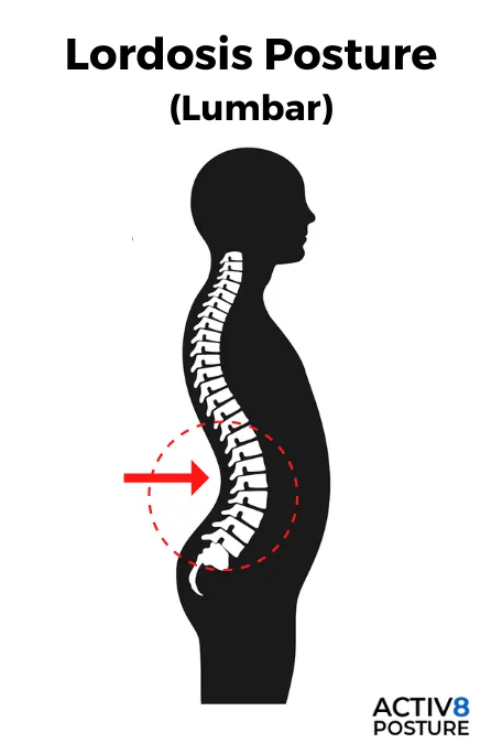
When does lumbar lordosis develop?
Lumbar lordosis develops when toddlers begin standing and walking
What are the two major parts of a typical vertebra?
The vertebral body anteriorly and the vertebral arch posteriorly.
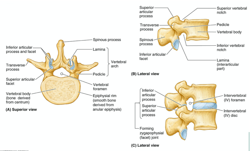
List the components recognized when a typical vertebra is dismantled.
Body
Vertebral arc
Articular processes
Transverse processes
Spinous process
Pedicles
Laminae
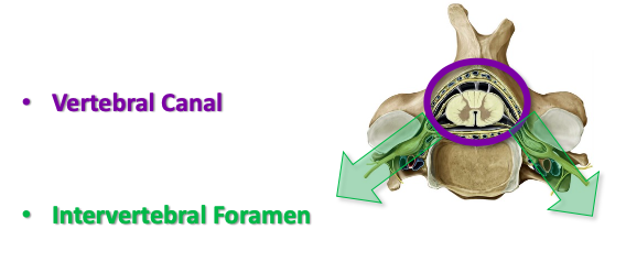
What is the vertebral foramen, and what does it house?
The vertebral foramen is an opening between the body and the vertebral arch, which houses the spinal cord.
What is the intervertebral foramen, and what is its function?
The intervertebral foramen is formed between the pedicles of adjacent vertebrae, and it protects exiting spinal nerves.
Describe the "Body" feature of a general vertebra.
It is an anterior projection and the largest bony segment, which stacks up to provide column height and shock absorption.
Describe the "Transverse Processes" feature of a general vertebra.
There are two (right and left) lateral projections that extend out from the vertebra in the transverse plane, serving as attachments for ribs and muscles.
Describe the "Spinous Process" feature of a general vertebra.
It extends posteriorly from the vertebrae in the sagittal plane and serves as an attachment for ribs and muscles.
Describe the "Articular Processes" feature of a general vertebra.
There are four (2 superior, 2 inferior for every segment) that extend superiorly and inferiorly from the vertebrae, allowing them to form joints with each other.
What are "Pedicles" and what do they connect?
Pedicles (right and left) connect the body of the vertebrae with the transverse processes (TVPS)
What are "Lamina" and what do they connect?
Lamina (right and left) connect the transverse processes (TVPS) of the vertebrae with the spinous processes (SPS).
How many pairs of spinal nerves are there, and how do they exit the spinal cord?
There are 31 pairs of spinal nerves exiting off the spinal cord and entering the periphery by passing through the intervertebral foramen
What is another name for the vertebral canal, and what is its function?
It is also known as the spinal canal and protects the spinal cord
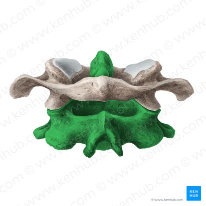
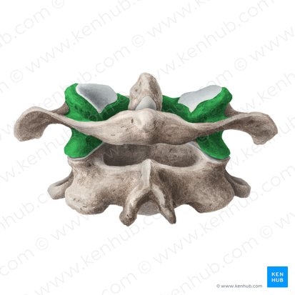
How many vertebrae are in the cervical region, and what type of curvature do they form?
There are 7 vertebrae in the cervical region, forming a lordotic curvature
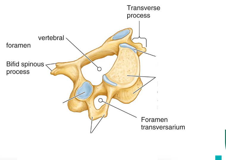
What are the main common features of a typical cervical vertebra?
Small body
Transverse foramen found in the transverse process
Bifurcated spinous process
What are the special features that make the Atlas (C1) different from a typical cervical vertebra?
Atlas (C1) has no body, no laminae, no pedicles, no SP
What additional anatomical features does the Atlas (C1) possess?
Anterior and posterior arches.
What is found on the posterior surface of the anterior arch of the Atlas (C1)?
An articular surface for the odontoid process of the axis (Dens of axis)
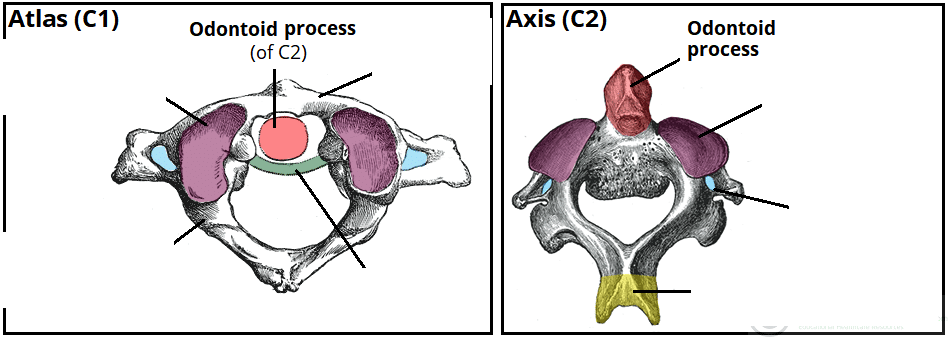
Describe the lateral mass of the Atlas (C1).
It is on either side of the vertebrae and contains articular surfaces on the superior and inferior surfaces
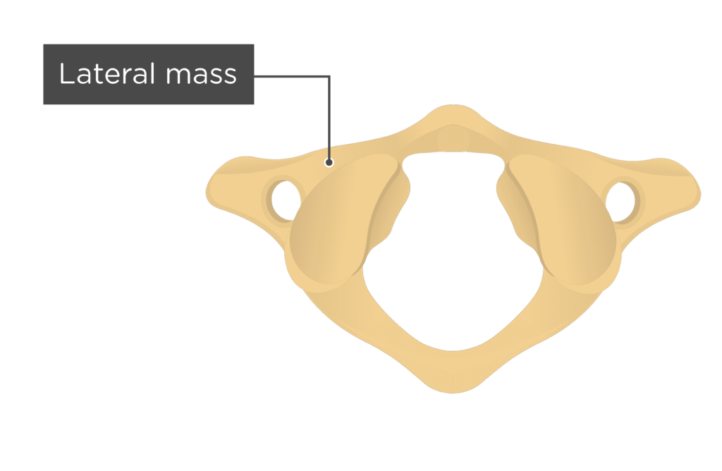
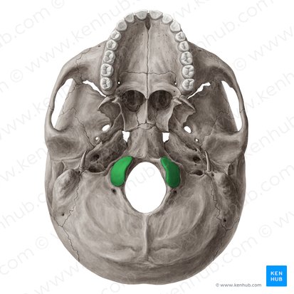
What does the superior articular surface of the Atlas (C1) articulate with?
The occipital condyles of the skull
What does the inferior articular surface of the Atlas (C1) articulate with?
The axis (C2).
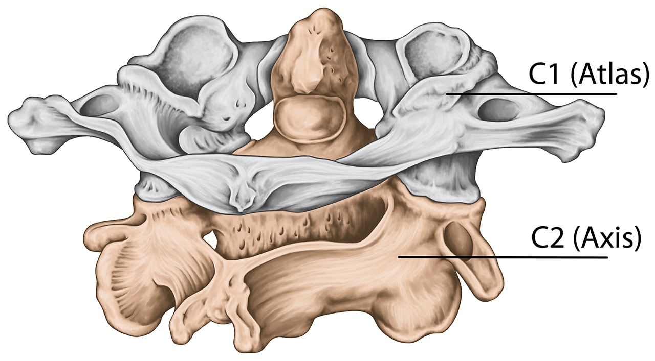
What is the exceptional feature of the Axis (C2)?
It possesses a large odontoid process (dens axis) which is attached to the superior surface of its body
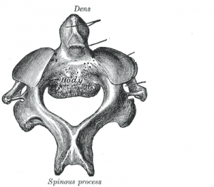
What is the function of the odontoid process (dens axis)?
This process acts as an axis (pivot) and permits the rotation of the atlas on the axis bone
What articular feature is found on the anterior surface of the dens axis on C2?
An articular facet matches up with the facet on the posterior surface of the anterior arch of C1 (atlas).

How is the "medial Atlantoaxial joint" formed?
It is formed by the articular facet on the anterior surface of the dens and the facet on the posterior surface of the anterior arch of C1 (atlas)
How is the "lateral atlantoaxial joint" formed?
It is formed between the superior articular facet of the axis and the inferior articular facet on the lateral mass of the atlas.
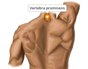
What is "Vertebra Prominens"?
Vertebra prominens is the proper name for the 7th cervical vertebra
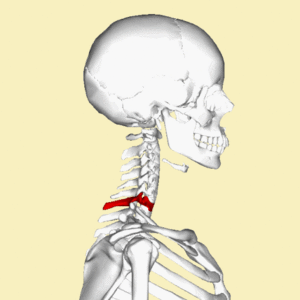
What is the most distinctive characteristic of the Vertebra Prominens?
The existence of a long and prominent spinous process which is palpable from the skin surface
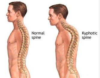
What type of curvature does the thoracic part of the vertebral column form, and how many vertebrae are in this region?
A kyphotic curvature
Made up of 12 vertebrae
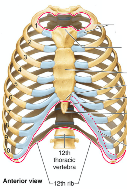
What are the main characteristics of typical thoracic vertebrae regarding rib articulation?
Costal facets are present on the sides of the bodies (demi-facet or semi-facet or hemi-facet) where the heads of the ribs articulate.
Costal facets on the transverse processes (called transverse costal facet) for articulation with the tubercles of the ribs (note: T11 and T12 have no facets on the transverse processes)
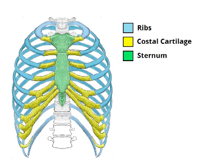
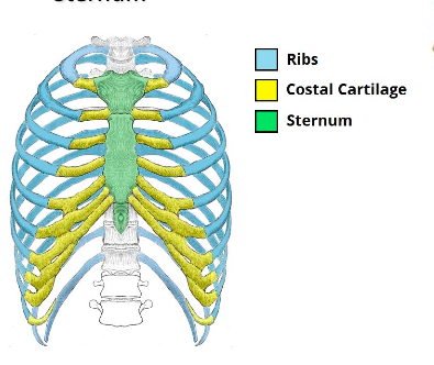
What are the components of the thoracic cage?
12 thoracic vertebrae + intervertebral discs
12 pairs of ribs + costal cartilages,
Sternum
List four functions of the thoracic cage.
Attachment for upper limbs
Attachments for many muscles of upper limbs, neck, abdomen, back, and muscles of respiration
Functions to protect the thoracic and abdominal organs
Attachment for mammary glands of breast tissue
How are ribs classified based on their attachment to the sternum?
TRUE ribs (1-7) – direct attachment to the sternum
FALSE ribs (8-10) – indirect attachment to the sternum
FREE / FLOATING (11 and 12) – no attachment to the sternum
How do "FALSE ribs" (8-10) attach to the sternum?
They attach/fuse to the costal cartilage of rib 7 to form an indirect attachment.

What are "intercostal spaces" and what do they contain?
The spaces between the ribs on each side of the thoracic cage, filled by the intercostal muscles and carrying the intercostal nerve and vessels.
Describe a typical rib.
A typical rib is a long, twisted, flat bone having a superior border and an inferior border
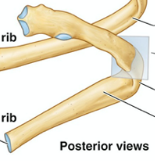
What is the costal groove of a rib, and what does it accommodate?
The inferior border overhangs and forms the costal groove, which accommodates the intercostal vessels and nerve.
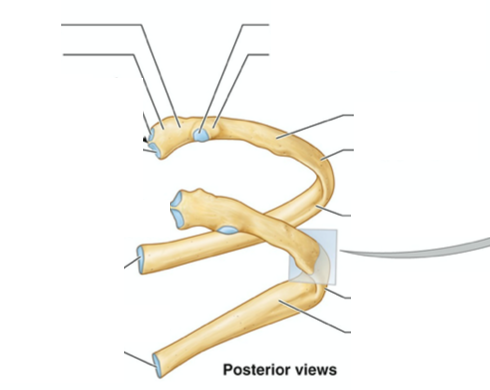
Name the main parts of a typical rib
Head
Neck
Tubercle
Shaft
Angle
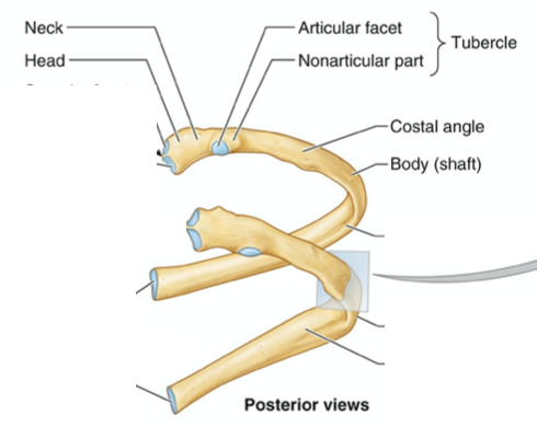
Describe the head of a typical rib.
The head has two semi-facets for articulation with the numerically corresponding vertebral body and that of the vertebra immediately above (separated by an inter-articular crest)
Where is the neck of a typical rib located?
The neck is a constricted portion situated between the head and the tubercle.
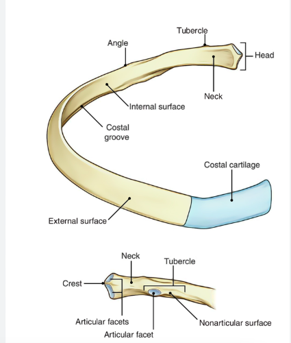
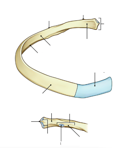
Describe the tubercle of a typical rib, including its articular and non-articular portions.
The tubercle is a prominence on the outer surface of the rib at the junction of the neck with the shaft.
Its articular portion has a facet for articulation with the transverse process of the numerically corresponding vertebra, while the non-articular portion gives attachment to ligaments
Describe the shaft (body) of a typical rib.
The shaft or body is thin, flattened, and twisted on its long axis. Its inferior border has the costal groove
What is the angle of a typical rib?
The angle is where the shaft of the rib bends sharply forward.
What is attached to the anterior end of each rib?
The corresponding costal cartilage.
What makes the first rib atypical?
It is flattened and wider than the rest of the ribs. It has a tubercle on the inner (medial) border, known as the scalene tubercle, for the insertion of the anterior scalene muscle.
This rib neither has an angle nor a costal groove, and its head only bears one articular surface for the body of T1
What are the atypical characteristics of the second rib?
The body of the second rib has similar characteristics to typical ribs, except it lacks the costal groove
What makes the 10th rib atypical?
It has only a single articular facet on its head.
What makes the 11th and 12th ribs atypical?
They have only a single articular facet on the head and no neck or tubercle. The 11th rib has a slight angle and shallow costal groove, but the 12th rib has no angle or costal groove
List the Atypical Ribs
1st Rib
2nd Rib
10th Rib
11th Rib
12th Rib
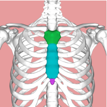
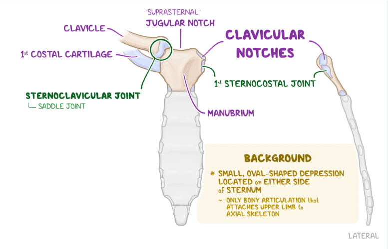
What is the sternum, and what are its 3 parts?
The sternum is a flat bone that may be divided into 3 parts:
Manubrium
The body
Xiphoid process

Describe the manubrium of the sternum and its articulations.
It is the upper part of the sternum that articulates with the clavicles and the first and upper part of the second costal cartilages on each side.
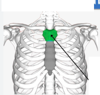
What is the suprasternal notch (jugular notch)?
It is the large, visible dip where the clavicle AND first rib join the sternum.
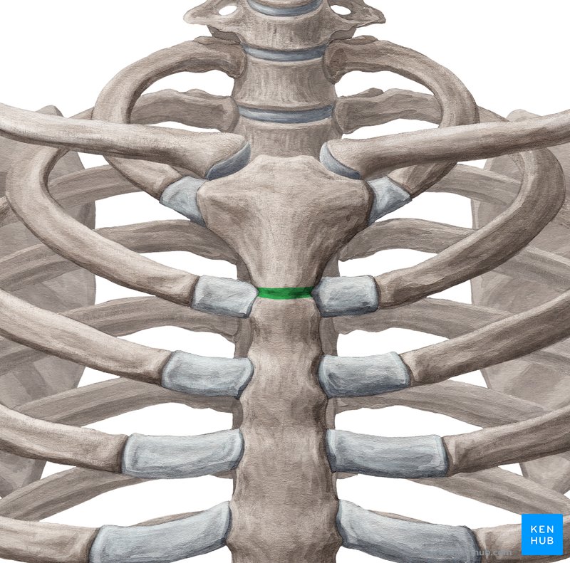
Describe the body of the sternum and its articulations.
It articulates above with the manubrium and below with the xiphoid process.
On each side are notches for articulation with the lower part of the second costal cartilage and the attachment of ribs 3-6 via their costal cartilages.
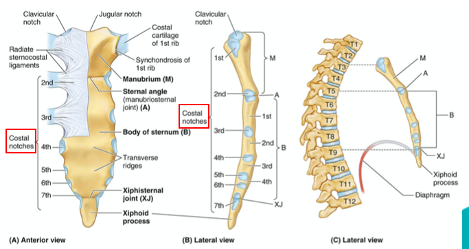
What is the sternal angle (Angle of Louis, manubriosternal joint)?
It is the angle formed by the junction of the manubrium and the body of the sternum in the form of a cartilaginous joint (which later becomes bony).
What is the clinical significance of the sternal angle?
It is a palpable clinical landmark that marks the approximate level of the 2nd pair of costal cartilages and the level of the intervertebral disc between T4 and T5.
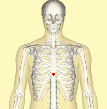
Describe the xiphoid process of the sternum.
It is the lowest and smallest part of the sternum.
What is the xiphi-sternal joint, and what does it attach?
It is demarcated by a transverse ridge and serves as an attachment for rib 7 costal cartilage

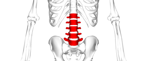
What type of curvature does the lumbar part of the vertebral column have, and how many vertebrae are in this region?
The lumbar part of the vertebral column is convex anteriorly/ concave posteriorly and consists of 5 vertebrae
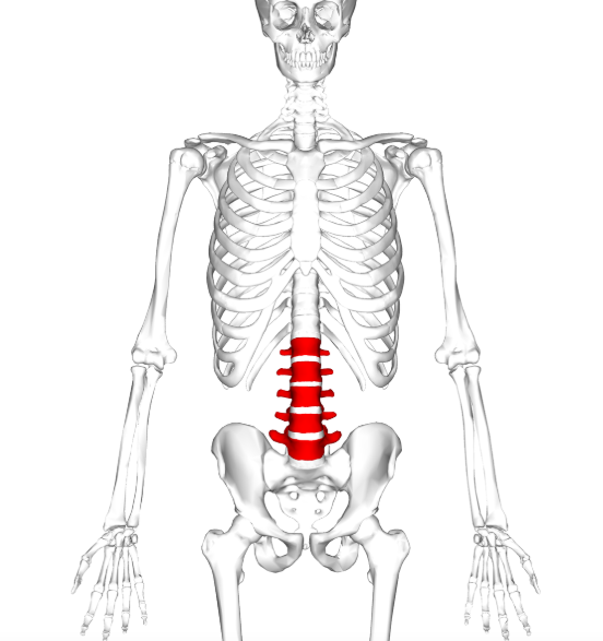
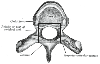
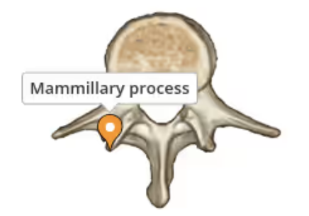
What are the key characteristics of lumbar vertebrae?
They have a massive and
Kidney-shaped body
Strong pedicles
Thick laminae
Long and slender transverse processes
A mammillary process found at the posterior margin of the superior articular processes
What features are notably absent in lumbar vertebrae?
They have no facets for articulation with ribs and no foramina in the transverse processes
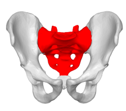
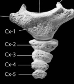
Describe the composition and shape of the sacrum.
It consists of 5 rudimentary vertebrae fused together to form a wedge-shaped bone, which is concave anteriorly.
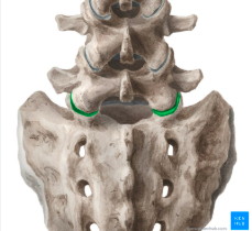
What does the upper border (base) of the sacrum articulate with?
The 5th lumbar vertebra.
What does the narrow inferior border of the sacrum articulate with?
The coccyx
How does the sacrum articulate laterally?
The sacrum articulates with the two hip bones to form the sacroiliac joints (in auricular surfaces)
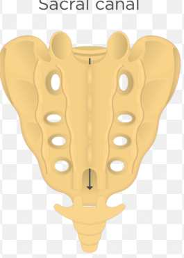
What canal is formed by the vertebral foramina within the sacrum?
The Sacral Canal
What bony ridges are formed on the posterior surface of the sacrum?
The spinous, articular, and transverse processes of the sacral vertebrae fuse together to form bony ridges known as sacral crests.
How many pairs of posterior/anterior sacral foramina are there, and what is the appearance of the anterior surface of the sacrum?
There are 4 pairs of foramina on the anterior surface (called anterior sacral foramina)
4 pairs on the posterior surface (called posterior sacral foramina)
So total 8 foramina on each side and 16 in total
The anterior surface of the sacrum is concave
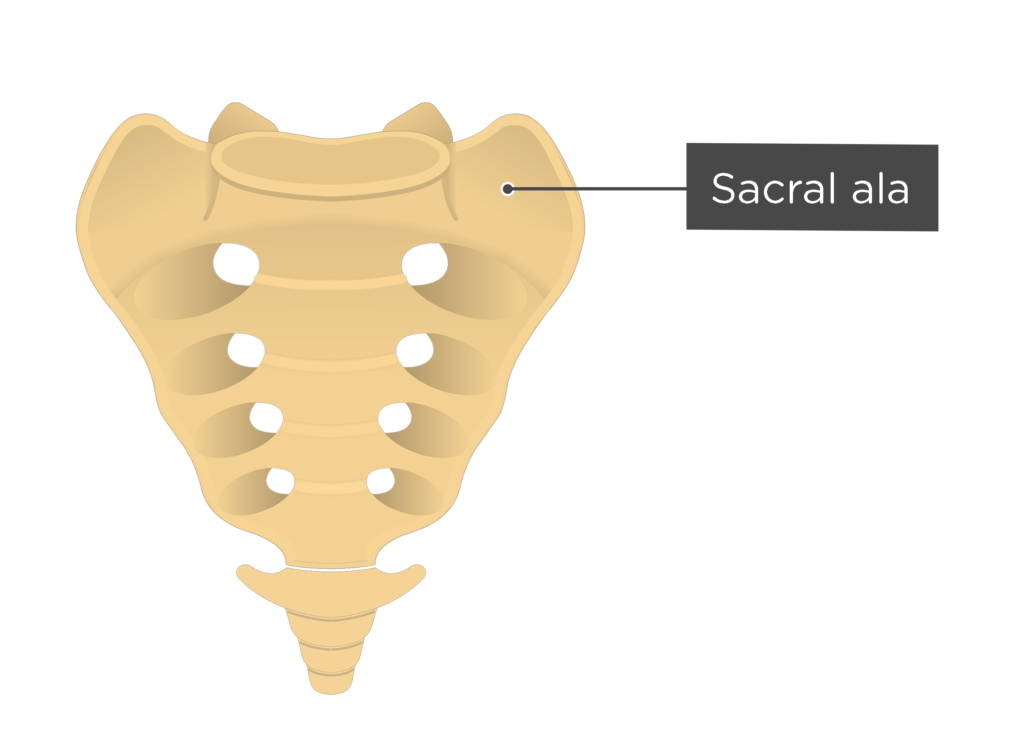
What are the Sacral Ala?
2 triangular flat surfaces found on the sides of the first sacral vertebra.
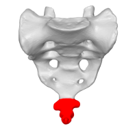
Describe the coccyx and its articulation.
The coccyx consists of 4 vertebrae fused together to form a small triangular bone, which articulates at its base with the apex of the sacrum.
Where are intervertebral discs located?
Between most of the vertebrae
What proportion of the vertebral column's length do intervertebral discs account for?
They make up approximately ¼ (25%) of the length of our vertebral column (the other ¾ is formed by the vertebrae)
What are the main functions of intervertebral discs?
Strong attachment between vertebrae for support and protection
Weight bearing
Shock absorption
What are the 2 main components of intervertebral discs?
Annulus fibrosus
Nucleus pulposus.
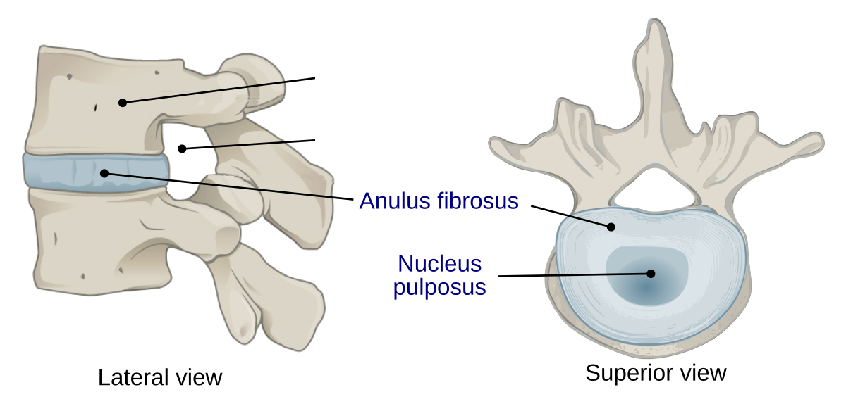
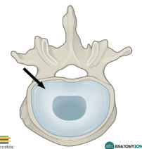
Describe the Annulus Fibrosus.
Concentric rings of fibrocartilage that connect adjacent vertebral bodies and secure the nucleus in position
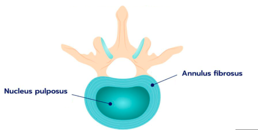
Describe the Nucleus Pulposus.
A semi-gelatinous mass that acts as a shock absorber.
Where are intervertebral discs not found?
No discs are found between the atlas and skull, atlas and dens, sacral segments, and coccygeal segments