Sexual reproduction in Humans

0.0(0)
studied byStudied by 0 people
learnLearn
examPractice Test
spaced repetitionSpaced Repetition
heart puzzleMatch
flashcardsFlashcards
Card Sorting

1/10

flashcard set

Earn XP

Description and Tags

1-6 (Male Reproduction system)

Study Analytics
Name
Mastery
Learn
Test
Matching
Spaced

No study sessions yet.

11 Terms

1
New cards

How do sperms move during ejaculation?

Before ejaculation:

Seminiferous tubules (production site) → collect in vasa efferentia tubes → carried to head of epididymis (where they become motile)

Ejaculation:

Head of epididymis → carried by vas deferens (and mixed with secretions) → penis

<p>Before ejaculation: </p><p><mark data-color="yellow" style="background-color: yellow; color: inherit">Seminiferous tubules</mark> (production site) → collect in <mark data-color="yellow" style="background-color: yellow; color: inherit">vasa efferentia tubes</mark> → carried to <mark data-color="yellow" style="background-color: yellow; color: inherit">head of epididymis </mark>(where they become motile)</p><p></p><p>Ejaculation:</p><p><mark data-color="yellow" style="background-color: yellow; color: inherit">Head of epididymis</mark> →  carried by <mark data-color="yellow" style="background-color: yellow; color: inherit">vas deferens</mark> (and mixed with secretions) → <mark data-color="yellow" style="background-color: yellow; color: inherit">penis </mark></p><p></p><p></p>
2
New cards

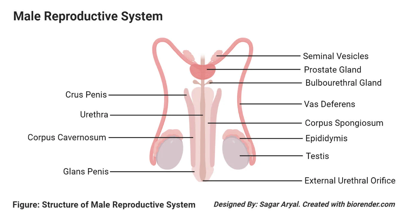
What are the accessory glands and their secretions (male reproductive system)?

  • Seminal vesicle (mucus)

    - secretes mucus in vas deferens

    - mucus secreted contain mixture of chemicals including fructose (respired by sperm)

  • Prostate gland (zinc-containing prostate fluid)

    Purpose of these secretions:

    • both alkaline → neutralises acidity in urine in urethra and the acidity of vaginal tract

    • provides nutrients for sperm - fructose, amino acids, zinc ions

3
New cards

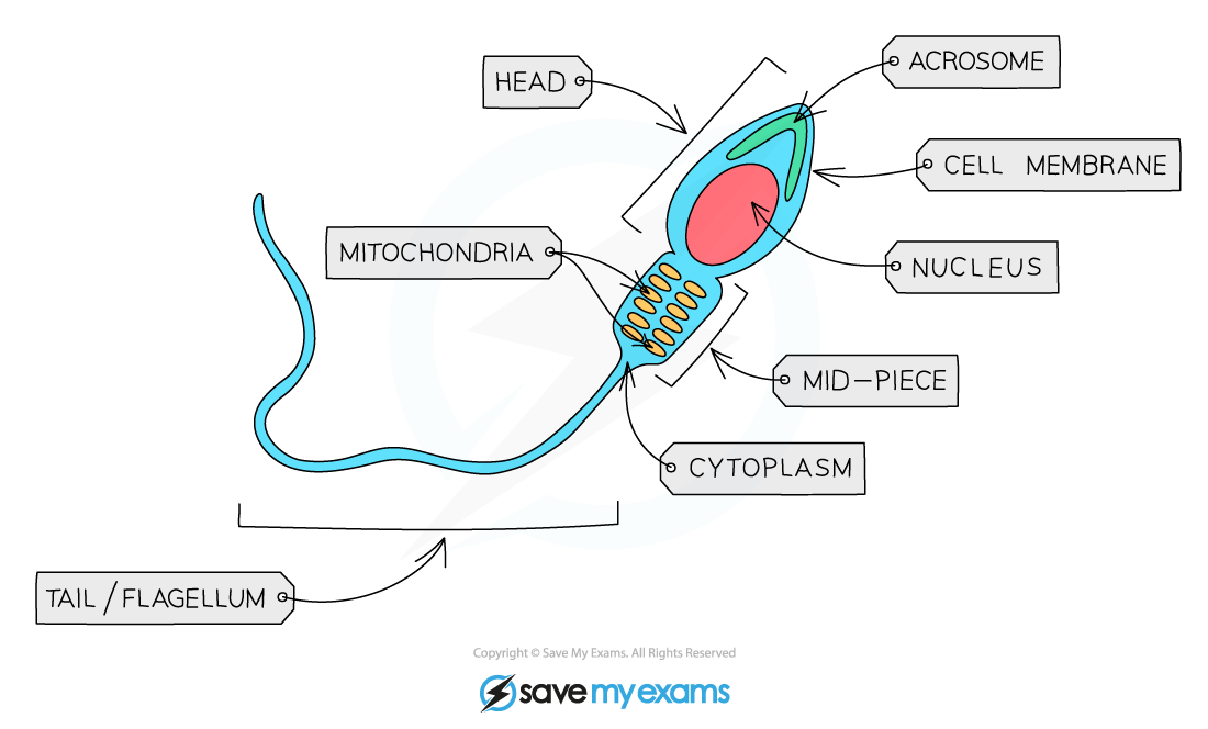
How does the structure of sperm look like?

mitochondria - provides ATP for movement

head - contains haploid nucleus

acrosome - contains lysosome enzymes used at fertilisation

tail - makes lashing movements that move sperm

<p>mitochondria - provides ATP for movement</p><p>head - contains haploid nucleus</p><p>acrosome - contains lysosome enzymes used at fertilisation</p><p>tail - makes lashing movements that move sperm</p>
4
New cards

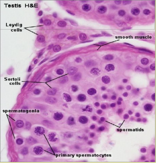
Histological examination of testis

Secondary spermatocytes are not seen easily as they progress quickly to spermatids

Adjacent to the lumen, spermatozoa can be seen

  • Between strands of developing spermatids are → Sertoli cells (columnar large oval nucleus and dense nucleolus)

    • secrete fluid which i) nourishes the developing sperm and ii) protects them from immune system

  • Cells between seminiferous tubules called → Leydig cells which secrete testosterone

    • essential in sperm formation and maturation aswell as development of male secondary sexual characteristics

<p>Secondary spermatocytes are not seen easily as they progress quickly to spermatids</p><p>Adjacent to the lumen, spermatozoa can be seen</p><ul><li><p>Between strands of developing spermatids are → <u>Sertoli cells</u> (columnar large oval nucleus and dense nucleolus)</p><ul><li><p>secrete fluid which i) <mark data-color="yellow" style="background-color: yellow; color: inherit">nourishes the developing sperm</mark> and ii) <mark data-color="yellow" style="background-color: yellow; color: inherit">protects them from immune system</mark></p></li></ul></li><li><p>Cells between seminiferous tubules called → <u>Leydig cells</u> which secrete <mark data-color="yellow" style="background-color: yellow; color: inherit">testosterone</mark></p><ul><li><p><mark data-color="yellow" style="background-color: yellow; color: inherit">essential in sperm formation and maturation</mark> aswell as development of male secondary sexual characteristics</p></li></ul></li></ul><p></p><p></p><p></p>
5
New cards

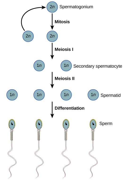
What is the sequence that occurs during spermatogenesis starting from epithelium germ cell?

1) Germinal epithelium cell → 2) spermatogonia → 3) primary spermatocyte → 4) secondary spermatocyte → 5) spermatids → 6) spermatozoa

6
New cards

Explain the process in spermatogenesis?

  1. Germinal epithelium (diploid) divides by mitosis to make diploid spermatogonia and more germinal epithelium cells.

  2. Spermatogonia divides many times by mitosis making more spermatagonia, some enlarge, making diploid primary spermatocytes

  3. Primary spermatocytes undergo meiosis I, making haploid secondary spermatocytes.

  4. Secondary spermatocytes undergo meiosis II, making haploid spermatids

  5. Spermatids mature into spermatozoa/sperm

<ol><li><p>Germinal epithelium (<strong><u>diploid</u></strong>) divides by <strong><u>mitosis</u></strong> to make diploid <mark data-color="yellow" style="background-color: yellow; color: inherit">spermatogonia</mark> and more <mark data-color="yellow" style="background-color: yellow; color: inherit">germinal epithelium cells.</mark></p></li><li><p>Spermatogonia divides many times by <strong><u>mitosis</u></strong> making <mark data-color="yellow" style="background-color: yellow; color: inherit">more spermatagonia</mark>, some enlarge, making <mark data-color="yellow" style="background-color: yellow; color: inherit">diploid primary spermatocytes</mark></p></li><li><p>Primary spermatocytes undergo <strong><u>meiosis I</u></strong>, making <mark data-color="yellow" style="background-color: yellow; color: inherit">haploid secondary spermatocytes.</mark></p></li><li><p>Secondary spermatocytes undergo <strong><u>meiosis II</u></strong>, making <mark data-color="yellow" style="background-color: yellow; color: inherit">haploid spermatids</mark></p></li><li><p><mark data-color="yellow" style="background-color: yellow; color: inherit">Spermatids</mark> mature into <mark data-color="yellow" style="background-color: yellow; color: inherit">spermatozoa/sperm</mark></p></li></ol><p></p>
7
New cards

How do oocytes (developing egg) move during ovulation?

Oocytes mature/derive from cells in the germinal epithelium (around periphery of ovary)

Mature follicles then migrate to surface of ovary (alternate monthly) → secondary oocyte is released from ovulation

Cilia at entrance of oviduct funnel sweep the 2ndary oocyte into fallopian tube

→ ciliated epithelial cells lining the fallopian tube convey the secondary oocyte to the uterus

8
New cards

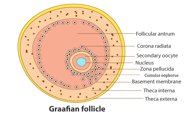
How does a mature/graafian follicle look like?

i) Zona pellucida - glycoprotein protein surrounding cell membrane of 2ndary oocyte.

ii) Corona radiata - cellular layer providing protection to them and provide nutrients

iii) Cumulus cells - contribute to radiata cells

iv) Theca - cells surrounding graafian follicle which release oestrogen.

<p>i) <strong><u>Zona pellucida</u></strong> - glycoprotein protein surrounding cell membrane of 2ndary oocyte.</p><p>ii) <strong><u>Corona radiata</u></strong> - cellular layer providing protection to them and provide nutrients</p><p>iii) <strong><u>Cumulus cells</u></strong> - contribute to radiata cells</p><p>iv) <strong><u>Theca</u></strong> - cells surrounding graafian follicle which release oestrogen.</p>
9
New cards

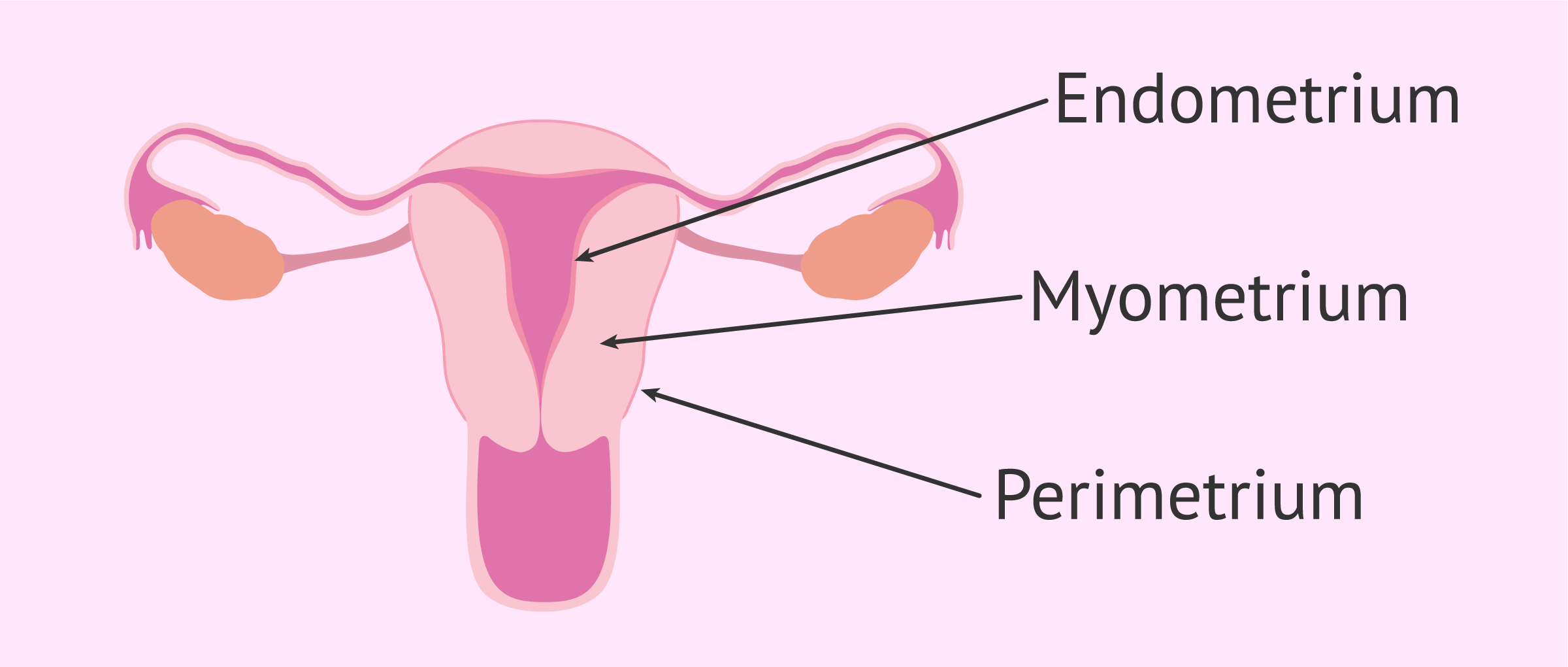
What are the 3 layers of the uterus wall?

Perimetrium - thin layer around the outside

Myometrium - muscle layer in the middle

Endometrium - innermost layer, mucous membrane well supplied with blood

→ layer that builds up and is shed in a monthly cycle (unless fertilisation occurs)

<p>Perimetrium - thin layer around the outside</p><p>Myometrium - muscle layer in the middle</p><p>Endometrium - innermost layer, mucous membrane well supplied with blood</p><p>→ layer that builds up and is shed in a monthly cycle (unless fertilisation occurs)</p>
10
New cards

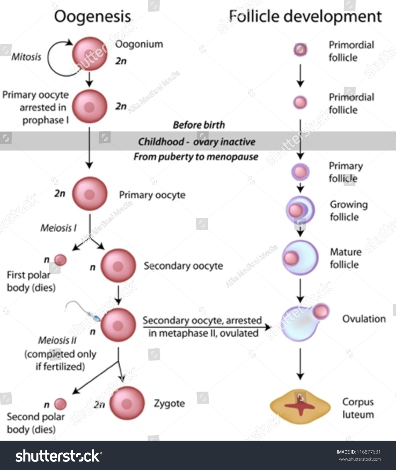
What is the sequence that occurs during oogenesis starting from epithelium germ cell?

1) Germinal epithelium cells → 2) Oogonium → 3) Primary oocyte + first polar body → 4) Secondary oocyte → 5) Ovum + second polar body

11
New cards

What is the process of oogenesis?

BEFORE BIRTH

  • Germinal epithelium cells divide repeatedly in ovary by mitosis → makes diploid oogonia + more germinal epithelium

  • Oogonia divide by mitosis and enlarge → diploid primary oocytes + more oogonia

  • Primary oocytes begin meiosis I (arrested at Prophase I)

    → millions of primary oocytes at Prophase I in newborn

AFTER BIRTH

  • After puberty, hormones stimulate development → b4 ovulation, primary oocyte completes meiosis I making secondary + first polar (disintegrates)

  • Secondary oocyte begins meiosis II, but arrested at metaphase II unless fertilisation occurs (would produce ovum)

<p>BEFORE BIRTH</p><ul><li><p>Germinal epithelium cells divide repeatedly in ovary by <strong><u>mitosis</u></strong> → makes <mark data-color="yellow" style="background-color: yellow; color: inherit">diploid oogonia </mark>+ <mark data-color="yellow" style="background-color: yellow; color: inherit">more germinal epithelium</mark></p></li><li><p>Oogonia divide by <strong><u>mitosis</u></strong> and enlarge → <mark data-color="yellow" style="background-color: yellow; color: inherit">diploid primary oocytes + more oogonia</mark></p></li><li><p><mark data-color="yellow" style="background-color: yellow; color: inherit">Primary oocytes</mark> begin <strong><u>meiosis I</u></strong> (arrested at <u>Prophase I</u>)</p><p>→ millions of primary oocytes at Prophase I in newborn</p></li></ul><p>AFTER BIRTH</p><ul><li><p>After puberty, hormones stimulate development → b4 ovulation, primary oocyte completes <strong><u>meiosis I</u></strong> making <mark data-color="yellow" style="background-color: yellow; color: inherit">secondary + first polar (disintegrates)</mark></p></li><li><p><mark data-color="yellow" style="background-color: yellow; color: inherit">Secondary oocyte</mark> begins <strong><u>meiosis II</u></strong>, but arrested at <u>metaphase II</u> unless fertilisation occurs (would produce <mark data-color="yellow" style="background-color: yellow; color: inherit">ovum</mark>)</p></li></ul><p></p>