Exam 2C- Anatomy - Hand and wrist
 Call Kai
 Learn
Learn Practice Test
 Spaced Repetition
 Match
Match Flashcards
 Knowt Play
1/64

There's no tags or description
Looks like no tags are added yet.
Name | Mastery | Learn | Test | Matching | Spaced | Call with Kai | Chat |
|---|
No analytics yet
Send a link to your students to track their progress
65 Terms
DeQuervain Tenosynovitis
- Washer-woman's sprain
- inflammation of sheath
- compartment 1 = abductor pollicis longus and extensor pollicis brevis
- now called "texters thumb"
ganglion cysts
- "bible bumps"
- cyst derived from tendon sheath of a tendon: common firm swelling on dorsum of hand/wrist
- can occur anywhere, but most common at scapho-lunate joint
Palmar carpal ligament
more proximal and superficial than the transverse carpal ligament; attaches to the styloid processes of the radius and ulna, and crosses over the flexor muscles
guyon's canal/ulnar canal
- between hamate and pisiform
- superficial to flexor retinaculum
- roof is the palmar carpal ligament, floor is flexor retinaculum
- ulnar a. and n.
carpal tunnel syndrome
- neurogenic (compression of median n.)
- symptoms: burning, tingling, itching, numbness in palm & fingers
distal radioulnar joint
pivot joint: head of ulna to ulnar notch of radius
- fibrocartilaginous radioulnar disk separates ulna from proximal carpals and holds ulna to distal radius- triquetral & lunate
radioulnar disk
- what unites radius and ulna during pronation/supination
- disk with distal radioulnar articulation's shape forms synovial cavity separate from radiocarpal joint's cavity
radiocarpal joint
wrist joint
- distal radius, fibrocartilaginous radioulnar disk, proximal row of carpals (radius to scaphoid & lunate)
- reinforced by radial collateral lig. of wrist and ulnar collateral lig. of wrist
radial collateral ligament
syloid process of radius to radial side of scaphoid
ulnar collateral ligament
syloid process of ulna to medial triquetrum and pisiform/transverse carpal ligament
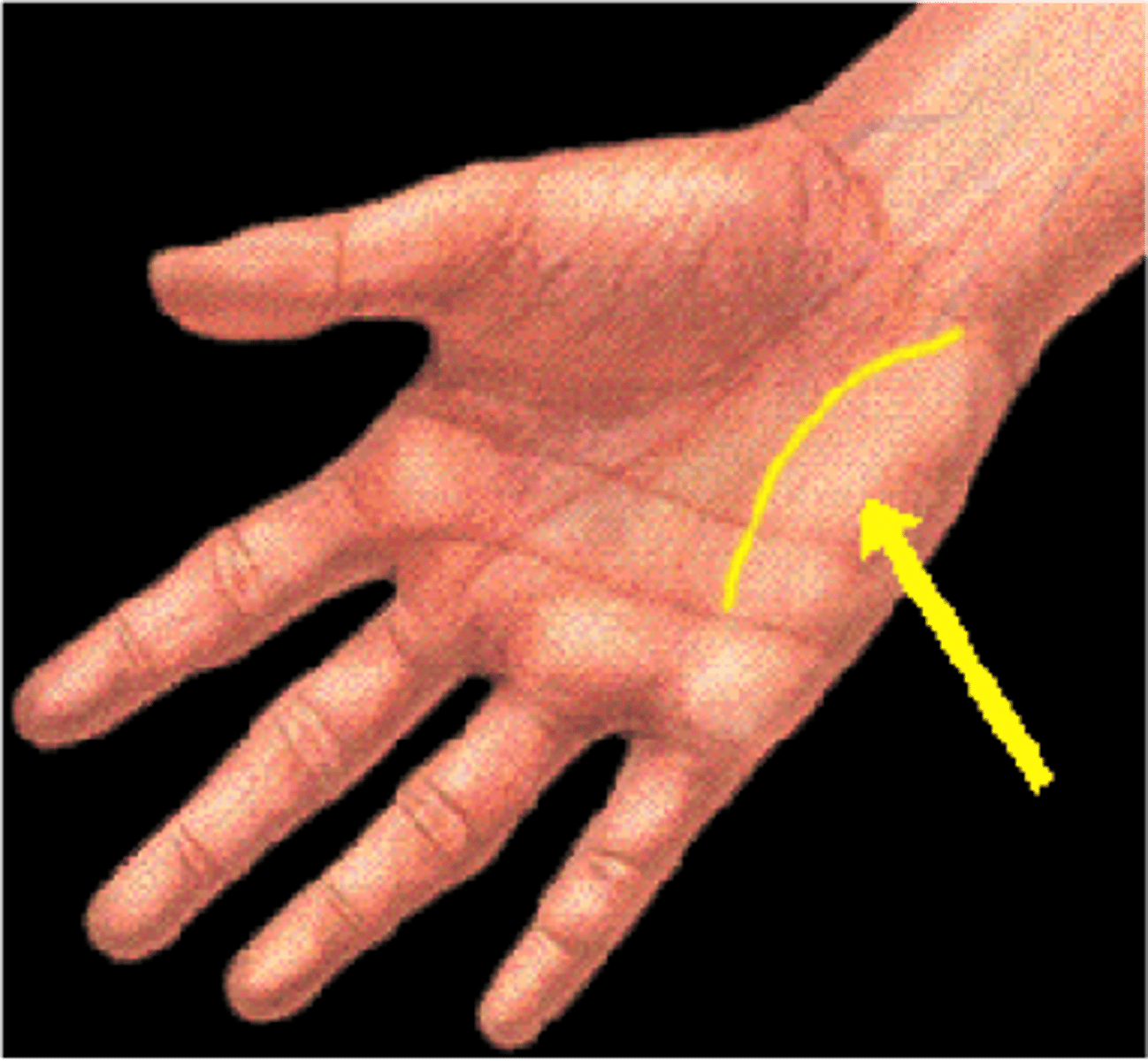
thenar eminence
thumb
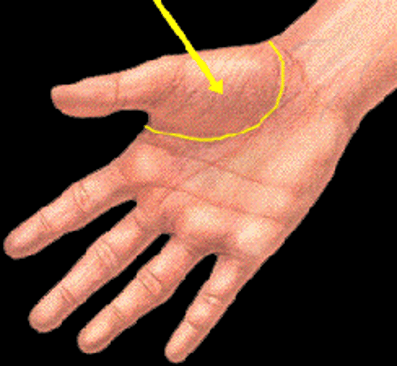
hypothenar eminence
little finger
cutaneous nerves
- palmar branch of median n.
- palmar branch of ulnar n.
- digital branches of median n.
- digital branches of ulnar n.
- superficial branch of radial n.
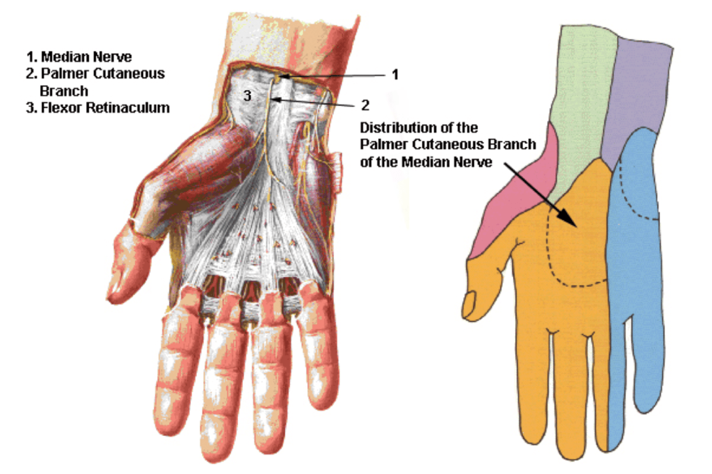
palmar branch of median n.
- begins before median n. enters carpal tunnel
- to mid palm
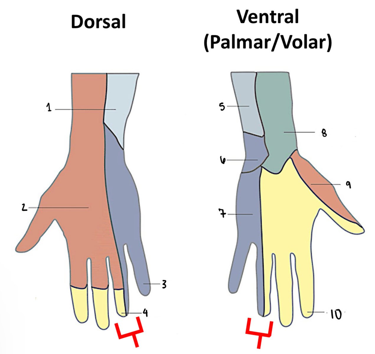
palmar branch of ulnar n.
- begins in wrist
- to medial palm
the hand
- entirely wrapped in fascia
- palmar and dorsal fascia continuous on sides
- thin, except at mid-palm
palmar aponeurosis
- thick fascia between thenar and hypothenar eminences. Reinforced by aponeuroses of palmaris longus muscles and divides into 4 slips that enter the fingers
- septum dives deep to attach to 3rd metacarpal (midpalmar septum)
midpalmar septum
divides the tendons of the flexor digitorum muscles, the thenar muscles, and the deeper lying interossei into 2 compartments:
1. the thenar space (deep to the thenar muscles
2. the midpalmar space (deep to the long tendons
midpalmar septum- clinical note
infections are restricted to the space in which it occurred (midpalmar or thenar), and numerous surgical approaches were described to open the space and drain the infected area
dupuytren''s contracture
- longitudinal slips of palmar aponeurosis develop cysts
- slips contract and produce passive flexion of affected fingers
palmaris brevis PA
palmar aponeurosis
palmaris brevis DA
skin over hypothenar eminence
palmaris brevis a & i
- action: increase palm concavity on hypothenar side
- innervation: ulnar n.
abductor pollicis brevis PA
trapezium and flexor retinaculum
abductor pollicis brevis DA
base of proximal phalanx
abductor pollicis brevis a & i
- action: abduct thumb, assist flexion at MP joint
- innervation: recurrent branch of median n.
flexor pollicis brevis PA
trapezium and flexor retinaculum
flexor pollicis brevis DA
base of proximal phalanx
flexor pollicis brevis a & i
- action: flex MP joint
- innervation: recurrent branch of median n.
opponens pollicis PA
trapezium and flexor retinaculum
opponens pollicis DA
shaft of 1st metacarpal
opponens pollicis a & i
- action: medially pulls and rotates 1st metacarpal into apposition position with another digit
- innervation: recurrent branch of median n.
adductor pollicis PA
- oblique: bases of 2nd and 3rd metacarpals
- transverse: shaft of 3rd metacarpal
adductor pollicis DA
base of proximal phalanx of thumb
adductor pollicis a & i
- action: adduct pollux
- innervation: deep branch of ulnar n.
abductor digiti minimi PA
pisiform and flexor retinaculum
abductor digiti minimi DA
base of proximal phalanx of digiti minimi
abductor digiti minimi a & i
- action: abduct pinky finger
- innervation: deep branch of ulnar n.
flexor digiti minimi brevis PA
hamate and flexor retinaculum
flexor digiti minimi brevis DA
base of proximal phalanx
flexor digiti minimi brevis a & i
- action: Flex MP joint
- innervation: deep branch of ulnar n.
opponens digiti minimi PA
Hook of hamate and flexor retinaculum
opponens digiti minimi DA
shaft of 5th metacarpal
opponens digiti minimi a & i
- action: pull 5th metacarpal toward midline and rotate into apposition position with thumb
- innervation: deep branch of ulnar n.
lumbricals PA
sides of tendons of flexor digitorum profundus
- 3 and 4 between tendons (unipennate 1 & 2) vs. bipennate (3 &4)
lumbricals DA
radial side of dorsal expansion of next highest numbered digit
- ex. 1st lumbrical goes to 2nd digit
lumbricals a & i
- action: flex MP and extend IP joints of fingers
- innervation: 1 & 2 = digital branches of median n. 3 & 4 = deep branch of ulnar n.
interossei in general
- palmar = 3
- dorsal = 4
- all are innervated by deep branch of ulnar n.
- All are able to extend PIP & DIP joints while stimultaneously flexing MP joints
palmar interossei PA
all attach to metacarpals
- #1 to medial side of 2nd, #2 to lateral 4th, #3 to lateral 5th
palmar interossei DA
dorsal expansion of same finger side as PA
palmar interossei a & i
- action: all adduct fingers and extend PIP and DIP joints while simultaneously flexing MP (PAD)
- innervation: deep branch of ulnar n.
dorsal interossei PA
surface of adjacent metacarpals
dorsal interossei DA
dorsal expansion
- 1st = index, 2nd & 3rd = either side of the middle finger, 4th = ring
dorsal interossei a & i
- action: abduct finger OR extend PIP and DIP joints while simultaneously flexing MP (DAB)
- innervation: deep branch of ulnar n.
radial n. in hand
- superficial branch of radial n. = prior, followed radial a.
- lateral branch = dorsal digital nerves (1)
- medial branch = dorsal digital nerves (2,3, radial side of 4)
median n. in hand
passes through carpal tunnel
- but the palmar cutaneous branch does not go through cubital tunnel
- after "tunnel", nerve becomes enlarged and flattened. gives off recurrent branch to thenar group.
- also called million dollar nerve: american football players have insurance on it because cleats stepping on the palm of the hand
common palmar digital nerves
- proper palmar digital branches: to pollux, index, middle fingers and radial half of ring finger (palmar and distal dorsal surfaces)
ulnar n. in hand
passes through guyon's canal and splits
1. superficial branch: terminal branch, cutaneous to ulnar portion of dorsal and palmar hand.
- common palmar digital nerve: proper palmar digital nerves to 5th finger and ulnar side of ring finger
2. deep branch: other terminal branch. to most intrinsic muscles of the hand
- traves with deep palmar branch of radial a. and ends at adductor pollicis
ulnar a.
- superficial branch: is the major supplier to- superficial palmar arch, gives off digital branches. Minor radial a. contributions
- deep branch: given off near pisiform and adds to deep palmar arch
radial a.
- dorsal carpal branch adds to dorsal carpal arch: gives off branches toward digits. princeps pollicis a., radialis indicis a.
- superficial palmar branch: to superior palmar arch- common palmar digital arteries, proper palmar digital arteries. terminates in deep palmar arch
intrinsic plus
- MP joints flexed and IP joints extended (nerves intact)
- safe position for hand splinting: collateral ligaments of MP and IP joints are taut (lengthened). Ideal for immobilization of most hand injuries = prevent contractures
intrinsic minus hand
IP joints flexed and MP joints extended
- injury to ulnar n. at wrist: if injury occurs at elbow- cubital tunnel syndrome, "claw" is less pronounced as deep flexors of 4th and 5th fingers are also paralyzed.
due to flexor digitorum profundas
intermetacarpal joints
deep transverse metacarpal ligaments
- transversely running ligamentous bands between distal metacarpal bones
- continuous with fibrous digital sheaths at each finger
- helps to stop hyperextension at PIP joints!!!!!!!!
swan neck deformity
PIP hyperextension with DIP flexion
- in worst casae, PIP hyperextension may lead to avulsion of ligaments
jammed finger
finger joint pain and swelling due to impact injury
mallet inger
a tear in dorsal expansion's connection to base of distal phalanx (unable to fully extend at DIP)