Special Sense Lecture 3 - Hearing/Vision
 Call Kai
 Learn
Learn Practice Test
 Spaced Repetition
 Match
Match Flashcards
 Knowt Play
1/46

There's no tags or description
Looks like no tags are added yet.
Name | Mastery | Learn | Test | Matching | Spaced | Call with Kai | Chat |
|---|
No analytics yet
Send a link to your students to track their progress
47 Terms
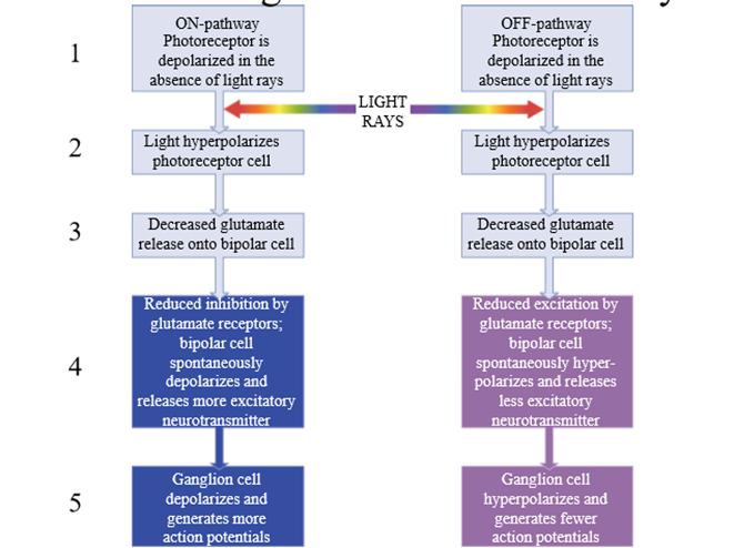
Similarities between On and OFF pathway of vision
Steps 1-3 are the same - Photoreceptor is depolarized in absence of light rays, Light hyperpolarizes photoreceptor, decreased glutamate release on bipolar cell
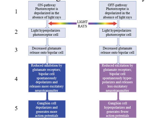
Differences between ON and OFF pathway of vision
Pathways respond opposite to light
In ON pathway - reduce inhibition causes ON bipolar cell to depolarize and release glutamate to ganglion cell
In OFF pathway - decreased glutamate from bipolar cell causes inhibition, preventing action potentials
Where is the visual cortex located
In the occipital lobe
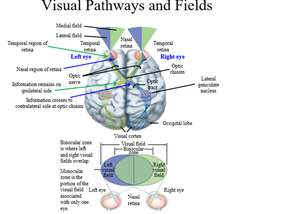
Information pathway of lateral vision
1. Lateral information hits nasal region of retina
2. Information travels via retinal ganglion axons towards the optic chiasm (where optic nerve meets brain base)
3. Information from nasal retina crosses to the contralateral side at the optic chiasm
4. Information synapses on to the lateral geniculate nucleus
5. Neurons take information to the visual cortex
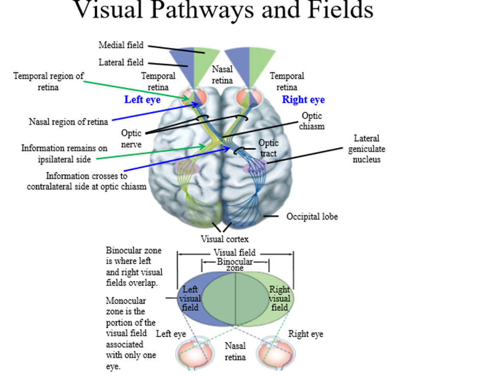
Information pathway of medial vision
1. Medial information hits the temporal region of the retina
2. Information travels via retinal ganglion axons towards optic chiasm
3. Information stays on the ipsilateral side of the optic chiasm (same side)
4. Information travels through the medial geniculate nucleus to the visual cortex
Use of the coexistence of the ON and OFF pathways
Improves image resolution by increasing the brain's ability to perceive contrast at edges or borders
How does hearing work
Based on the physics of sound, physiology of the ear, nerves in the brain, and the brain processing of acoustic information
Pinna
The physical/external ear that resides outside the head
Temporal lobe
Region of brain containing the auditory cortex
Most common medium in which we hear sound energy
Air
How is sound created
Movement of an object displaces air particles
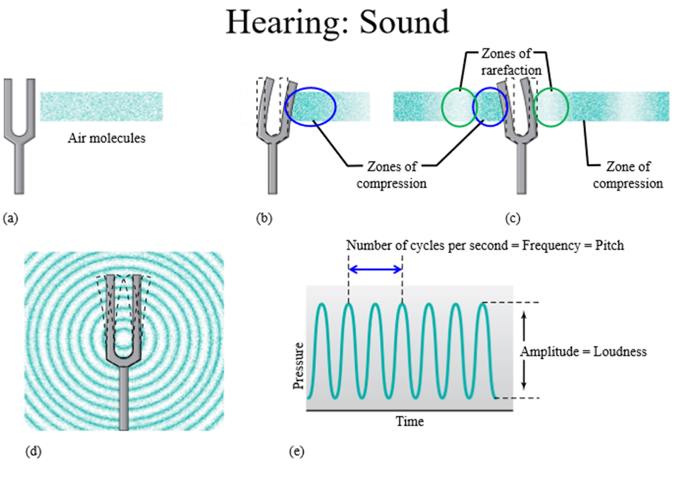
Zones of compression
Regions where air molecules that are tightly packed or close together
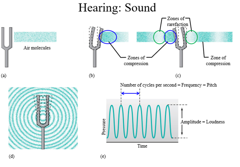
Zone of rarefaction
Regions with low amount of air molecules
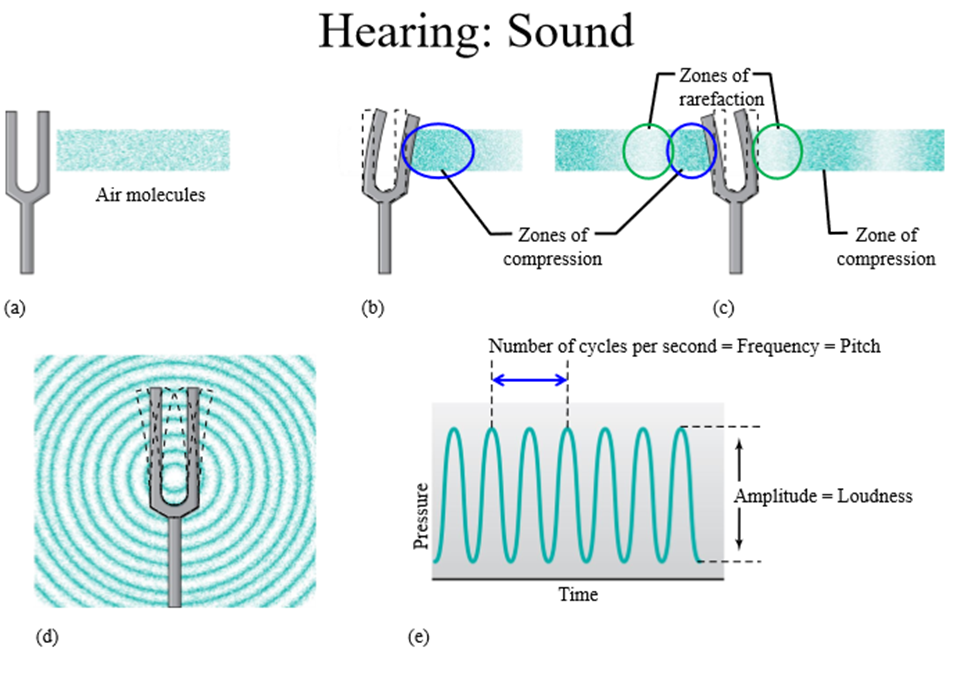
How is amplitude (volume) determined
Number of air molecules located within zone of compression
How is frequency (pitch) determined
The distance between zones of compression/rarefaction in a given time
The faster the vibration...
The higher the pitch
How does our ear hear sounds
Zones of compression and rarefaction hit the ear
Role of the outer ear and external auditory canal
Funnel the zones of compression and rarefaction in towards the middle and inner ear
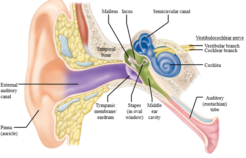
Tympanic membrane
Vibrates in and out at the same amplitude and frequency consistent with the features of the sound you are listening to
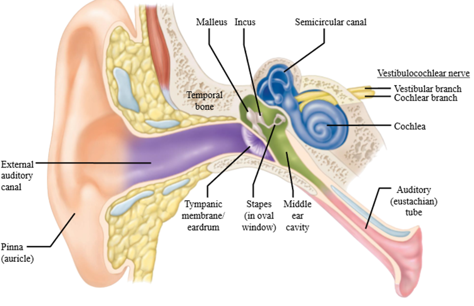
Cochlea
Inner ear
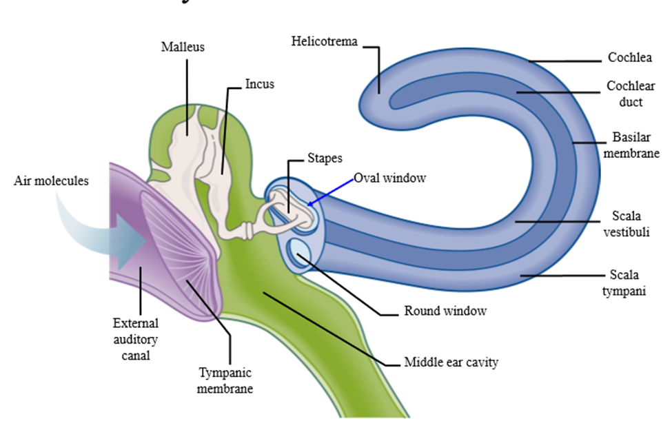
Three bones of the middle ear
Malleus, incus, stapes
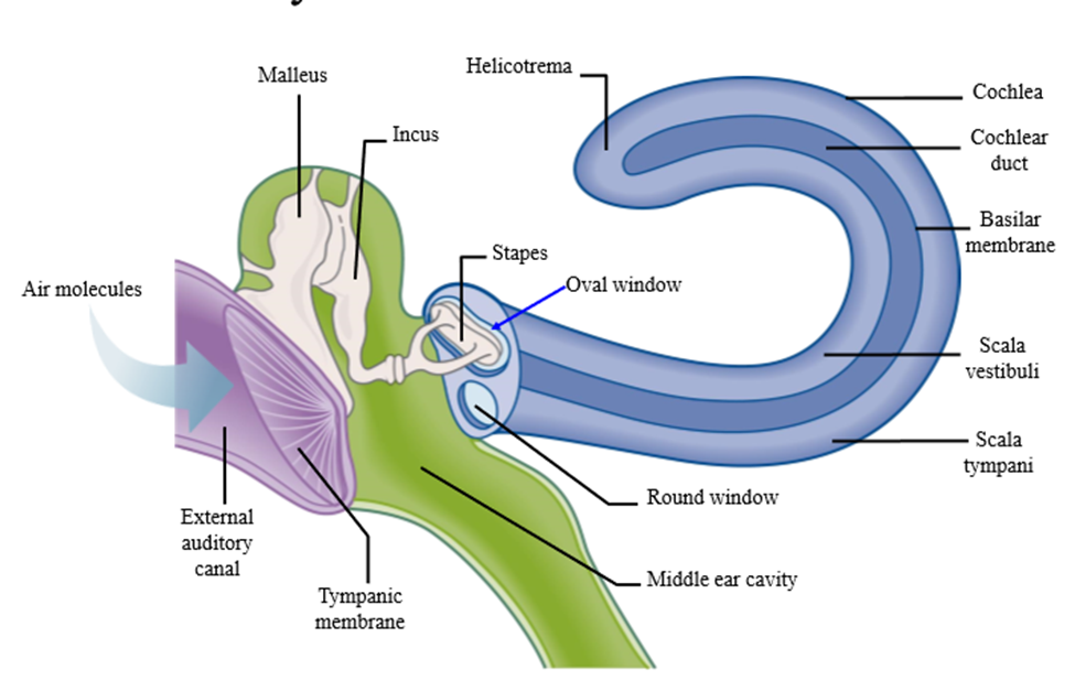
Role of the ear bones
Amplify sound 15-20X
Attachment of the bones
Tympanic membrane --> Malleus --> Incus --> Stapes
Why is amplification via the ear bones necessary
Outer/middle ear are filled with air, the inner ear is filled with fluid
Role of the ear skeletal muscles
Contract to dampen movement of the bones, reducing impact on the inner ear in response to loud, consistent sounds
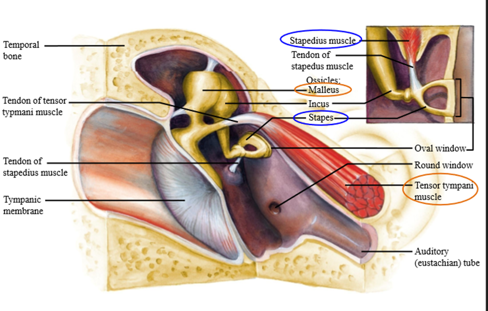
Muscle attached to the malleus
Tensor tympani muscle
Muscle attached to the stapes
Stapedius muscle
Role of the stapes
Terminates on the inner ear onto the oval window. Pushes fluid forward into the middle ear (causing movement)
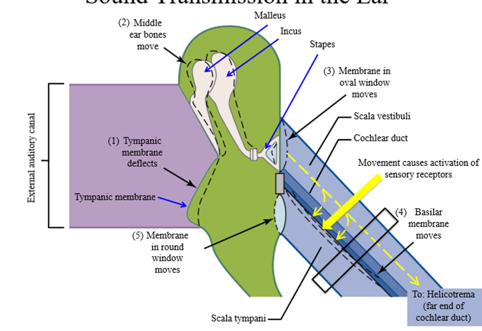
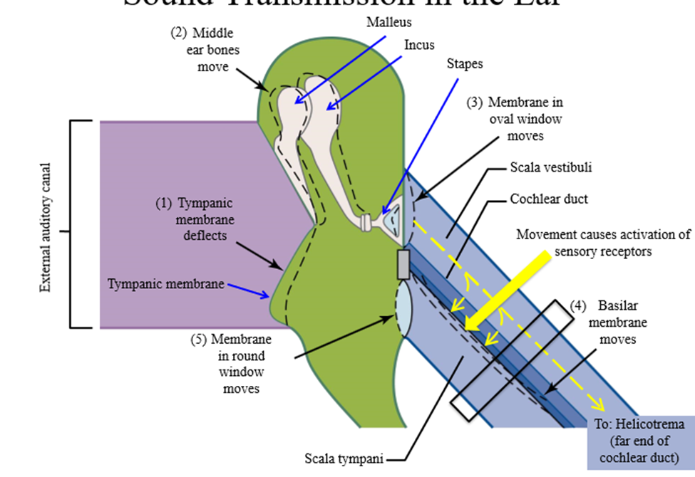
3 components of the cochelea
Scala vestibuli, scala tympani, cochlear duct
Scala vestibuli
Top of the cochlea - contains perilymph (same composition of CSF)
Scala tympani
Middle of the cochlea - contains perilymph
Cochlear duct
Bottom of the cochlea - contains endolymph and contains the sensory receptors of the auditory system
Pathway of sound from the external auditory canal
1. Sound waves come through he external auditory canal
2. Soundwaves move tympanic membrane
3. Tympanic membrane moves bones of the middle ear
4. Stapes pushes against oval window
5. Oval window movement causes movement of perilymph down across the cochlear duct, moving from scala vestibuli towards scala tympany
6. Movement of fluid causes activation of sensory receptors of the cochlear duct
Hair cells
Specialized auditory receptor of the cochlear duct that allow for sound transduction of vibrations into neural signals. Takes place on the organ of corti
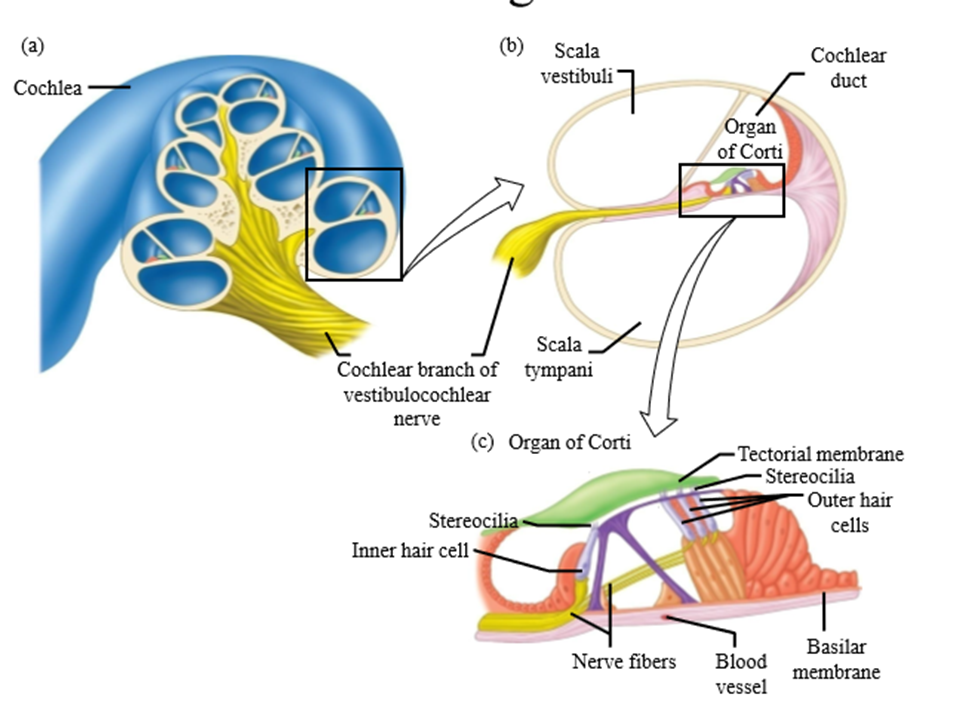
2 groups of hair cells
Single row of inner hair cells and three rows of outer hair cells
Role of the single row of inner hair cells
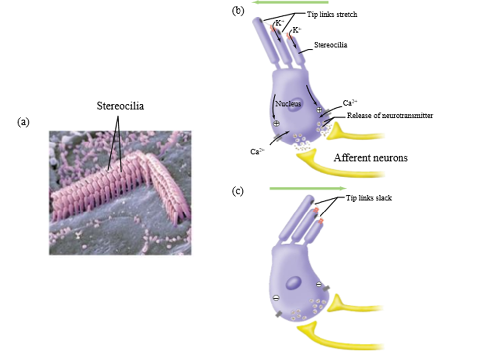
Stereocilia extend into endolymph and transduce pressure waves caused by fluid movement into receptor potentials
Role of the 3 rows of outer cell hairs
Attach to the basilar membrane - different regions of the basilar membrane vibrate at different frequencies
How sound reaches the CNS
Hair cells in the region of the most vibration undergo the most mechanical deformation. This information is sent to the CNS, interpreting the hair cell stimulation as a particular frequency
Does the organ of corti detect different sound frequencies
Yes. Different parts of the basilar membrane correspond to different frequencies
Vestibucoclear nerve
Takes auditory information from the ear towards the brain
Pathway of sound from the eardrum to the hair cells
1. Tympanic membrane vibrates based on the characteristics of the sound
2. activates the malleus, incus and stapes in the middle ear → pushes against the oval window and amplifies the movements
3. pushes the perilymph down across the cochlear duct
4. when fluid is pushed down, the basilar membrane moves up and down based on the amplitude and the frequency (volume and the pitch) of the sounds
5. the hair cells move back and forth when basilar membrane moves up and down
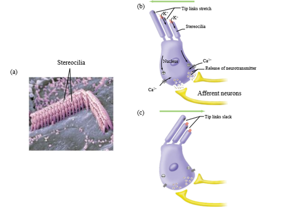
Receptor pathway activation of hair cell movement
1. Stereocilia are bent by vibrations
2. Mechanically-gated cation channels open, allowing potassium to flow into the cell (down the concentration gradient)
3. Hair cell depolarizes, generated graded potentials
4. Glutamate is released from hair cell onto the afferent neurons (the vestibulocochlear nerve)
5. Signal travels from the afferent neurons to the brain
6. Hair cells are bent in opposite direction closes channels, allowing cells to repolarize
Two ways to restore hearing
Hearing aids, cochlear implants
When are hearing aids used
When the auditory of the ear is not as sensitive
Hearing aid function
Amplifier placed in auditory canal to amplify existing sounds
When are cochlear implants used
When someone has permanent damage to the outer, middle, or inner ear (machinery does not work)
Cochlear implant function
Speaker put on the outside of the head converts sound to electrical impulses. Impulses are sent to the vestibulocochlear nerve, bypassing the ear machinery