Histology and Pathology Week 1
 Call Kai
 Learn
Learn Practice Test
 Spaced Repetition
 Match
Match Flashcards
 Knowt Play
1/43

There's no tags or description
Looks like no tags are added yet.
Name | Mastery | Learn | Test | Matching | Spaced | Call with Kai | Chat |
|---|
No analytics yet
Send a link to your students to track their progress
44 Terms
4 main types of tissue
connective, epithelial, muscle, nerve
Epithelial tissue
Medium stain, outer cell/contact with world, 9 main types; name by shape, layering, and special features
Simple epithelium
All cells contact basal laminate
Basal lamina
Layer of extracellular matrix secreted by epithelial cells, makes up basement membrane
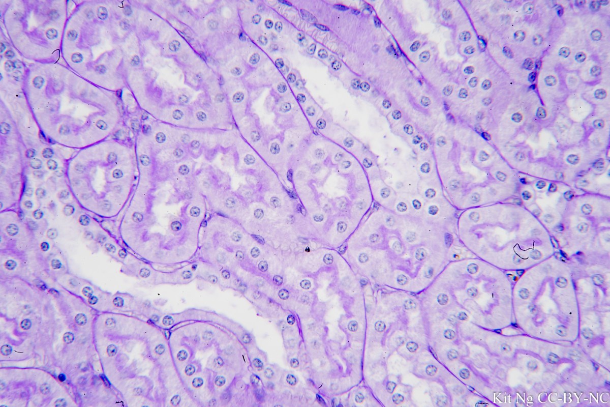
Simple cuboidal epithelium
Square shape, secrete products in glands and ducts, look for them surrounding follicles and glands
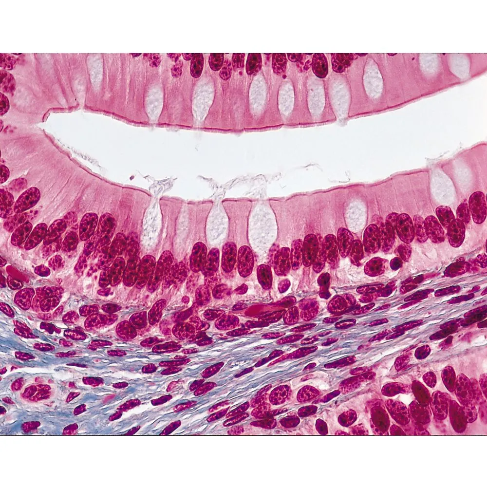
Simple columnar epithelium
Tall and thin but round nucleus, can be near goblet cells; found in digestive tract, airways, and other tubes (uterine tubes, epididymis, ductus deferens)
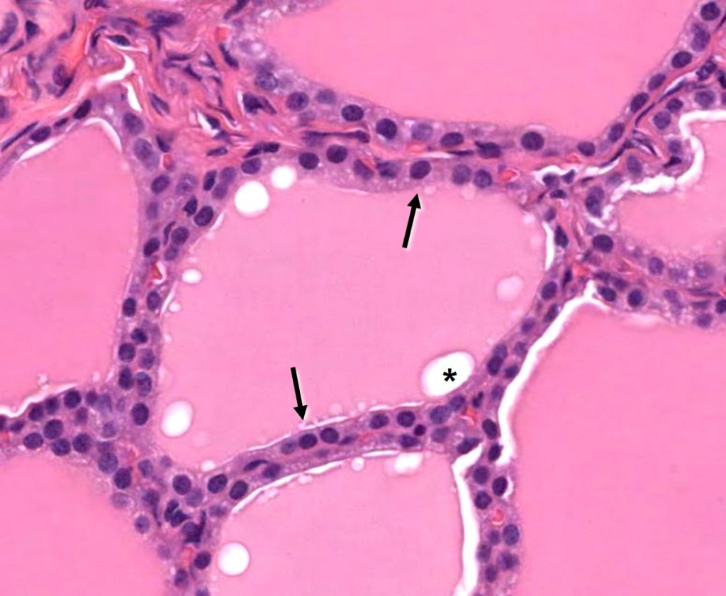
Simple low epithelium
AKA squamous or thin, scale like pattern, squished nucleus
Stratified squamous epithelium
Layers of low epithelium, either has surface terminal cells (vagina, esophagus) or keratin layers (skin)
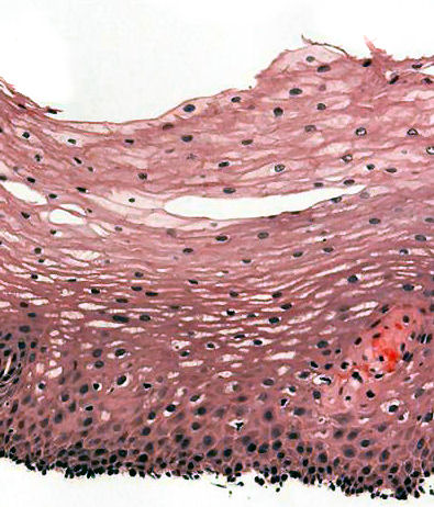
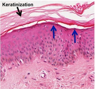
Keritanized epithelium
Found in skin
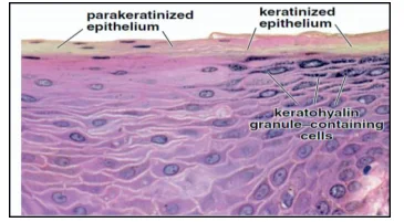
Parakeratinized epithelia
Can be found in masticatory mucosa
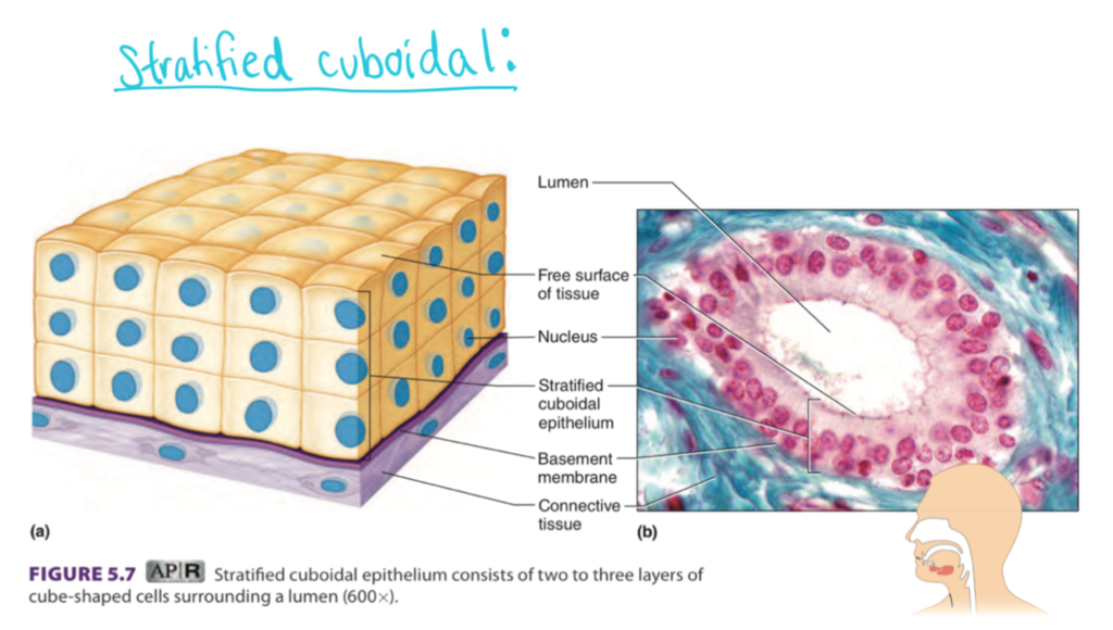
Stratified cuboidal
Layers of cubes
Stratified columnar
Can be found in gland ducts
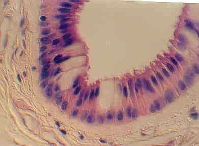
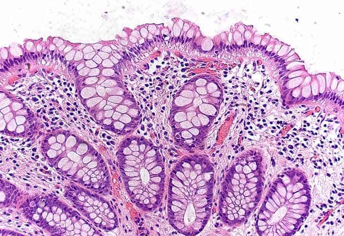
Goblet cell
Produces and stores mucus. stains poorly
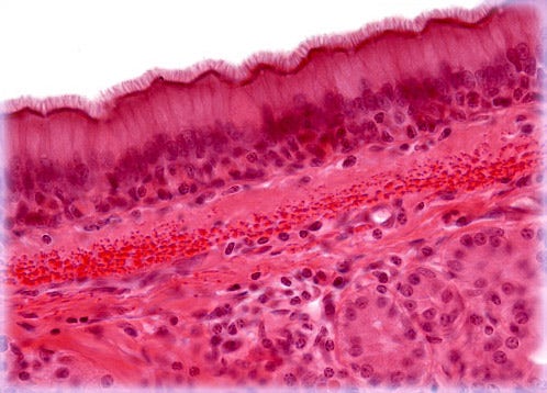
Ciliated columnar epithelium
Airway and uterine tubes, spread mucus and remove debris
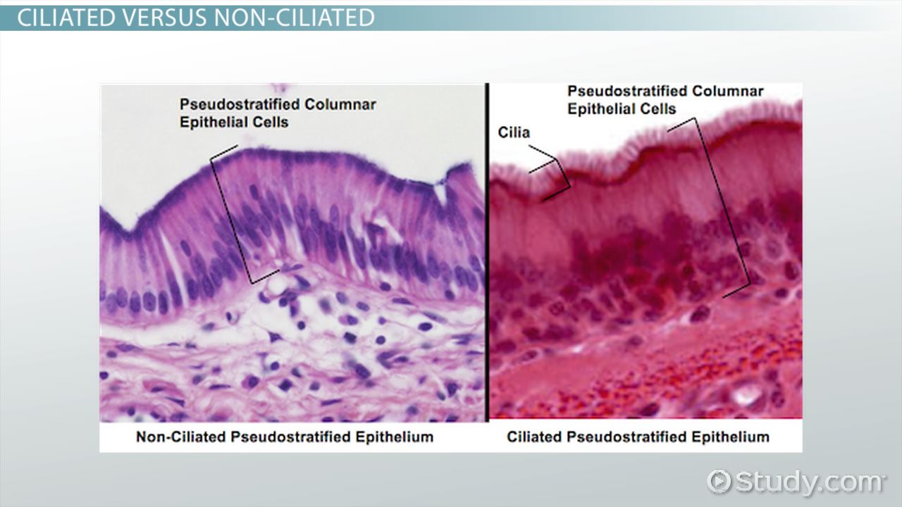
Pseudostratified ciliated columnar epithelium
Respiratory epithelium, appear stratified but are all in contact with basal lamina
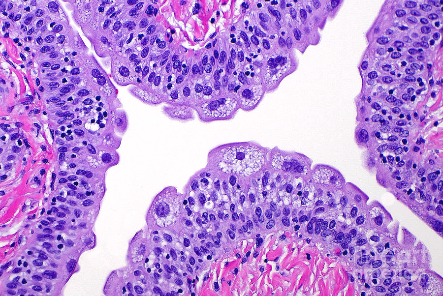
Urothelium
Many shapes, appear stratified (pseudostratified), might see multinucleated cells (umbrella cells) at surface; only in ureter, bladder, and urethra (look for lumen)
Connective tissue
Medium to light stain; includes adipose tissue, bone, cartilage, tendons, and more
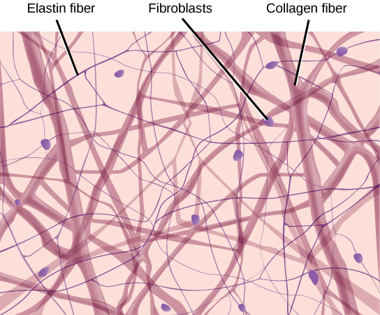
Loose connective tissue
More neurons, cells make up more mass than collagen fibers (if you can see fibers and a decent amount of nuclei, loose is indicated - think zoom level)
Dense irregular connective tissue
Cells widely separated by thick bands of collagen, few nuclei; thick bands of type I collagen, but fibers randomly oriented
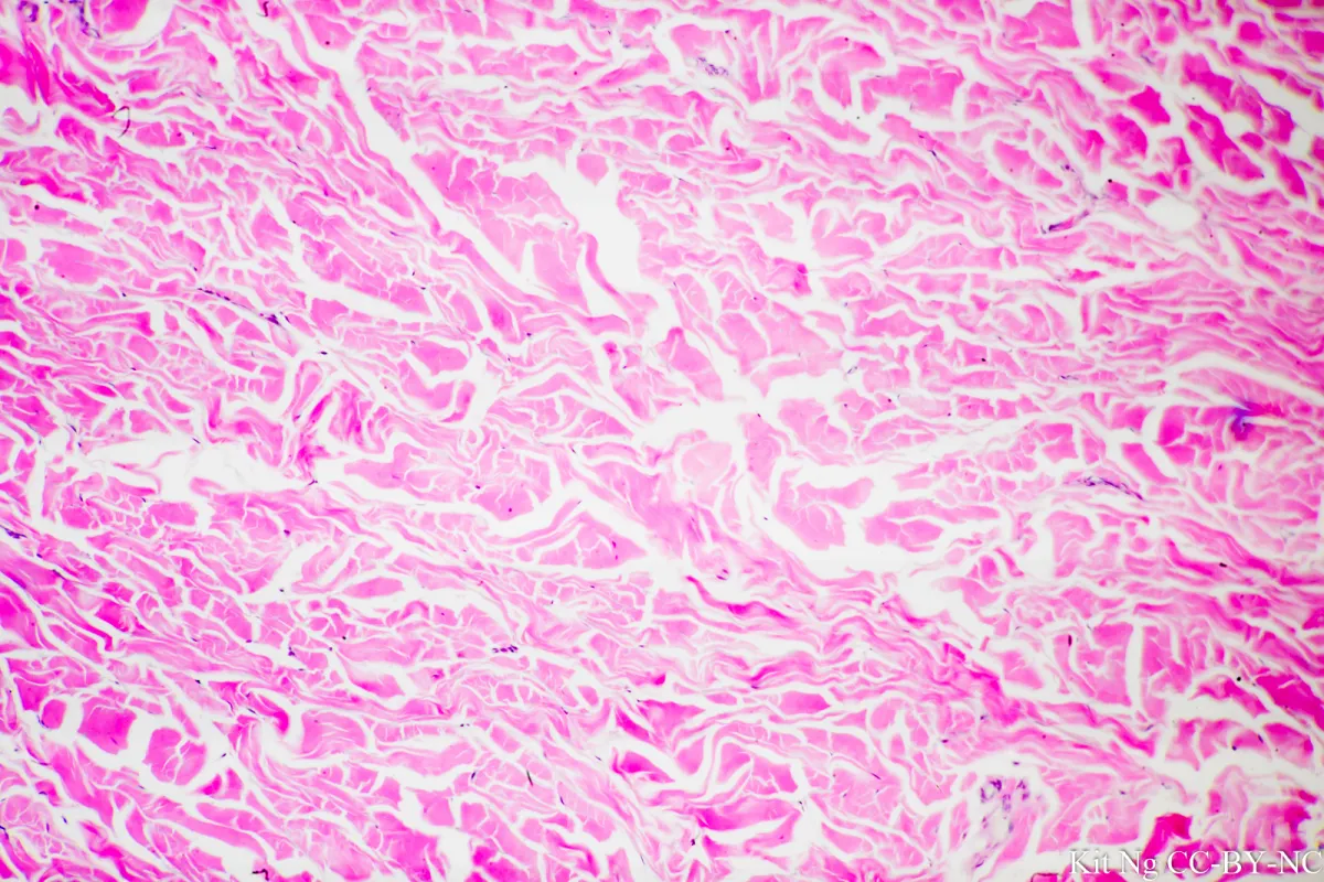
Dense regular connective tissue
Patterned collagen fibers, tendons and ligaments
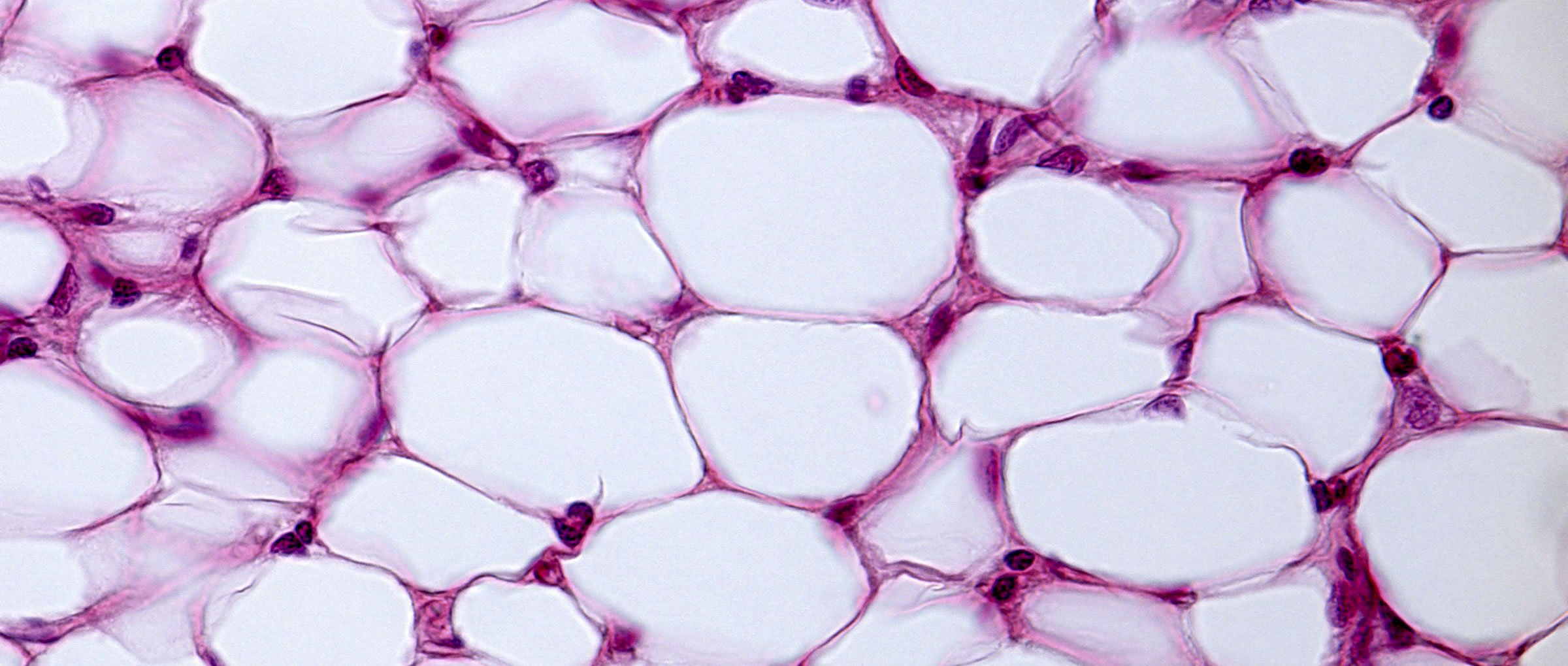
Adipocytes
Connective tissue proper, lipid store is removed in fixation process leaving empty bubble; nucleus often flattened, thin cytoplasm ring around bubble
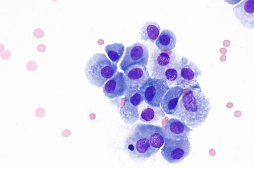
Mast cells
store and release substances for inflammatory response (heparin, histamines, leukotrienes; stain darkly due to granules (small circles), found in dermis, respiratory tract, digestive tract
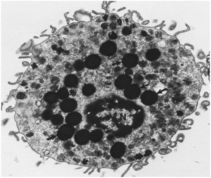
Macrophages
Attack cells that are found in connective tissue; very dark center with lighter surrounding bubbly body (from lysosomes)
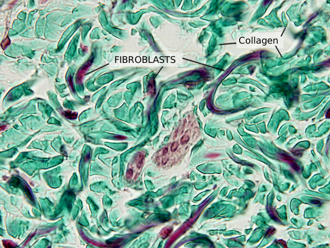
Fibroblasts
Help create supporting fibers (collagen), overactivity can cause fibrosis (scarring); small, dark, football like shape
Muscle tissue
Darker stain, differentiate between cardiac, smooth, and skeletal
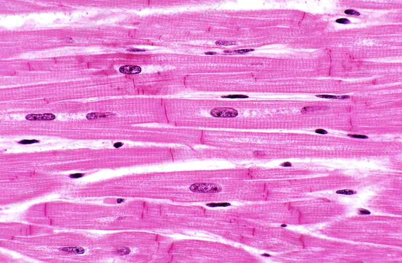
Cardiac muscle
Only found in heart, branched, has intercalcated disc, lightly striated, 1-2 nuclei per cell
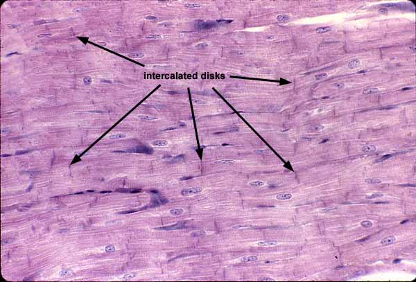
Intercalcated disc
Dark lines where cardiac cells join, have gap junctions for coordinated contractions, fascia adherens and desmosomes
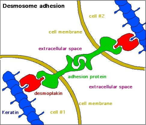
Desmosomes
Like welding points holding cells together
Skeletal muscle
Striated, many peripheral nuclei, geometric shape, large, arranged into fascicles
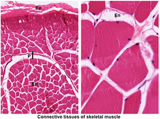
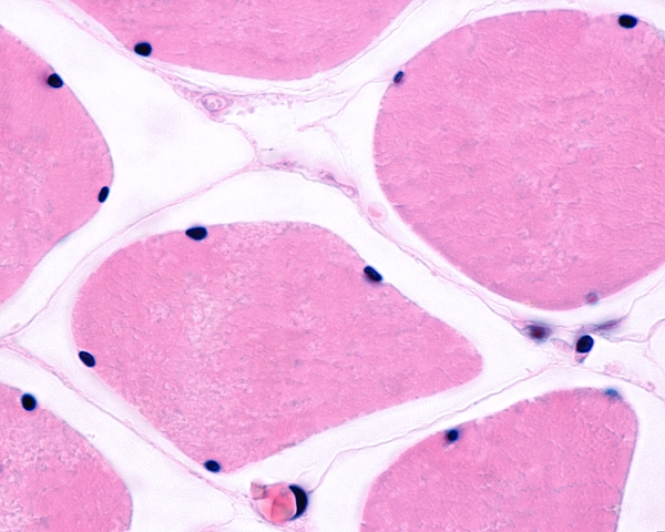
Endomysium
Innermost surrounding layer of muscles, basal lamina and reticular fibers; surrounds single myocyte/myofiber
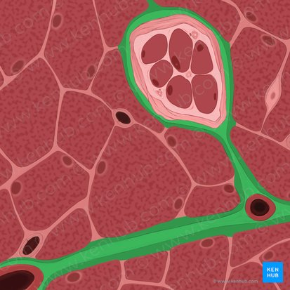
Perimysium
Surrounds fascicle
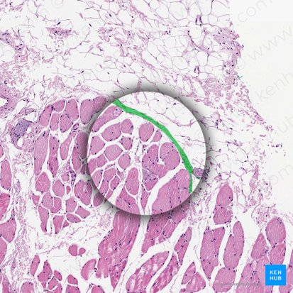
Epimysium
Surrounds whole muscle, fascia, continuous with tendons, dense irregular connective tissue
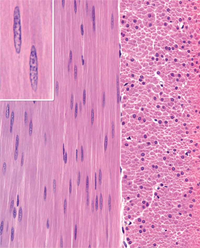
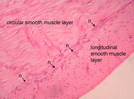
Smooth muscle
Involuntary muscles, found in blood vessels and digestive system, not striated; small, irregular, pointed/spindle shaped, irregular or even corkscrew nucleus
Nervous tissue
Light stain, differentiate CNS vs PNS, white matter vs grey matter
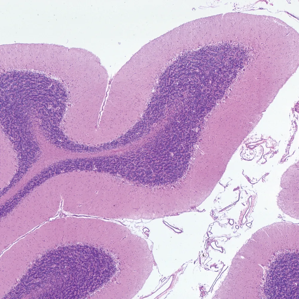
Cerebellum
Distinct dendritic pattern, near 4th ventricle and choroid plexus
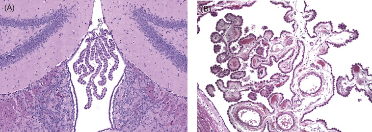
Choroid plexus
Ependymal cells, makes CSF, found in ventricles
Neuron cell bodies
Found in grey matter, usually only cell body stains; dendrites and axons do not stain well unless alternate method is used
Glial cells
Fill gaps between neurons in CNS, nuclei seen in white matter
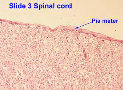
Pia mater
Looks similar to thin keratin or collagen, thin layer around grey matter in brain and white matter in spine
Peripheral nerve
Typically no neuron bodies, nuclei usually Schwann cells; similar look to connective tissue and smooth muscle but lighter stain, look for PNS connective tissue/organization
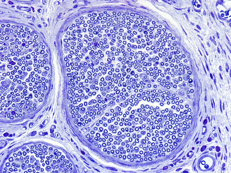
Epineurium
Outer cover, surrounds entire nerve, dense irregular connective tissue
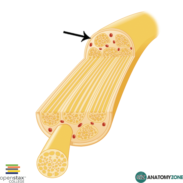
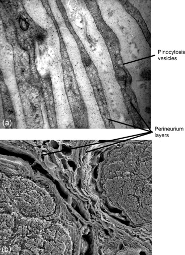
Perineurium
Middle layer, surrounds bundles of axons
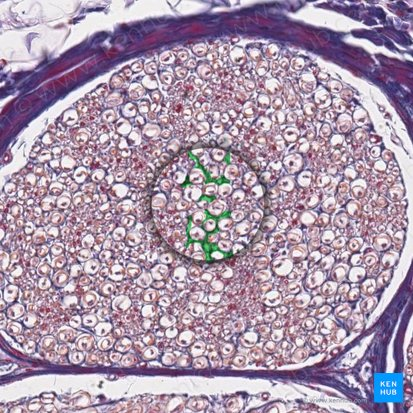
Endoneurium
Inner layer, around individual axons and Schwann cells
Enteric ganglia
Autonomic ganglia found in digestive tract, help regulate gut movement; look for bundles of nerves between layers of smooth muscle and connective tissue