Secondary messenger signalling
1/35
There's no tags or description
Looks like no tags are added yet.
Name | Mastery | Learn | Test | Matching | Spaced |
|---|
No study sessions yet.
36 Terms
What type of agonist is ACh?
-non-selective muscarinic/nicotinic agonist
Does sliding filament theory apply to all types of muscle (smooth, skeletal, cardiac)?
YES
What happens in a non-asthmatic person?
-the muscles around the bronchial tubes are relaxed and the tissue thin allowing for easy airflow
What is diffuse pain?
-pain that's spread across the body, or in many areas. It can change in location and intensity. Hard to pinpoint
What are ligand-gated ion channels? (LGICs) Describe its structure
-Contain both the ligand binding site (ligands bind to them, causing the ion channel to open) and a permeable ion channel within the same protein complex
-also known as ionotropic receptor
-contains 5 subunits. Each subunit spans the membrane 4 times and the subunits are arranged to form a pore
Describe the structure of a GPCR (3). Give an example of a GPCR
-It has an extracellular domain that binds to the ligand
-it has an intracellular domain that interacts with the G protein
-it has a transmembrane domain, which is the single polypeptide chain that spans the membrane 7 times
-a1,a2, b1 and b2 receptors
Give 2 examples of a LGIC receptor
-nicotinic ACh receptor
-GABAA receptor
Give an example of a tyrosine kinase receptor
-insulin receptor
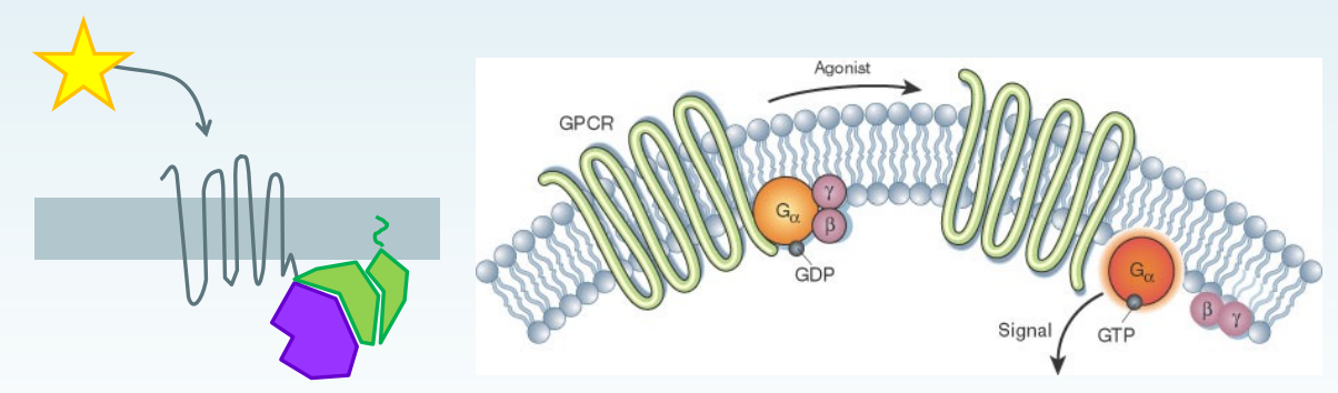
Explain how a 7TM GPCR works generally (5)
-7TM receptor coupled with G protein. G protein has 3 subunits: Ga (alpha), GB (beta) and Gy
7TM= 7-transmembrane receptors. They span the cell membrane 7 times. Most 7TM receptors are GPCRs.
star=ligand e.g. a hormone or neurotransmitter. The ligand binds to the 7TM receptor. This causes a conformational change of the receptor, and a signal is transmitted to the intracellular domain, leading to G protein activation.
once the G protein is activated, GDP is converted into GTP on the Ga subunit. This sends signals inside the cell, which triggers a response.
The G protein then dissociates from the 7TM receptor and the Ga subunit dissociates from the GB and Gy. The Ga and GBy subunits can interact with the effectors. After the G subunits have dissociated:
the Ga subunit activates enzymes like adenylyl cyclase, which converts ATP to cAMP, a secondary messenger.
GBy subunits can regulate ion channels or other effectors.
The Ga subunit hydrolyses GTP (active state) to GDP (inactive state). This inactivates G-protein signalling
Once this hydrolysis occurs, the Ga subunit is no longer active, so it rebinds to the GBy subunit, forming the heterotrimeric G-protein complex again, ready for the next signalling cycle.
-The effectors can go on to activate other proteins. 1 7TM receptor activated can lead to an effector activating many target proteins (this is intracellular signal cascades)
Are ACh receptors LGIC?
yes
Explain this image, but the first panel only
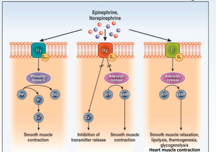
-the 3 broad/main classes of 7TM receptors are:
-α1-Adrenergic Receptor
-α2-Adrenergic Receptor
-β-Adrenergic Receptors
-α1-Adrenergic Receptor (Left):
Activated by: Epinephrine or norepinephrine AKA adrenaline or noradrenaline.
Coupled to Gq protein:
Once activated, it activates the Gq protein, which then activates Phospholipase C (PLC).
PLC cleaves PIP2 into:
IP3 (inositol 1,4,5-trisphosphate) : Releases calcium ions from the sarcoplasmic reticulum. good because action potential not needed to release them now.
DAG (diacylglycerol): activates Protein Kinase C (PKC), which is involved in cell regulation
Effect: Smooth muscle contraction (e.g., in blood vessels) due to IP3 releasing Ca2+ ions, which can then cause vasoconstriction

Explain the second panel
α2-Adrenergic Receptor (Middle Panel):
Activated by: Epinephrine or norepinephrine.
Coupled to Gi protein:
Once activated by the ligand, it activates the Gi protein, which then inhibits adenylyl cyclase. This means less ATP being converted to cAMP, reducing the production of cAMP, increasing smooth muscle contraction.
-remember cAMP causes downstream signalling

Explain the third panel
-β1 and 2-Adrenergic Receptors (Right Panel):
Activated by: Epinephrine or norepinephrine.
Coupled to Gs protein:
ligand activates the receptor, which activates the Gs protein, which then activates adenylyl cyclase, increasing the production of cAMP.
cAMP activates Protein Kinase A (PKA), which leads to several downstream effects.
Effects:
Smooth muscle relaxation (e.g., bronchodilation in B2 receptors).
increased glucose levels (in b2 receptors)
Increased heart muscle contraction (in β1-adrenergic receptors).
-PKA for 3rd panel, PKC for first
-q,i,s
What are the 3 main types of G-proteins that regulate signalling and what are their roles?
Gq: Activates phospholipase C for calcium and PKC signaling.
Gi/o: Inhibits adenylyl cyclase, reducing cAMP levels. i=inhibit
Gs: Stimulates adenylyl cyclase, increasing cAMP levels. S=stimulate
Compare the difference between GPCR responses and ionotropic receptors in terms of rate
-GPCR-Mediated Responses:
Slower response: GPCR signalling involves multiple steps inside the cell (they involve intracellular signaling cascades).
Ion Channel Receptor Responses:
Faster response: Ligand-gated ion channels directly open or close in response to ligand binding, allowing ions (e.g., Na⁺, K⁺, Ca²⁺) to flow across the membrane. No need for intermediary signalling steps.
What is the clinical implication of GPCR vs ion channels?
-Functional Role:
GPCRs regulate complex processes like metabolism, gene transcription, and long-term cellular responses.
Ion channels are critical for rapid events like nerve impulse transmission and muscle contraction.
Clinical Implication:
Drugs targeting GPCRs may have delayed effects, while ion channel modulators have faster physiological responses.
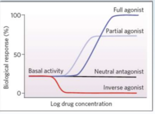
What is basal activity?
-inherent, resting level of activity exhibited by a receptor, enzyme, or biological system in the absence of any external stimulation triggering it (e.g., ligand binding or agonist interaction).
Do GPCR pathways have basal activity?
yes, many of them do
What % of drug targets are GPCR’s?
estimated 30%
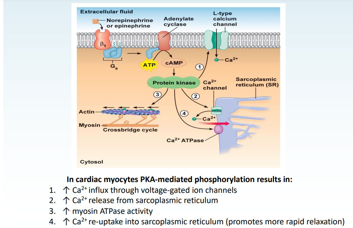
In beta receptors, it was mentioned that the protein kinase A is activated, which has several downstream effects. Explain those effects in cardiomyocytes (i.e. from B1 receptors)
This is an image of the signal transduction pathway involving β₁-adrenergic receptors (a type of G-protein-coupled receptor) in cardiac myocytes, and how it leads to increased cardiac muscle contraction and relaxation.
-PKA is involved in calcium handling and muscle contraction. It does 4 things:
Phosphorylation Targets (Effects of PKA):
L-type Calcium Channels (1): PKA increases Ca²⁺ influx through the channel and into the cell from the extracellular fluid.
Sarcoplasmic Reticulum (2 ):PKA also enhances Ca²⁺ release from the sarcoplasmic reticulum into the cytoplasm, further increasing intracellular Ca²⁺ concentration.
Myosin ATPase (3): PKA increases the activity of myosin ATPase, which is critical for cross-bridge cycling and muscle contraction.
SERCA (4): PKA enhances the activity of the sarcoplasmic reticulum Ca²⁺ ATPase (SERCA). This leads to more (rapid) reuptake of Ca²⁺ into the sarcoplasmic reticulum, promoting faster relaxation of the cardiac muscle.
Overall Outcomes:
Increased Contraction: more intracellular Ca²⁺ boosts actin-myosin cross-bridge cycling, enhancing cardiac muscle contraction.
Faster Relaxation: Increased Ca²⁺ reuptake into the sarcoplasmic reticulum allows the heart to relax more quickly, preparing for the next contraction.
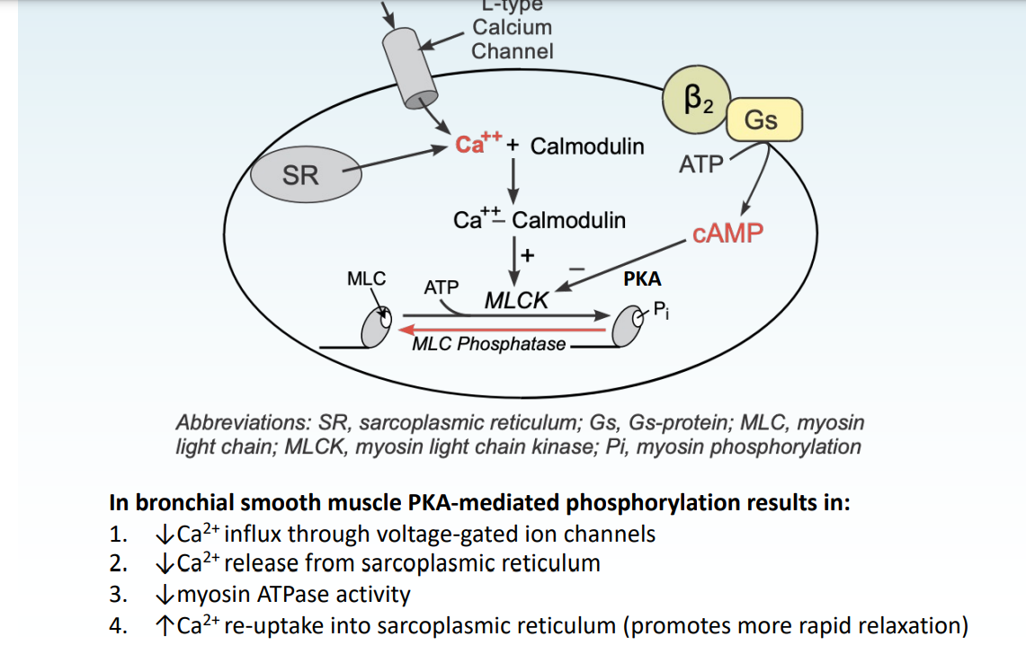
Explain the effects of PKA on bronchial muscle (from activation of B2 receptors)
- β₂-adrenergic receptor activation leads to relaxation of bronchial smooth muscle via PKA-mediated phosphorylation. The PKA has 4 main effects:
Decreased Ca²⁺ Influx:
PKA decreases Ca²⁺ entry into the cell by reducing the activity of L-type calcium channels, leading to less calcium available for contraction.
Decreased Ca²⁺ Release from the SR:
Reduced Ca²⁺ release from the sarcoplasmic reticulum (SR) further decreases the intracellular Ca²⁺ concentration.
Inhibition of Myosin Light Chain Kinase (MLCK):
PKA phosphorylates and therefore inactivates MLCK. Without MLCK activity, smooth muscle contraction is inhibited.
Increased Ca²⁺ Reuptake:
PKA enhances the reuptake of Ca²⁺ into the SR via the sarcoplasmic reticulum Ca²⁺ ATPase (SERCA). This promotes faster muscle relaxation by lowering cytosolic Ca²⁺ levels.
Result:
Relaxation of Bronchial Smooth Muscle (bronchodilation) in airways
Summary of Effects:
↓ Ca²⁺ influx through voltage-gated channels.
↓ Ca²⁺ release from the sarcoplasmic reticulum.
↓ Myosin ATPase activity (due to reduced MLCK activity).
↑ Ca²⁺ reuptake into the sarcoplasmic reticulum (promoting faster relaxation).
β-adrenergic receptors activate Adenylyl Cyclase which makes cAMP. If the cell is smooth muscle…
If the cell is cardiac muscle…
-If the cell is smooth muscle, a protein responds to cAMP by making myosin relax. relaxation
-If the cell is cardiac muscle, proteins inside respond to cAMP by activating myosin, and increase calcium concentration. contraction
α2-adrenergic receptors deactivate Adenylyl Cyclase, which stops making cAMP. If the cell is smooth muscle… If the cell is cardiac muscle…
-If the cell is smooth muscle, a protein that responds to cAMP by making myosin relax
-If the cell is cardiac muscle, proteins inside respond to cAMP by activating myosin, and increase calcium concentration
Explain what receptor-tyrosine kinases are and their structure
-single transmembrane proteins that dimerise when they bind ligands, and activate intracellular kinases, causing a range of signalling pathways
-they have an extracellular binding domain, a single transmembrane domain and an intracellular tyrosine kinase domain
-example of a RTK= ephrin
What can activation of a single type of cascade have and give an example?
-Different effects in different cells i.e. G5 leads to increased force of contraction in cardiac muscle but relaxation of smooth muscles
When inactive and bound to 7TM receptors how many subunits do G-Proteins have and what are they? What subunit binds to GDP?
-3 -> alpha, beta and gamma
-alpha
How are tyrosine kinase receptors different to GPCRs?
-They are slower with activity usually over hours
What are tyrosine kinase receptors activated by?
-a ligand e.g. some growth factors and insulin
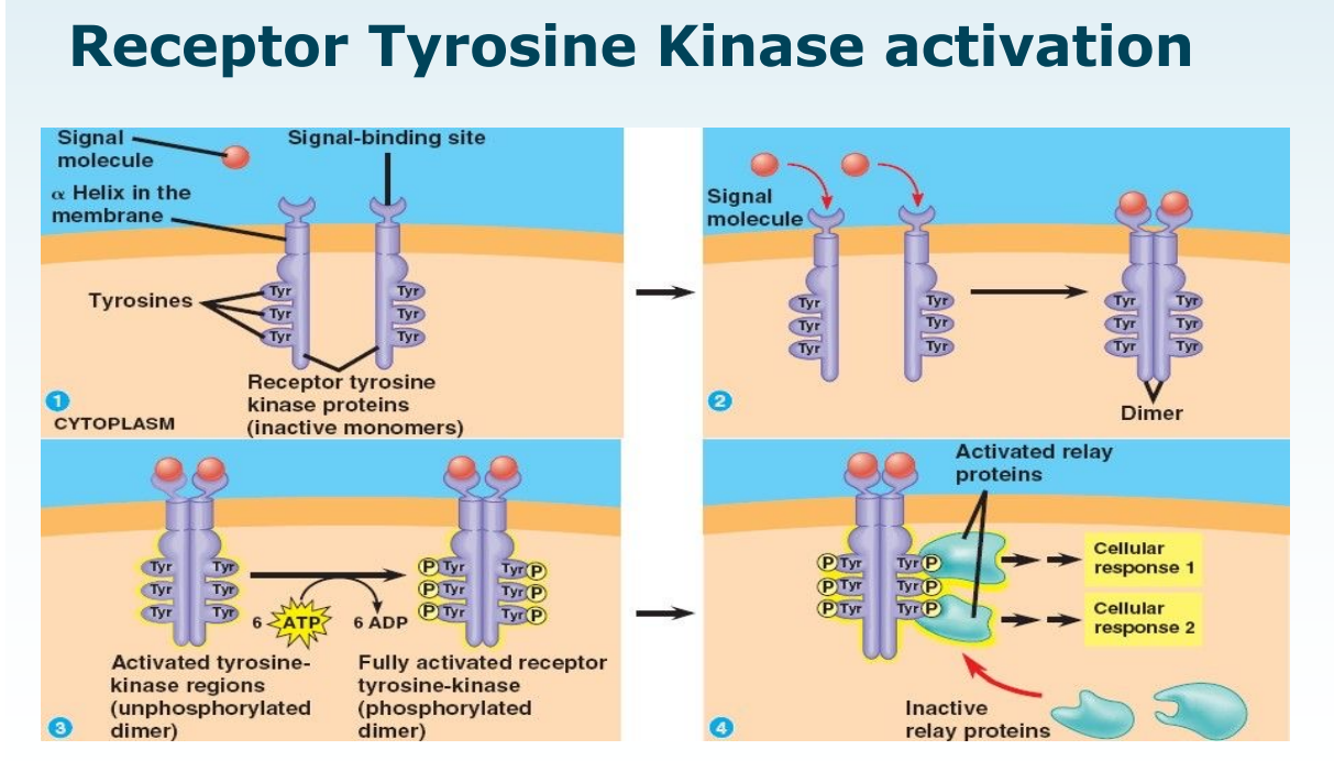
What happens on binding a ligand to tyrosine kinase receptors? (i.e. how do these receptors work?
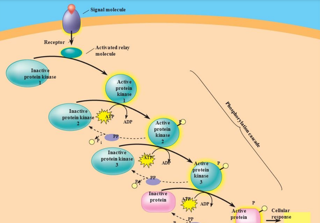
-Ligand binding causes receptor dimerisation and activation of intrinsic tyrosine kinase activity. This leads to autophosphorylation of the tyrosine residues on the receptor, which leads to them phosphorylating additional intracellular proteins including kinases which go on to phosphorylate and therefore activate more proteins so on and so fourth until it activates the effector protein and creates a cellular response
-amplification and cascade mechanism
-all this from 1 signal molecule
Are there many GPCR pathways activated even when there is no ligand present and what does this mean?
-Yes this is basal activity -> so compounds can increase or decrease basal activity
Walk through the graph to show the types of agonists, antagonists and basal activity:
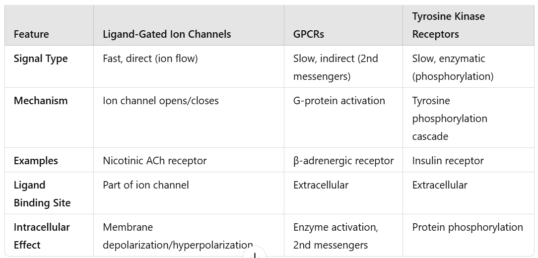
Explain the differences between ligand-gated ion channel receptors, Gprotein coupled receptors (GPCRs) and tyrosine kinase receptors (copare them)
1. Ligand-Gated Ion Channel Receptors 🛠
Structure:
LGIC= Composed of multiple subunits that form a pore in the membrane. Typically have extracellular ligand-binding domains and a transmembrane ion channel.
GPCR=Single polypeptide chain with 7 transmembrane α-helices (7-TM receptors).
TKR= Typically consist of an extracellular ligand-binding domain, a single transmembrane domain, and an intracellular tyrosine kinase domain.
Function:
LGIC=These receptors act as ion channels that open or close in response to the binding of a specific ligand. They allow ions to flow across the membrane, causing changes in membrane potential.
GPCR= activates G protein
TKR= autophosphorylation, amplification and cascade
Signaling:
LGIC= Direct and fast response; binding of the ligand directly triggers the ion channel to open or close.
GPCR= Indirect and slower than ligand-gated ion channels
TKR= Slow compared to ion channels
Give 3 examples of secondary messengers
-cAMP, IP3 and DAG.
Is epinephrine an agonist or antagonist?
-agonist
Can receptors be active i.e. doing something even if a ligand isn’t binded to them?
yes, some receptors can because they have basal activity
Explain how glycogen is broken down after the activation of the beta receptor
-give all steps of beta receptor activation
once PKA is activated, it activates and phosphorylates phosphorylase kinase. Then, phosphorylase kinase phosphorylates glycogen phosphorylase (specifically the "b" form into active "a" form)
Active glycogen phosphorylase a cleaves G1P from glycogen.
G1P is converted to G6P and in the liver, it's converted to free glucose and released into the blood (via glucose-6-phosphatase).