2. tubes and lines pt 2
1/49
Earn XP
Description and Tags
gastric tubes, chest tubes, pacemakers, respiratory tubes
Name | Mastery | Learn | Test | Matching | Spaced |
|---|
No study sessions yet.
50 Terms
when are NG/OG tubes used
when the pt is unable to swallow safely
NG/OG tubes are used when a pt cannot swallow safely. when might this be (5)
declining consciousness, stroke, aphasia, cognitive decline resulting in poor nutrition, oral/esophageal tumor resulting in obstruction
how do we use NG tubes during an overdose
NG tube is inserted to suction gastric contents, or provide a neutralizing agent (activated charcoal)
T or F: prior to initial NG tube use, a CXR is done to confirm placement
true
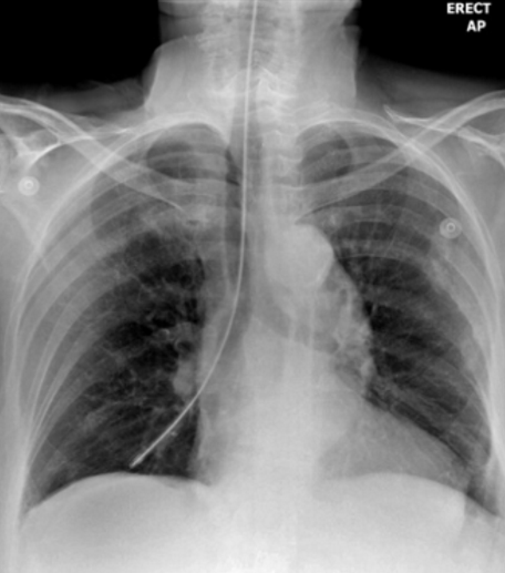
how do we see NG and OG tubes on a radiograph
they have a radiopaque strip
during xrays, if a pt has an NG/OG tube that is being used for a feeding pump, what must we not do + why
don’t place them supine; increases risk of aspiration
describe NG/OG tube insertion and handling
the approx length is externally marked prior to insertion, then tube is lightly secured, then CXR is done to confirm placements. once confirmed, the tube is firmly secured with tape or neck ties
ideal NG tube position
tip should be at least 10cm past the gastro-esophageal junction
on an xray, where will the end of the NG tube be
10cm below the diaphragm, aka over the gastric bubble
complications of NG tubes (6)
tissue trauma, esophageal/mediastinal perforation, pneumothorax, aspiration, hemorrhage, rarely death
list 2 common mispositions for NG tube placement
tip entering the right bronchus, or looping back towards the larynx
describe the optimal appearance of an NG tube on an xray (not just where it ends)
should be // to the spine and slightly left of the SPs, enters the stomach at the diaphragm level, curves with the shape of the stomach and passes the gastric bubble, the weighted tip should point downwards and be slightly angled L/R
describe the incorrect NG tube placement
ends in the right bronchial tree
describe the incorrect NG tube placement
loops back within the esophagus
describe the incorrect NG tube placement
insufficient insertion; needs to be longer
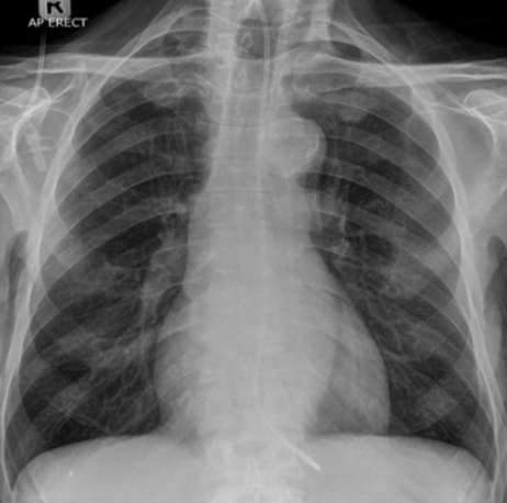
where do chest tubes go
into the pleural space
role of chest tubes
remove fluid or air
what types of pts need chest tubes
those from ICU, CCU, or trauma, or non-acute pt with chronic lung pathologies where fluid builds up in the chest
where are chest tubes inserted
5th intercostal space + slightly anterior to the midaxillary line
what are chest tubes connected to outside of the body
drainage receptables or mechanical suction units
list the two types of chest tubes
large bore, small bore
what are large bore chest tubes used for
pneumothorax (air removal)
what are small bore chest tubes used for
fluid drainage
how are we able to see chest tubes on radiographs
they have a radiopaque strip
where in the body will large bore chest tubes terminate + why
superior/anterior portions of the pleural cavity; air rises
where in the body will small bore chest tubes terminate + why
inferiorly/posteriorly; fluid sinks
before positioning a pt with a chest tube, how are chest tubes secured and how do we move with them
they’re secured to the mattress via kelly clamps, so we remove the clamps before positioning
complications of chest tube insertion (4)
pneumothorax, surgical emphysema, tension pneumothorax, hemorrhage
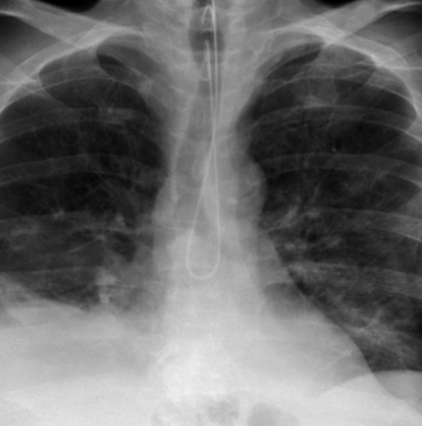
role of pacemakers
maintain adequate heart rate; used when the natural pacemaker isn’t fast enough or if there is a block in the electrical conduction system
how do we program modern pacemakers
externally programmable, the cardiologist can set optimum pace mode for each pt
what are temporary PMs used for + give examples
treat short term heart problems; slow heartbeat from a heart attack, surgery, or overdose
what are permanent PMs used for
long term heart rhythm problems
how does the pacemaker work
provides low levels of electrical stimulation to the heart muscle; it can sense the pts heart rate and provide stimulation when needed
describe internal pacemakers
surgically implanted inside the pt chest
how big is the generator component of an internal pacemaker
size of a matchbox, weighs 20g
describe external pacemakers
bulk remains in a pocket created under the skin, the leads are placed transvenously
rules for 24h post insertion for pacemakers
pt cannot abduct or lift their left arm
complications of pacemaker insertion
lead dislodgement, pneumothorax, lead migration, lead failure, generator failure
how long is the lifespan for the pacemaker’s generator
10 years
purpose of respiratory tubes
assist with airflow to the lungs by bypassing obstructions, preventing aspiration, or controlling breathing
list the 2 types of respiratory tubes
tracheostomy tubes, endotracheal tubes
where are ETTs inserted
mouth or nasal passage into the trachea
role of ETTs
help with respiratory collapse resulting from paralysis, pulmonary edema, or adult respiratory distress syndrome, or they’re used for airway management during procedures that use general anesthesia
describe the optimal location of ETTs
fixed at the mouth, the distal tip is in the trachea at 5-7cm superior to the carina
complications of ETT insertion
tip inserted too far = single lung ventilation or collapse on the contralateral side, esophageal insertion = abdomen ventilation, pneumomediastinum and surgical emphysema = air introduced to the surrounding area (ST/heart)
what is a trachesotomy
surgical opening in the anterior neck and into the trachea
ETT ventilation via tracheostomy is not recommended past which time frame
14-21 days
how are tracheostomy tubes secured
fixed to the skin by a fabric strip tied around the pts neck
optimal tip location for tracheostomy tubes
at the midpoint between the upper end of the tube and the carina (6cm above the carina)
complications of tracheostomy tube insertions
bleeding, pneumothorax, permanent damage to vocal cord nerve, accidental removal, infection of insertion site, malpositioning